章節導言
- 本章作者:Pratistadevi K. Ramdial、Boris C. Bastian、Jeffrey P. North、John Goodlad、John A. McGrath、Alexander J. Lazar。參考文獻與額外資料見 www.expertconsult.com。
- 涵蓋主題:檢體固定/取材送檢/處理/包埋/切片、常規與特殊染色、免疫組織化學 (immunohistochemistry) 與疑難排解、免疫螢光 (immunofluorescence)、電子顯微鏡 (electron microscopy)、冷凍切片、診斷性細胞病理技術、遺傳性皮膚疾病診斷,以及分子技術(染色體核型分析、等位基因不平衡、FISH、CGH、PCR、NGS)與淋巴瘤診斷(TCR 與 IG 基因重排分析)。
檢體固定 (Fixation)
- 目的:維持病灶特徵清晰一致,並保存組織於適合各種染色與輔助技術的最佳狀態。
- 方法:多數採液態試劑的化學固定 (chemical fixation);亦可用物理法(加熱、微波、冷凍乾燥、freeze substitution)或化學法(coagulant 與 cross-linking)。
- 最常用固定劑:10% 中性緩衝福馬林 (10% neutral-buffered formalin),pH 7.2–7.4,可避免組織切片中形成 formalin pigment。
- 固定速率約 每小時 1 mm;固定劑體積理想上至少為檢體體積的 10 倍。大型檢體(如腫瘤)可切成 5-mm 厚切片,覆以浸固定劑的紗布並隔夜固定。
- 影響固定品質的因素:檢體大小、固定時間與溫度、pH、濃度、滲透壓 (osmolality)、固定劑離子組成與添加物。
組織處理、包埋與切片 (Processing, Embedding, Sectioning)
- 組織處理 (tissue processing):去除可萃取水分以獲得最佳診斷品質切片,步驟為固定 → 脫水 (dehydration) → 透明化 (clearing) → 滲透 (infiltration) → 包埋 (embedding)。
- 處理設備:carousel 旋轉式、self-contained 真空滲透處理機(圖 2-2)、微波處理。多數實驗室採隔夜流程;微波輔助可縮短至 1–2 小時。
- paraffin(石蠟) 為最常用滲透/包埋介質,適用多數常規與特殊染色。
- 包埋原則:皮膚樣本方向應對刀片提供最小阻力(圖 2-3);通常以與表皮 (epidermis) 成直角的平面切取,以減少壓縮與變形。
取材送檢與切片類型 (Grossing / Put-through)
- 切片類型:小型切取式(shave、core、punch)與切除性檢體 (excisional)。
- 需評估邊緣的切除性檢體在送檢前應 inked;若有定位縫線,採四象限四色塗繪或兩色塗繪半側(圖 2-1)。
- Shave biopsy:可二/三等分後 on edge 包埋;對小型 melanoma 的削切切除,邊緣包埋至關重要,以同時量化侵犯的寬度與深度。
- Core/punch biopsy:直徑一般 2–8 mm,供較大病灶的診斷性取樣。>4 mm 應二等分、切面朝下包埋(避免遺漏病灶);<4 mm 整個 (in toto) 送檢。
技術性人工假象 (Technical Artifacts)
- 處理不佳 → 切片不完整、於水浴中膨脹或崩解;包埋不正確 → 方向不良的不完整切片。
- microtome 機構故障、刀片鬆動/鈍化/損壞、clearance angle 不準 → 厚薄不均、folds(圖 2-4)、holes(圖 2-5)、scores(圖 2-6)、chatter。
- 組織內鈣化區、縫線及刀片缺口 (nicks) → chatter 或在與刀緣成直角方向裂開。
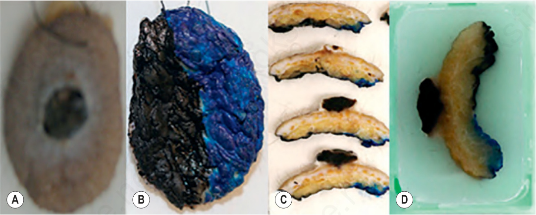
圖 2-1:化膿性肉芽腫 (pyogenic granuloma) 的大體呈現 (A),並對其下表面進行兩色塗繪 (B)。2-mm 厚大體切片顯示送檢時 (C) 與石蠟塊中 (D) 的黑、藍塗繪。
Fig. 2.1 Gross representation of pyogenic granuloma (A), with two-color painting of the inferior surface (B). 2-mm–thick gross sections demonstrating the black and blue painting at put-through (C) and in paraffin blocks (D).
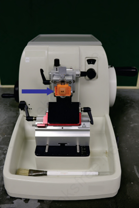
圖 2-3:微切片機 (microtome) 上含有皮膚組織(箭頭)的石蠟塊 (paraffin block)。
Fig. 2.3 Paraffin block containing skin tissue (arrow) on microtome.