臨床特徵 (Clinical Features)
- 神經鞘黏液瘤 (nerve sheath myxoma / neurothekeoma) 最常發生於四肢,主要見於手部/手指、膝部/脛骨前區 (pretibial region) 及踝部/足部,好發於生命的第四個十年,並對男性有偏好。
- 罕見病例曾報告於嬰兒。
- 較小比例的病例出現在頭頸部,包括口腔。
- 顱內 (intracranial)、眼眶 (orbital)、脊椎旁 (paravertebral) 及縱膈腔 (mediastinum) 為罕見的發生部位。
- 甲下 (subungual) 病例極為罕見。
- 曾描述過一例與多發性血管黏液瘤 (multiple angiomyxomas) 相關的個案。
- 本病與神經纖維瘤病 (neurofibromatosis) 無關聯,典型上腫瘤表現為單一、長期存在、無症狀、隆起、膚色的結節,病程長短不一,直徑小於 3 cm。
- 局部復發(有時為多發性)可見於高達百分之四十七的病人。此腫瘤無明顯的惡性潛能。
組織病理特徵 (Histologic Features)
- 其外觀具特徵性;為一界線分明 (well-defined)、多結節 (multinodular) 或多小葉 (multilobular) 的無被膜 (unencapsulated) 腫塊,主要位於真皮 (dermis) 與皮下 (subcutis)(Figs 35.362–35.364)。
1801 Benign neural tumors
- 雖然後者具有不同的組織學特徵與免疫組化表現,並不支持神經鞘 (nerve sheath) 起源,但它們仍被稱為細胞型「neurothekeoma (cellular neurothekeoma)」(見第 1769 頁)。
鑑別診斷 (Differential Diagnosis)
- 與淺表性血管黏液瘤 (superficial angiomyxoma) 的區別,根據後者缺乏界限分明性 (circumscription)、血管不明顯 (inconspicuous blood vessels)、散在發炎細胞的存在,以及常見的上皮成分 (epithelial elements)。
- 黏液樣神經纖維瘤 (myxoid neurofibroma) 界限不清,且缺乏小葉狀結構 (lobular architecture)。
- 真皮黏液瘤 (dermal myxomas) 細胞稀少 (hypocellular),血管少見,且基質黏液 (stromal mucin) 豐富。
- 當界限分明的柵欄狀神經瘤 (circumscribed palisaded neuroma) 出現顯著黏液樣變化 (myxoid change) 時,與之區別可能非常困難。
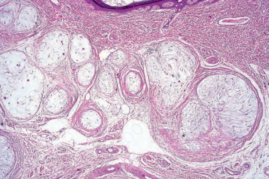
圖 35-362:Neurothekeoma:腫瘤由被纖維間隔 (fibrous septa) 分隔的離散小葉 (discrete lobules) 所組成。
Fig. 35.362 Neurothekeoma: the tumor is composed of discrete lobules separated by fibrous septa.
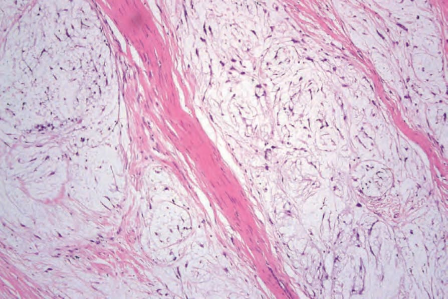
圖 35-363:Neurothekeoma:小葉由分散於黏液樣基質 (myxoid stroma) 中的纖細梭形細胞 (delicate spindled cells) 所構成。
Fig. 35.363 Neurothekeoma: the lobules are composed of delicate spindled cells dispersed in a myxoid stroma.
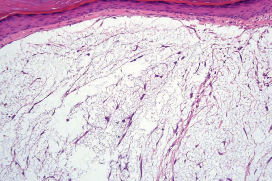
圖 35-364:Neurothekeoma:高倍視野顯示梭形 (fusiform) 與星狀 (stellate) 的腫瘤細胞。
Fig. 35.364 Neurothekeoma: high-power view showing fusiform and stellate tumor cells.
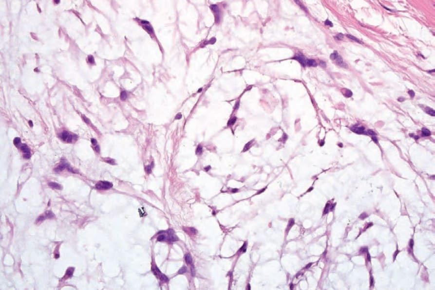
圖 35-365:Neurothekeoma:注意黏液樣基質 (myxoid stroma)。
Fig. 35.365 Neurothekeoma: note the myxoid stroma.
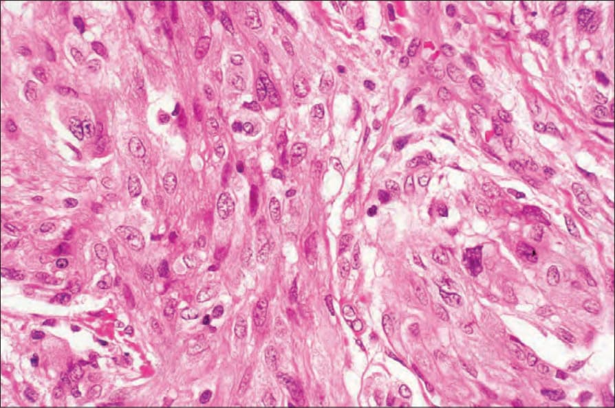
圖 35-366:Neurothekeoma:在此切片中,細胞呈上皮樣 (epithelioid),具豐富的細胞質與明顯的泡狀核 (vesicular nuclei)。
Fig. 35.366 Neurothekeoma: in this section, the cells are epithelioid with abundant cytoplasm and conspicuous vesicular nuclei.
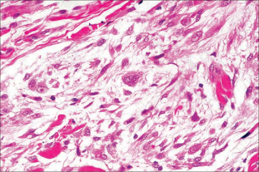
圖 35-367:Neurothekeoma:注意多核巨細胞 (multinucleate giant cell)。
Fig. 35.367 Neurothekeoma: note the multinucleate giant cell.
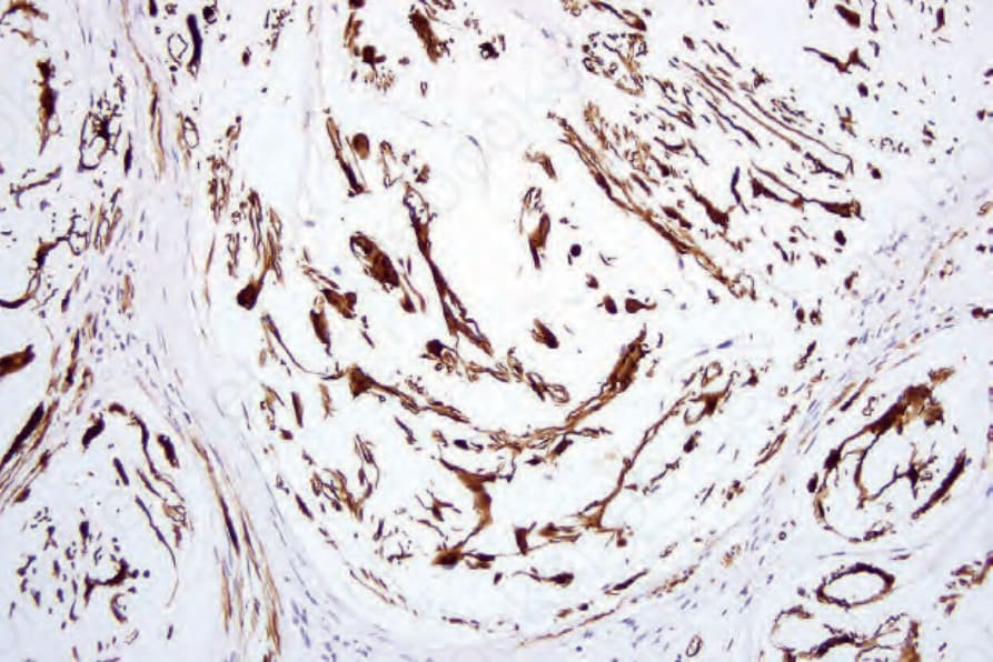
圖 35-368:Neurothekeoma:腫瘤細胞呈 S100 protein 陽性。
Fig. 35.368 Neurothekeoma: the tumor cells are S100 protein positive.
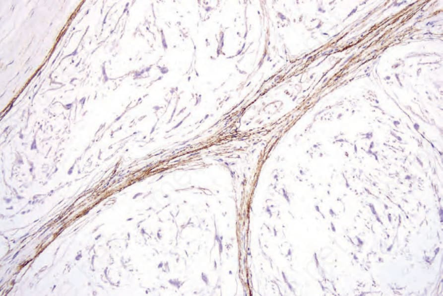
圖 35-369:Neurothekeoma:神經外膜 (perineurium) 以 EMA 免疫組化 (immunohistochemistry) 突顯。
Fig. 35.369 Neurothekeoma: the perineurium is highlighted with EMA immunohistochemistry.