Nerve sheath myxoma (neurothekeoma)
Nerve sheath myxoma (neurothekeoma)
Clinical features Nerve sheath myxoma (neurothekeoma) arises most often on the extremities, mainly the hand/fingers, knee/pretibial region and ankle/foot, in the fourth decade of life and shows a predilection for males.1–8 Rare cases have been reported in infants.9 Much smaller percentage of cases presents in the head and neck including the oral cavity.10–17 Intracranial, orbital, paravertebral, and mediastinum are rare sites of occurence.18–22 Subungual cases are exceptional.23,24 A single case associated with multiple angiomyxomas has been described.25 There is no association with neurofibromatosis and typically the tumor presents as a solitary, long-standing, asymptomatic, raised, skin-colored nodule of variable duration measuring less than 3 cm in diameter.1–7 Local recurrences, sometimes multiple, may be seen in up to 47% of patients. This tumor has no evident malignant potential.7
Histologic features The appearances are distinctive; it is a well-defined, multinodular or multilobular unencapsulated mass situated predominantly in the dermis and subcutis (Figs 35.362–35.364).
1801 Benign neural tumors
Although the latter have different histologic features and immunohistochemistry that do not support a nerve sheath origin, they are nevertheless still known as cellular ‘neurothekeoma’ (see page 1769).
Differential diagnosis Distinction from superficial angiomyxoma is based on the lack of circumscription, inconspicuous blood vessels, presence of scattered inflammatory cells and the common occurrence of epithelial elements in the latter tumor. Myxoid neurofibroma is poorly circumscribed and lacks a lobular architecture. Dermal myxomas are hypocellular and show few blood vessels and abundant stromal mucin. Distinction from circumscribed palisaded neuroma may be very difficult when the latter has prominent myxoid change.30
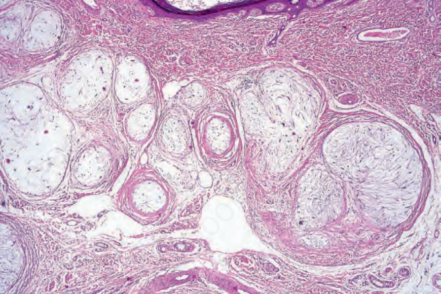
Fig. 35.362 Neurothekeoma: the tumor is composed of discrete lobules separated by fibrous septa.
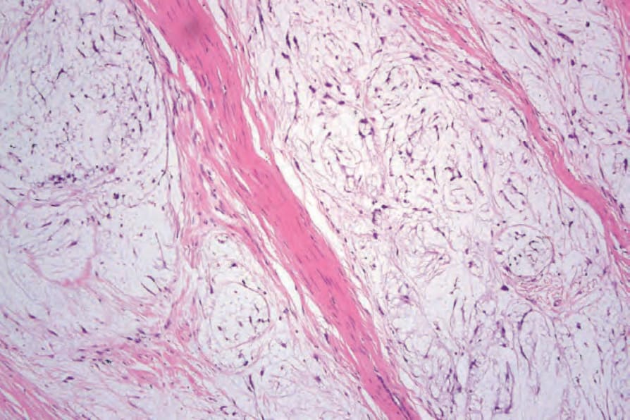
Fig. 35.363 Neurothekeoma: the lobules are composed of delicate spindled cells dispersed in a myxoid stroma.
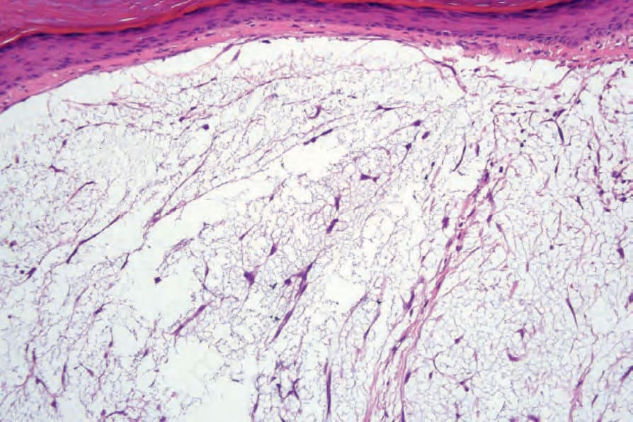
Fig. 35.364 Neurothekeoma: high-power view showing fusiform and stellate tumor cells.
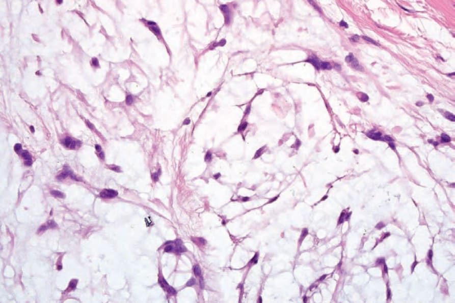
Fig. 35.365 Neurothekeoma: note the myxoid stroma.
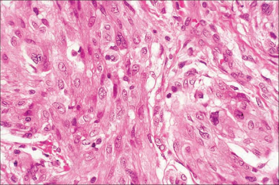
Fig. 35.366 Neurothekeoma: in this section, the cells are epithelioid with abundant cytoplasm and conspicuous vesicular nuclei.
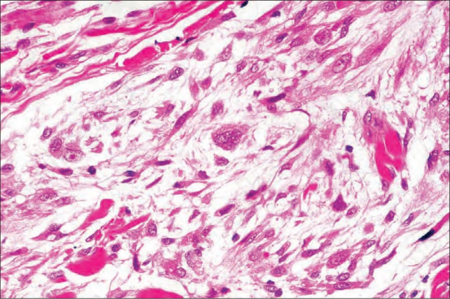
Fig. 35.367 Neurothekeoma: note the multinucleate giant cell.
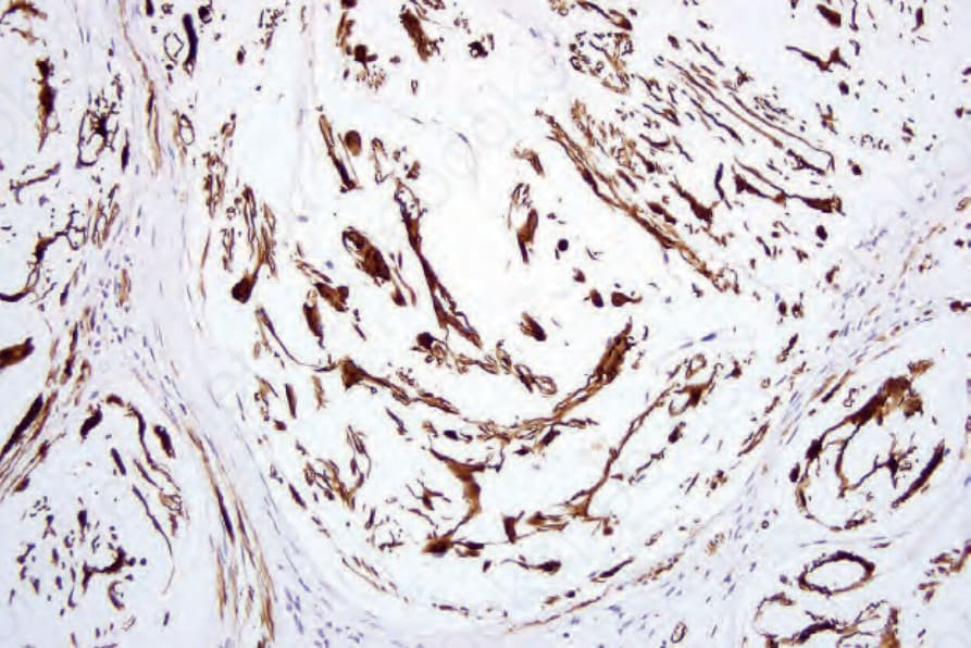
Fig. 35.368 Neurothekeoma: the tumor cells are S100 protein positive.
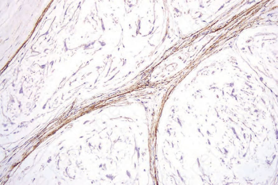
Fig. 35.369 Neurothekeoma: the perineurium is highlighted with EMA immunohistochemistry.