血管瘤樣纖維組織細胞瘤 (Angiomatoid Fibrous Histiocytoma)
血管瘤樣纖維組織細胞瘤 (angiomatoid fibrous histiocytoma)
臨床特徵 (Clinical Features)
- 血管瘤樣纖維組織細胞瘤 (angiomatoid fibrous histiocytoma)(過去稱為 angiomatoid malignant fibrous histiocytoma)是一種罕見的腫瘤。
- 它通常發生於皮下組織,僅在極少數情況下發生於兒童或任一性別之年輕成人之四肢或軀幹的真皮。1–7
- 出現於頭頸部者屬例外情形。8
- 不尋常的發生部位包括肺、腦、縱膈、外陰、後腹膜及卵巢。9–14
- 發生於較深部軟組織者較為少見。
- 曾有一例先天性個案被記載。15
- 亦曾報導發生於慢性放射性皮膚炎 (chronic radiodermatitis) 背景下的個案、一名 HIV 陽性兒童所發生的腫瘤,以及一名罹患神經母細胞瘤 (neuroblastoma) 兒童的個案。16–18
- 大多數腫瘤生長緩慢,直徑小於 2 cm。
- 病人有時會出現全身性症狀,包括發燒、體重〔下〕…
免疫組化與特殊染色 (Immunohistochemistry & Special Stains)
- 在免疫組化方面,腫瘤細胞約 50% 的個案對 desmin 及 muscle actin (HHF-35) 呈陽性,但對 smooth muscle actin 呈陰性(見 Fig. 35.203)。4
- 對 EMA、CD68 及 CD99 的陽性反應,通常見於高達 50% 的個案。5,6
- 腫瘤細胞對 S100 protein、keratins 及血管標記 (vascular markers) 呈陰性。
- 結合形態學與免疫表型,這些病灶有可能呈現 myoid(可能為 myofibroblastic)分化,雖然 EMA 與 desmin 並存的情形並不尋常。4
- 超微結構研究顯示各式各樣的細胞,包括 fibroblastic、myofibroblastic、histiocyte-like 或未分化 (undifferentiated) 的型態。3,15,25
鑑別診斷 (Differential Diagnosis)
- 與 aneurysmal fibrous histiocytoma 的區分,於後者該條目下討論。
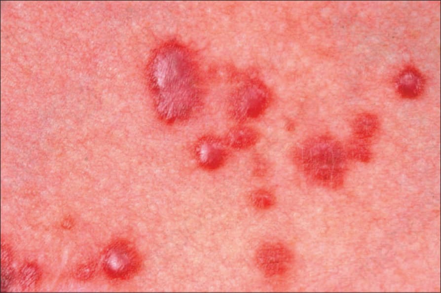
圖 35.203:多核細胞血管組織細胞瘤 (multinucleate cell angiohistiocytoma):在此例中,丘疹呈現出血性外觀 (hemorrhagic)。承蒙 Institute of Dermatology, London, UK 提供。
Fig. 35.203 Multinucleate cell angiohistiocytoma: in this example, the papules appear hemorrhagic. By courtesy of the Institute of Dermatology, London, UK.
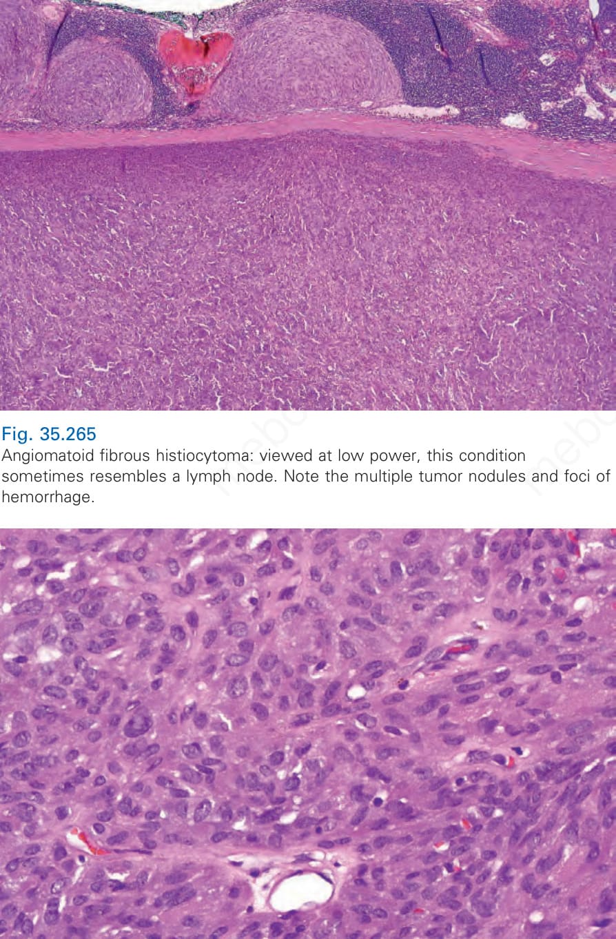
圖 35.265:血管瘤樣纖維組織細胞瘤 (angiomatoid fibrous histiocytoma):在低倍視野下,此病灶有時類似一個淋巴結 (lymph node)。注意多發性的腫瘤結節及出血灶 (foci of hemorrhage)。
Fig. 35.265 Angiomatoid fibrous histiocytoma: viewed at low power, this condition sometimes resembles a lymph node. Note the multiple tumor nodules and foci of hemorrhage.