Angiomatoid fibrous histiocytoma
Angiomatoid fibrous histiocytoma
Clinical features Angiomatoid fibrous histiocytoma (previously known as angiomatoid malignant fibrous histiocytoma) is a rare tumor. It usually arises in the subcutaneous tissues and only exceptionally in the dermis of the extremities or trunk in children or young adults of either sex.1–7 Presentation in the head and neck is exceptional.8 Unusual sites include the lung, brain, mediastinum, vulva, retroperitoneum and ovary.9–14 Lesions arising in deeper soft tissues are less common. A congenital example has been documented.15 A case arising in the background of chronic radiodermatitis, a tumor developing in an HIV-positive child and one in a child with neuroblastoma have also been reported.16–18 Most tumors are slow growing and less than 2 cm in diameter. Patients sometimes present with systemic symptoms including fever, weight
Immunohistochemically, tumor cells are positive in about 50% of cases for desmin and for muscle actin (HHF-35), but not for smooth muscle actin (see Fig. 35.203).4 Positivity for EMA, CD68 and CD99 is usually seen in
1774 Connective tissue tumors
up to 50% of cases.5,6 Tumor cells are negative for S100 protein, keratins and vascular markers. It is possible, combining morphology with immunophenotype, that these lesions show myoid (probably myofibroblastic) differentiation, although the combination of EMA and desmin is unusual.4
Ultrastructural studies show a variety of cells including fibroblastic, myofibroblastic, histiocyte-like or undifferentiated forms.3,15,25
Differential diagnosis Distinction from aneurysmal fibrous histiocytoma is discussed under the latter entity.
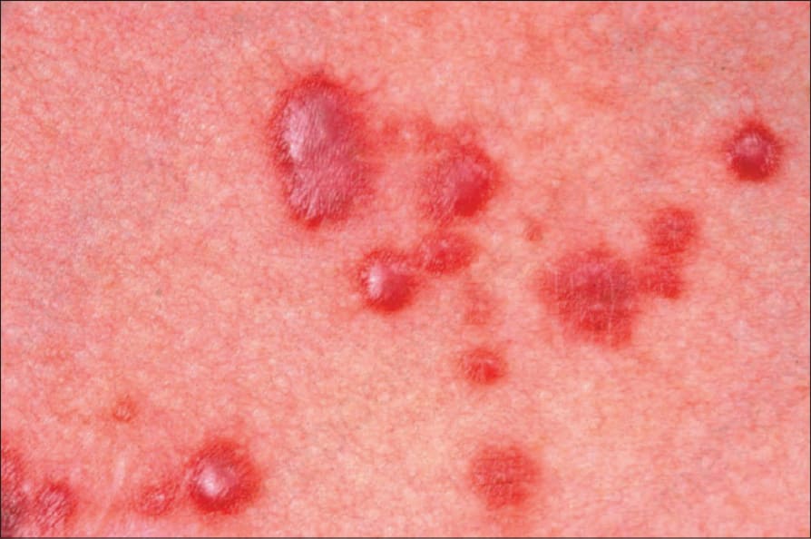
Fig. 35.203 Multinucleate cell angiohistiocytoma: in this example, the papules appear hemorrhagic. By courtesy of the Institute of Dermatology, London, UK.
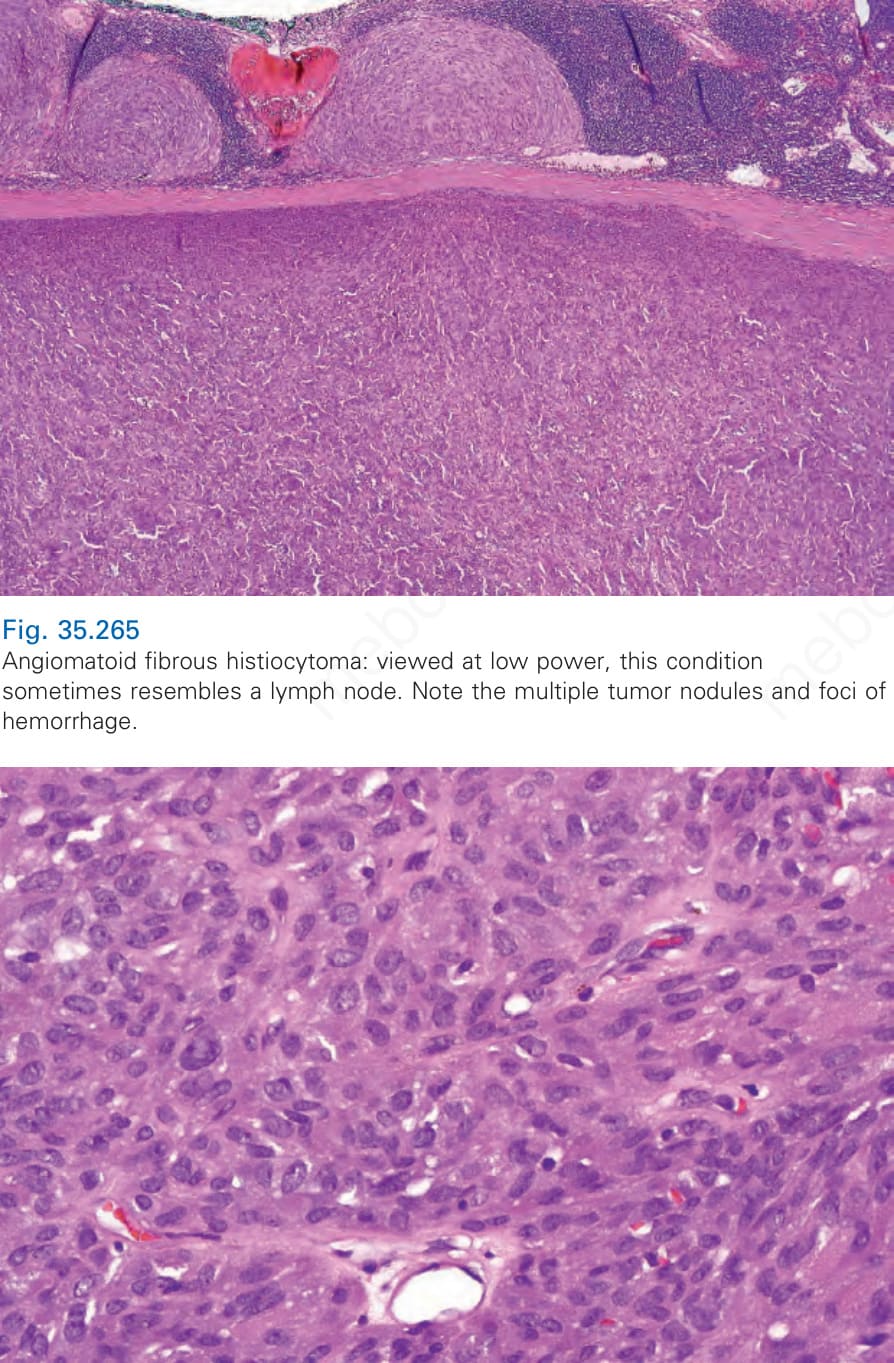
Fig. 35.265 Angiomatoid fibrous histiocytoma: viewed at low power, this condition sometimes resembles a lymph node. Note the multiple tumor nodules and foci of hemorrhage.