臨床特徵 (Clinical Features)
- 多核細胞血管組織球瘤 (multinucleate cell angiohistiocytoma) 是一種特殊的疾病,特徵為多發、局限性的血管瘤樣丘疹 (angiomatous papules),好發於中年女性的上肢與下肢 (Figs 35.201–35.203)。
- 大腿與手背是常見的侵犯部位,其次為臉部。
- 全身性 (generalized) 病灶非常罕見。
- 曾有一例於懷孕期間發生的報告。
- 病灶無症狀,且不傾向自發性消退。
- 曾有一例報告發生於口腔、另一例發生於陰道,還有一處病灶與醫源性 (iatrogenic) 動靜脈瘻 (arteriovenous fistula) 相關發生。
- 皮膚鏡 (dermoscopic) 特徵可能令人聯想到皮膚纖維瘤 (dermatofibroma)。
鑑別診斷 (Differential Diagnosis)
- 萎縮型皮膚纖維瘤 (atrophic dermatofibroma) 看起來可能與 multinucleate angiohistiocytoma 極為相似;然而前者呈現為單一病灶。
- 與卡波西氏肉瘤 (Kaposi sarcoma) 的區別在於:存在不規則、鋸齒狀、薄壁的血管腔道,缺乏多核巨細胞 (multinucleate giant cells),以及存在漿細胞 (plasma cells)。
- Multinucleate angiohistiocytoma 在免疫組織化學 (immunohistochemistry) 上缺乏 HHV8。
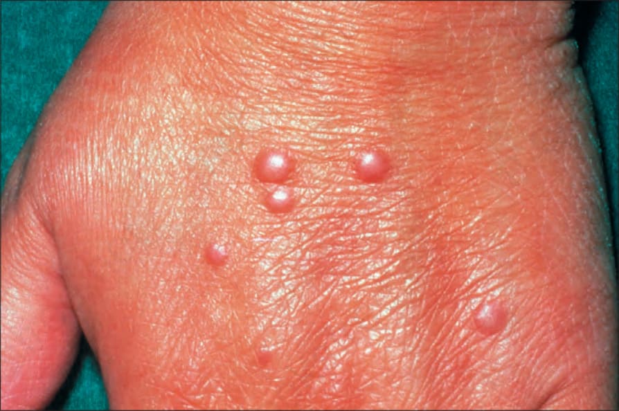
圖 35.201:多核細胞血管組織球瘤 (multinucleate cell angiohistiocytoma):可見多發性丘疹 (multiple papules)。
Fig. 35.201 Multinucleate cell angiohistiocytoma: multiple papules are present.
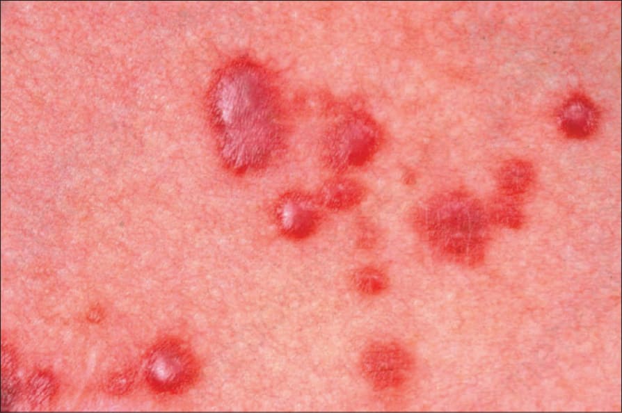
圖 35.203:多核細胞血管組織球瘤 (multinucleate cell angiohistiocytoma):在此例中,丘疹呈現出血性 (hemorrhagic)。承蒙英國倫敦皮膚科學研究所 (Institute of Dermatology, London, UK) 提供。
Fig. 35.203 Multinucleate cell angiohistiocytoma: in this example, the papules appear hemorrhagic. By courtesy of the Institute of Dermatology, London, UK.
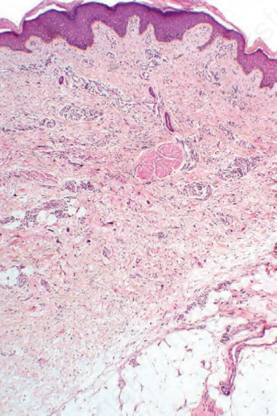
圖 35.204:多核細胞血管組織球瘤 (multinucleate cell angiohistiocytoma):真皮 (dermis) 內為一血管性與膠原性 (vascular and collagenous) 的增生性病灶,伴隨明顯的多核巨細胞 (multinucleate giant cells)。
Fig. 35.204 Multinucleate cell angiohistiocytoma: within the dermis is a vascular and collagenous proliferative lesion with conspicuous multinucleate giant cells.
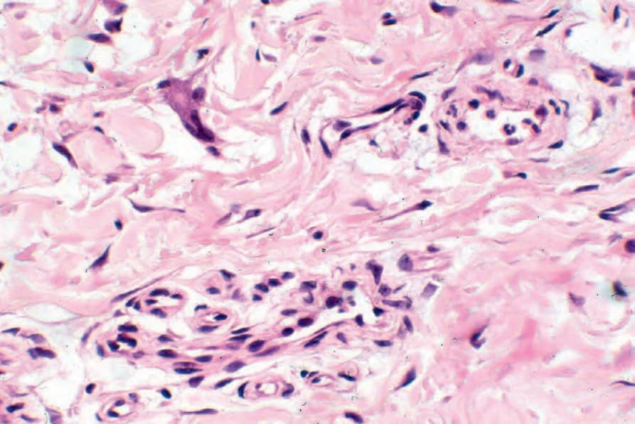
圖 35.206:多核細胞血管組織球瘤 (multinucleate cell angiohistiocytoma):巨細胞 (giant cell) 的高倍視野。
Fig. 35.206 Multinucleate cell angiohistiocytoma: high-power view of giant cell.