Multinucleate cell angiohistiocytoma
Multinucleate cell angiohistiocytoma
Clinical features Multinucleate cell angiohistiocytoma is a distinctive condition characterized by multiple, localized, angiomatous papules with predilection for the upper and lower limbs of middle-aged women (Figs 35.201–35.203).1–6 The thigh and dorsum of the hands are frequent sites of involvement followed by face.7 Generalized lesions are very rare.8–11 A case developing during pregnancy has been reported.12
The lesions are asymptomatic and do not tend to regress spontaneously.13 A case has been reported in the oral cavity another in vagina and a further lesion occurred in association with an iatrogenic arteriovenous fistula.14–16 Dermoscopic features may be reminiscent of dermatofibroma.17,18
1759 Benign fibrohistiocytic tumors and tumorlike lesions
Differential diagnosis An atrophic dermatofibroma can look remarkably similar to multinucleate angiohistiocytoma; however, the former presents as a single lesion. Distinction from Kaposi sarcoma is based on the presence of irregular, jagged, thin-walled vascular channels, absence of multinucleate giant cells and presence of plasma cells. Multinucleate angiohistiocytoma lacks HHV8 on immunohistochemistry.22
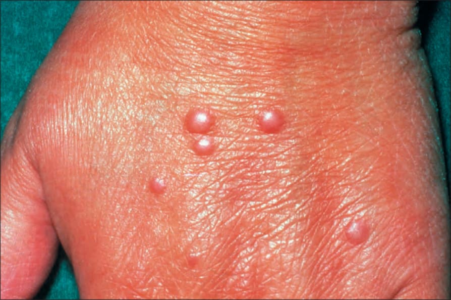
Fig. 35.201 Multinucleate cell angiohistiocytoma: multiple papules are present.
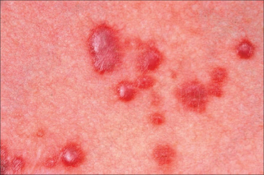
Fig. 35.203 Multinucleate cell angiohistiocytoma: in this example, the papules appear hemorrhagic. By courtesy of the Institute of Dermatology, London, UK.
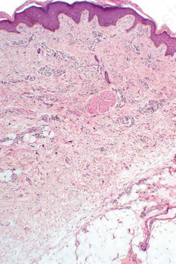
Fig. 35.204 Multinucleate cell angiohistiocytoma: within the dermis is a vascular and collagenous proliferative lesion with conspicuous multinucleate giant cells.
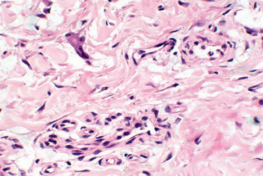
Fig. 35.206 Multinucleate cell angiohistiocytoma: high-power view of giant cell.