纖維性丘疹 (Fibrous Papule)
臨床特徵 (Clinical Features)
- 纖維性丘疹 (fibrous papule) 是一種非常常見的病灶,好發於中年成人的臉部,特別好發於鼻部。
致病機轉與組織學特徵 (Pathogenesis and Histologic Features)
- 過去曾有人提出 fibrous papule 代表一種陳舊性纖維化的痣 (old fibrosed nevus),但多項研究已證實事實並非如此。肥大細胞 (mast cells) 可能在 fibrous papule 的發生中扮演角色。近期一項研究顯示其有哺乳動物雷帕黴素標的路徑 (Mammalian Target of Rapamycin Pathway) 的活化,與結節性硬化症複合症相關的血管纖維瘤 (tuberous sclerosis complex-associated angiofibroma) 類似。
- 組織學上,病灶略為隆起、邊界清楚,位於淺層真皮。其由膠原性間質 (collagenous stroma) 組成,伴有增多的血管腔道 (vascular channels),以及散在的細胞,這些細胞型態由梭形 (spindle shaped) 到多核 (multinucleated) 不等 (Figs 35.199 and 35.200)。曾有報告描述一例具有多核神經節樣細胞 (multinucleated ganglion-like cells) 的病例。有絲分裂象 (mitotic figures) 極為罕見,部分細胞可能表現深染 (hyperchromatism)。亦曾有文獻描述具有顆粒細胞變化 (granular cell change)、透明細胞變化 (clear cell change) 及上皮樣細胞 (epithelioid cells) 的變異型。可見局部色素沉著 (focal pigmentation) 與發炎 (inflammation)。具散在多形性細胞 (pleomorphic cells) 的病灶與多形性纖維瘤 (pleomorphic fibroma) 有所重疊。其上覆的表皮 (overlying epidermis) 表現正常或略為扁平。
- 在免疫組織化學 (immunohistochemistry) 方面,病灶內的細胞對 factor XIIIa 呈陽性,亦可對 CD34 呈陽性。S100 protein 為陰性。具透明細胞 (clear cells) 的病灶對 NKI/C3 呈陽性。
- 在超微結構 (ultrastructurally) 上,這些細胞具有纖維母細胞 (fibroblasts) 的特徵。
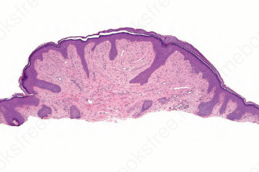
圖 35-199:纖維性丘疹 (fibrous papule):取自鼻樑病灶的削切式切片 (shave biopsy)。真皮顯示緻密的膠原性組織 (dense collagenous tissue)。
Fig. 35.199 Fibrous papule: shave biopsy from a lesion on the bridge of the nose. The dermis shows dense collagenous tissue.
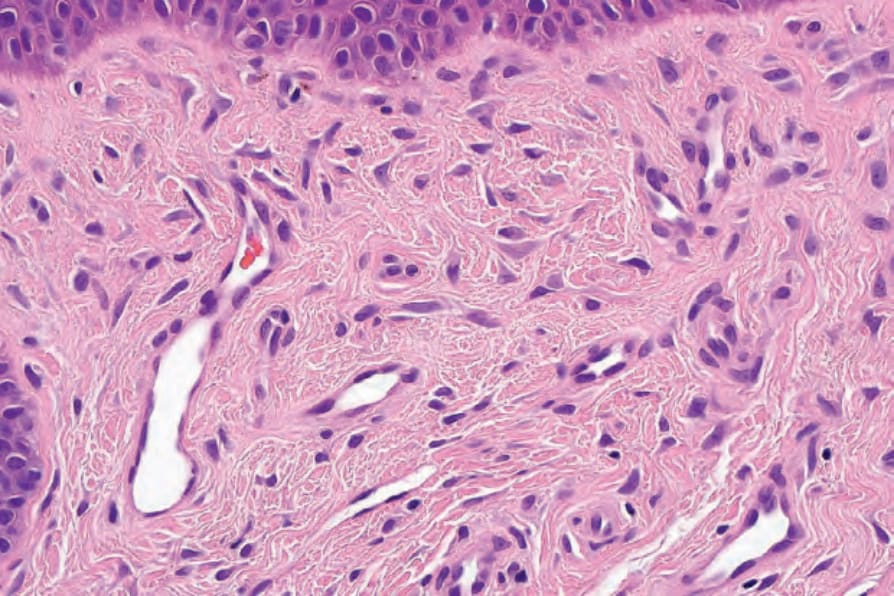
圖 35-200:纖維性丘疹 (fibrous papule):散在樹突狀細胞 (dendritic cells) 的高倍視野。
Fig. 35.200 Fibrous papule: high-power view of scattered dendritic cells.
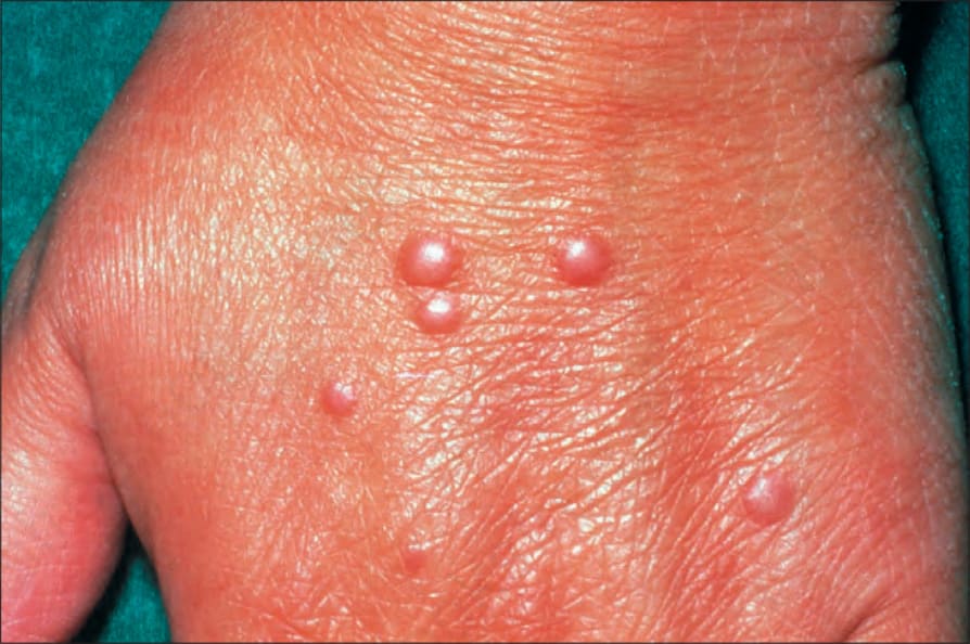
圖 35-201:多核細胞血管組織細胞瘤 (multinucleate cell angiohistiocytoma):可見多個丘疹 (papules)。
Fig. 35.201 Multinucleate cell angiohistiocytoma: multiple papules are present.

圖 35-202:多核細胞血管組織細胞瘤 (multinucleate cell angiohistiocytoma):手部為特徵性的好發部位。By courtesy of the Institute of Dermatology, London, UK.
Fig. 35.202 Multinucleate cell angiohistiocytoma: the hand is a characteristic site. By courtesy of the Institute of Dermatology, London, UK.