Fibrous papule
Fibrous papule
Clinical features Fibrous papule is a very common lesion that presents on the face of middle-aged adults, with predilection for the nose.1–3 It is usually
Pathogenesis and histologic features Although in the past it was suggested that fibrous papule represents an old fibrosed nevus, several studies have demonstrated that this is not the case.6–7 Mast cells may have a role in the development of fibrous papule.8 A recent study showed activation of the Mammalian Target of Rapamycin Pathway, similar to tuberous sclerosis complex-associated angiofibroma.9
Histologically, the lesion is slightly raised, well circumscribed and located in the superficial dermis. It is composed of a collagenous stroma with increased vascular channels and scattered cells varying from spindle shaped to multinucleated (Figs 35.199 and 35.200). A case with multinucleated ganglion-like cells has been reported.10 Mitotic figures are exceptional and some of the cells may show hyperchromatism. Variants with granular cell
1758 Connective tissue tumors
change, clear cell change and epithelioid cells have been described.11–16 Focal pigmentation and inflammation may be seen. Lesions with scattered pleomorphic cells overlap with pleomorphic fibroma. The overlying epidermis appears normal or slightly flattened.
By immunohistochemistry, the cells in the lesion are positive for factor XIIIa and may also be positive for CD34.17–20 S100 protein is negative. Lesions with clear cells are positive for NKI/C3.21
Ultrastructurally, the cells have features of fibroblasts.22,23
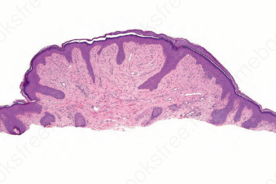
Fig. 35.199 Fibrous papule: shave biopsy from a lesion on the bridge of the nose. The dermis shows dense collagenous tissue.
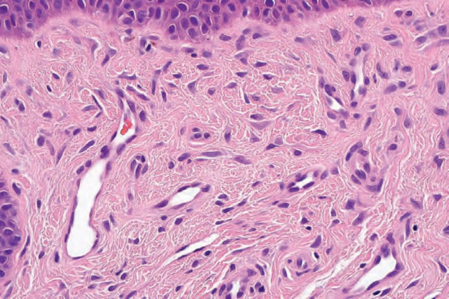
Fig. 35.200 Fibrous papule: high-power view of scattered dendritic cells.
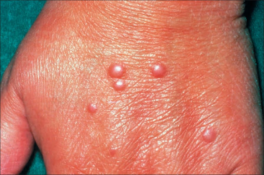
Fig. 35.201 Multinucleate cell angiohistiocytoma: multiple papules are present.

Fig. 35.202 Multinucleate cell angiohistiocytoma: the hand is a characteristic site. By courtesy of the Institute of Dermatology, London, UK.