巨細胞纖維母細胞瘤 (Giant Cell Fibroblastoma)
臨床特徵 (Clinical Features)
- 巨細胞纖維母細胞瘤 (giant cell fibroblastoma) 是一種罕見的真皮或皮下腫瘤,表現為緩慢生長的腫塊,直徑可達 6 cm。
- 它常見於 10 歲以下的兒童(將近三分之二的病例),尤以男性為多,但病灶亦可發生於年輕、中年及老年成人。
- 極少數情況下,腫瘤可為先天性 (congenital)。
- 它具有廣泛的解剖分布,好發於軀幹(背部、胸部與腹部),較少見於近端肢體。曾有發生於外陰 (vulva) 及陰莖 (penis) 的病例報告。
- 不完全切除後,局部復發 (local recurrence) 見於高達 50% 的病例。部分病例復發為 dermatofibrosarcoma protuberans(見下文),此腫瘤被視為該疾病的兒童型。
致病機轉與組織學特徵 (Pathogenesis and Histologic Features)
-
巨細胞纖維母細胞瘤具有與 dermatofibrosarcoma protuberans 相同的染色體異常(見下文)。存在 t(17;22)(q22;q13),導致 platelet-derived growth factor B-chain (PDGFB) 與膠原蛋白基因 COL1A1 融合。亦可見由 t(17;22) 所形成的環狀染色體 (ring chromosomes)。
-
顯微鏡下,巨細胞纖維母細胞瘤是一個界限不清的真皮及淺層皮下病灶,由溫和至中度多形性 (pleomorphic) 的梭形細胞與多核巨細胞(常呈花環樣,floret-like)組成,位於顯著疏鬆的黏液樣間質 (myxoid stroma) 中;此間質有時呈局部玻璃樣變 (hyalinized),並典型地含有不規則、開裂的竇狀腔隙 (sinusoidal spaces),類似血管腔 (Figs 35.135 and 35.136)。
-
然而,後者僅由深染的單核或多核巨細胞間斷性襯覆,而這些細胞並不表現血管標記 (vascular markers)(Fig. 35.137)。
-
核分裂象 (mitotic figures) 僅偶爾可見。局部出血及呈洋蔥皮樣 (onion-skin pattern) 的血管周圍淋巴球常見。
-
例外的腫瘤為純真皮性,少數情況下可見局部累及骨骼肌 (skeletal muscle)。
-
腫瘤細胞 CD34 陽性,亦對 CD99 陽性。
-
在超微結構 (ultrastructurally) 及免疫組織化學 (immunohistochemistry) 上,腫瘤細胞顯示纖維母細胞 (fibroblastic) 特徵。
-
巨細胞纖維母細胞瘤在組織發生學上與 dermatofibrosarcoma protuberans 密切相關,此點由以下事實所證明:部分病例同時呈現兩者的組織學特徵、兩種病灶中的腫瘤細胞均為 CD34 陽性,而更重要的是,兩種腫瘤具有相同的細胞遺傳學異常(見下文)(Figs 35.138 and 35.139)。
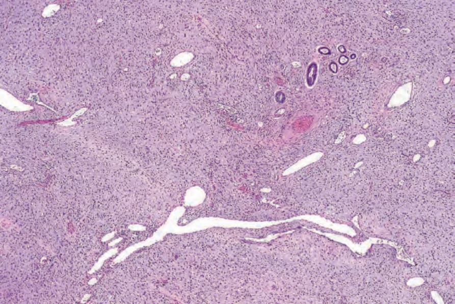
圖 35-135:巨細胞纖維母細胞瘤 (giant cell fibroblastoma):擴張的血管樣腔隙與混雜的梭形細胞及巨細胞共存於黏液樣間質 (myxoid stroma) 中是其特徵。
Fig. 35.135 Giant cell fibroblastoma: the admixture of dilated vessel-like spaces and mixed spindled and giant cells in a myxoid stroma is characteristic.
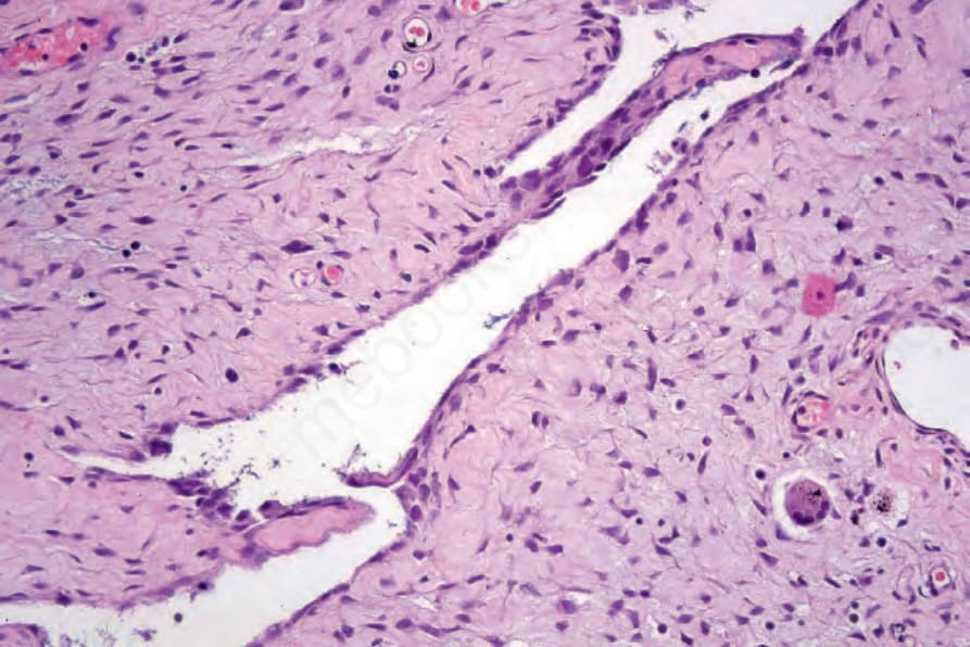
圖 35-136:巨細胞纖維母細胞瘤 (giant cell fibroblastoma):中倍視野顯示由腫瘤細胞襯覆的血管樣腔隙 (blood-vessel–like space)。
Fig. 35.136 Giant cell fibroblastoma: medium-power view showing blood-vessel–like space lined by tumor cells.
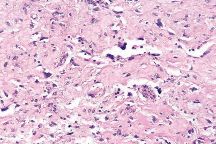
圖 35-137:巨細胞纖維母細胞瘤 (giant cell fibroblastoma):竇狀樣腔隙 (sinusoid-like spaces) 由多核巨細胞 (multinucleate giant cells) 襯覆。
Fig. 35.137 Giant cell fibroblastoma: the sinusoid-like spaces are lined by multinucleate giant cells.
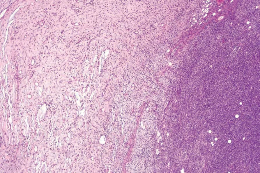
圖 35-138:巨細胞纖維母細胞瘤 (giant cell fibroblastoma):此病灶在視野右側與典型的 dermatofibrosarcoma protuberans 難以察覺地融合。
Fig. 35.138 Giant cell fibroblastoma: this lesion merges imperceptibly with typical dermatofibrosarcoma protuberans on the right side of the field.
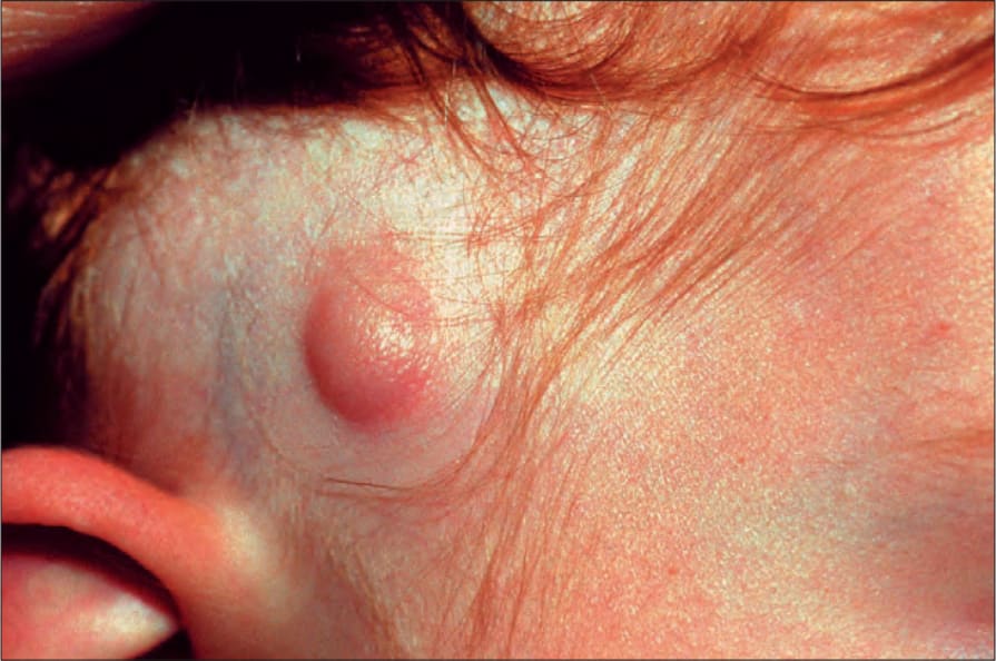
圖 35-140:隆突性皮膚纖維肉瘤 (dermatofibrosarcoma protuberans):如圖所示的先天性病灶極為罕見。圖片由英國倫敦 Institute of Dermatology 提供。
Fig. 35.140 Dermatofibrosarcoma protuberans: congenital lesions as shown here are exceedingly rare. By courtesy of the Institute of Dermatology, London, UK.