Giant cell fibroblastoma
Giant cell fibroblastoma
Clinical features Giant cell fibroblastoma is a rare dermal or subcutaneous tumor that presents as a slowly growing mass up to 6 cm in diameter.1–7 It commonly affects children under 10 years of age (almost two-thirds of cases), especially males, but lesions may also occur in young, middle-aged and elderly adults.7 Exceptionally, a tumor may be congenital.8 It has a wide anatomical distribution with predilection for the trunk (back, chest and abdomen) and (less commonly) the proximal extremities.1–4 A tumor in the vulva and one in the penis have been reported.9,10 Local recurrence is seen in up to 50% of cases after incomplete excision.1–4 Some cases recur as dermatofibrosarcoma protuberans (see below) and this tumor is considered a pediatric form of this disease.
Pathogenesis and histologic features Giant cell fibroblastoma shares the same chromosomal abnormalities as those of dermatofibrosarcoma protuberans (see below). There is a t(17;22) (q22;q13) resulting in fusion of the platelet-derived growth factor B-chain (PDGFB) and the collagen gene COL1A1. Ring chromosomes resulting from the t(17;22) are also seen.11–14 Microscopically, giant cell fibroblastoma is a poorly circumscribed dermal and superficial subcutaneous lesion composed of bland to moderately pleomorphic spindle and multinucleated giant cells (frequently floret-like) in a conspicuous loose myxoid stroma, which is sometimes focally hyalinized and typically contains irregular gaping sinusoidal spaces simulating vascular lumina (Figs 35.135 and 35.136). The latter, however, are only lined discontinuously by hyperchromatic mononuclear or multinucleated giant cells, which do not stain for vascular markers (Fig. 35.137). Mitotic figures are only rarely seen. Focal hemorrhage and perivascular lymphocytes in an onion-skin pattern are common.7 Exceptional tumors are purely dermal and rarely there is focal involvement of skeletal muscle.7 Tumor cells are positive for CD34 and also for CD99.15
Ultrastructurally, and by immunohistochemistry, the tumor cells display fibroblastic features.
Giant cell fibroblastoma is closely related histogenetically to dermatofibrosarcoma protuberans, as demonstrated by the fact that some cases present combined histologic features, tumor cells in both lesions are CD34 positive and, more importantly, both tumors share the same cytogenetic abnormalities (see below) (Figs 35.138 and 35.139).11–13,16,17 Cases of giant
1741 Intermediate (rarely metastasizing) fibroblastic and myofibroblastic tumors
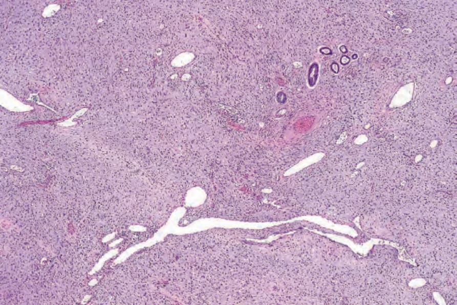
Fig. 35.135 Giant cell fibroblastoma: the admixture of dilated vessel-like spaces and mixed spindled and giant cells in a myxoid stroma is characteristic.
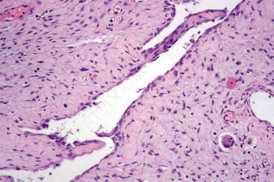
Fig. 35.136 Giant cell fibroblastoma: medium-power view showing blood-vessel–like space lined by tumor cells.
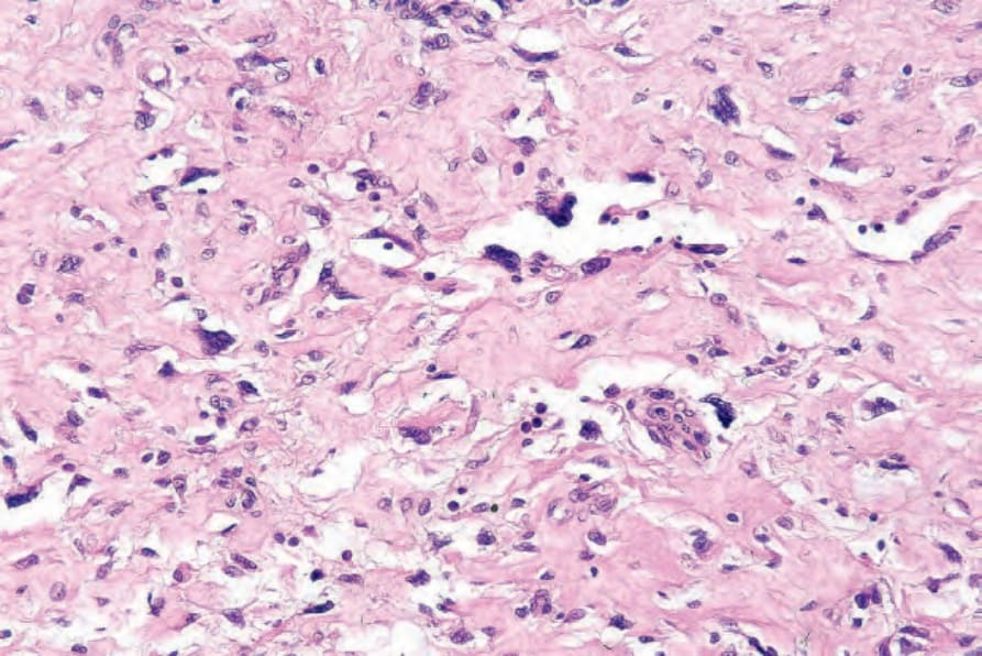
Fig. 35.137 Giant cell fibroblastoma: the sinusoid-like spaces are lined by multinucleate giant cells.
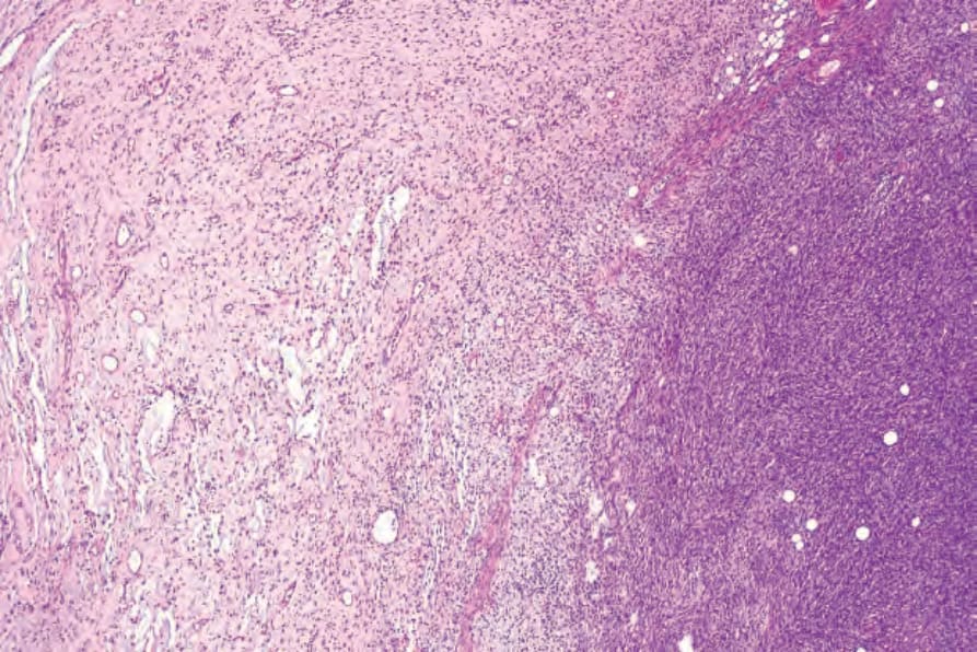
Fig. 35.138 Giant cell fibroblastoma: this lesion merges imperceptibly with typical dermatofibrosarcoma protuberans on the right side of the field.
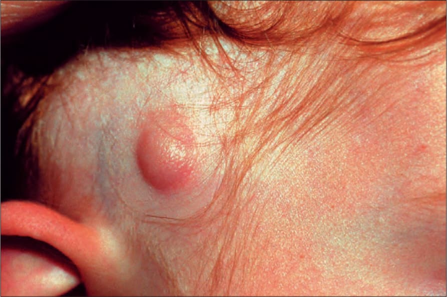
Fig. 35.140 Dermatofibrosarcoma protuberans: congenital lesions as shown here are exceedingly rare. By courtesy of the Institute of Dermatology, London, UK.