鈣化性腱膜纖維瘤 (Calcifying Aponeurotic Fibroma)
鈣化性腱膜纖維瘤 (calcifying aponeurotic fibroma)
臨床特徵 (Clinical Features)
鈣化性腱膜纖維瘤 (calcifying aponeurotic fibroma)(亦稱 juvenile aponeurotic fibroma)為一種極為罕見的病灶,主要見於生命的前二十年,且偏好男性。它表現為單一的小結節狀或浸潤性腫塊,最常位於足部或手部,尤其是手掌。侵犯其他部位(如頭頸部、背部、腹壁、下肢與上肢)非常罕見。多發性病灶屬例外情形。骨侵犯極為罕見。局部復發(特別是在較年輕的病人)相當常見,可發生於高達 50% 的病例。
組織病理特徵 (Histopathology)
在此腫瘤中已有報告指出 FN1-EGF 基因融合 (FN1-EGF gene fusion) 為其主要驅動突變 (driver mutation),此融合至今尚未在任何其他腫瘤中被偵測到。
1731 Benign fibrous and myofibroblastic tumors and tumorlike lesions
Calcifying aponeurotic fibroma 的特徵為形成一個由緻密、相當富細胞性 (cellular) 之纖維組織所構成的不規則腫塊,廣泛侵犯皮下與肌肉結構 (Fig. 35.107)。肌纖維母細胞 (myofibroblasts) 傾向於飽滿且具有顯著的細胞核。通常這些細胞呈線性或柵欄狀 (palisaded) 排列,並構成腫瘤的主體。隨著病灶成熟,鈣化區域會發生軟骨樣化生 (chondroid metaplasia) (Fig. 35.108)。偶可見髓外造血 (extramedullary hematopoiesis)。
它們表現為跨掌指關節 (metacarpophalangeal joint) 或近端指間關節 (proximal interphalangeal joint) 之相當邊界不清的纖維性增厚病灶,最常見於中年人 (Fig. 35.109)。它們可能為家族性,與 Dupuytren contracture 或 plantar fibromatosis 相關,繼發於反覆創傷,或為特發性 (idiopathic)。它們幾乎總是無症狀。指節墊樣 (knuckle pad-like) 病灶可發生於與 keratin 9 突變相關的 epidermolytic palmoplantar keratoderma。曾有一例指節墊 (knuckle pads) 合併白甲症 (leukonychia) 與耳聾 (deafness) 的紀錄。一例與 pseudoxanthoma elasticum 之少見關聯,可能屬巧合。
鑑別診斷 (Differential Diagnosis)
鑑別診斷包括 palmar fibromatosis(在年輕人中罕見,且傾向於相當局限)以及軟組織軟骨瘤 (soft tissue chondroma)(其缺乏緻密的肌纖維母細胞成分,僅由軟骨組織構成,而該軟骨組織常相當富細胞性)。
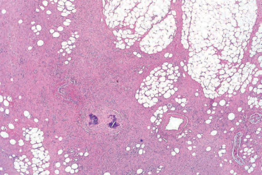
圖 35-107:鈣化性腱膜纖維瘤 (calcifying aponeurotic fibroma):皮下脂肪內可見一個由纖維母細胞性 (fibroblastic) 組織構成的不規則浸潤性腫塊。
Fig. 35.107 Calcifying aponeurotic fibroma: an irregular infiltrative mass of fibroblastic tissue is present in the subcutaneous fat.
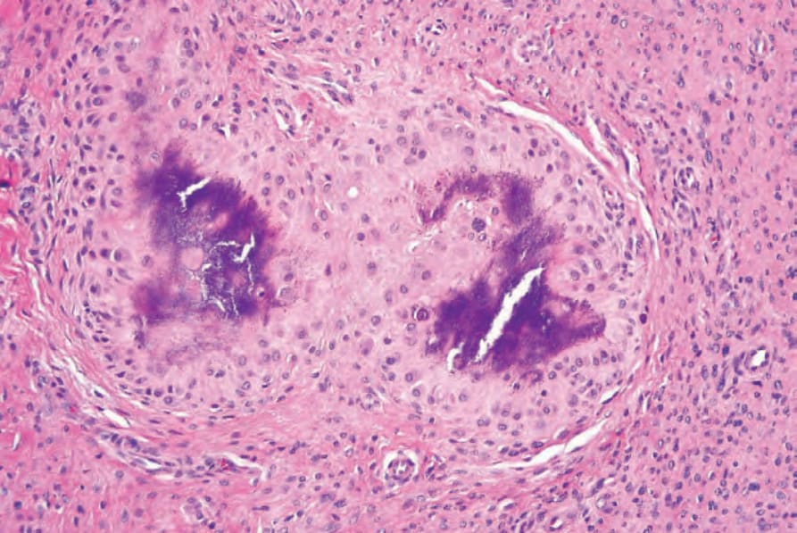
圖 35-108:鈣化性腱膜纖維瘤 (calcifying aponeurotic fibroma):在此切片中,可見軟骨樣化生 (chondroid metaplasia) 以及局部嗜鹼性鈣化 (focal basophilic calcification)。
Fig. 35.108 Calcifying aponeurotic fibroma: in this section, there is chondroid metaplasia and there is focal basophilic calcification.
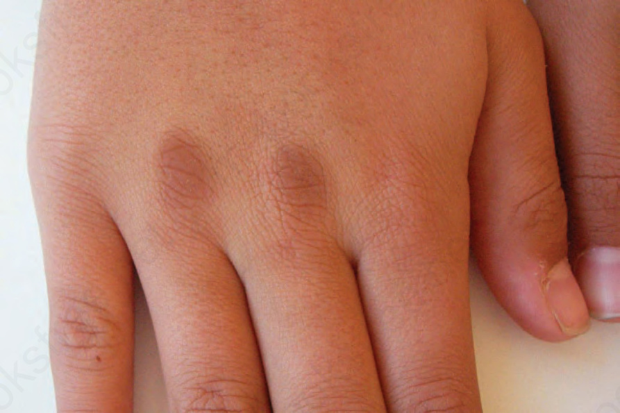
圖 35-109:指節墊 (knuckle pad):指間關節 (interphalangeal joints) 與第二掌指關節 (second metacarpophalangeal joint) 上方可見典型的增厚斑塊。By courtesy of M.M. Black, MD, Institute of Dermatology, London, UK.
Fig. 35.109 Knuckle pad: typical thickened plaques are present over the interphalangeal joints and second metacarpophalangeal joint. By courtesy of M.M. Black, MD, Institute of Dermatology, London, UK.
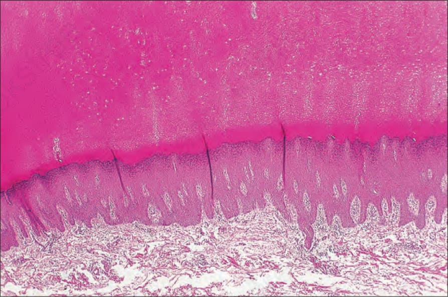
圖 35-110:指節墊 (knuckle pad):在此切片中可見大量角化過度 (hyperkeratosis) 與棘層肥厚 (acanthosis)。其外觀為非特異性。
Fig. 35.110 Knuckle pad: in this section there is massive hyperkeratosis and acanthosis. The appearances are non-specific.