臨床特徵 (Clinical Features)
- 蟹足腫 (keloid) 是一種常見的反應性病灶,代表過度旺盛的疤痕形成 (exuberant scar formation)。它通常會超出原始損傷的部位向外延伸。
- 雖然 keloid 偶爾看似自發出現,但一般認為大多數是局部創傷的直接結果,即使該創傷輕微或未被察覺 (Figs 35.52–35.54)。
- Keloid 也可因發炎而形成,例如尋常性痤瘡 (acne vulgaris) 等病況。isotretinoin 的使用也曾被連結到 keloid 的發生。與 aromatase 相關的爆發性 keloid(eruptive keloids)。
- Keloid 通常發生於頭頸部(尤其是耳部)、上胸部及上臂,但幾乎可見於任何皮膚部位,不過如手足及生殖器等區域則極少受到影響。
- 其特徵為隆起、邊界清楚、相當光滑的病灶,隨時間進展逐漸變得更加硬化 (indurated)。它們偶爾會搔癢或觸痛,並可能為多發性,這再次反映了個體對其發生的易感性 (susceptibility)。無論使用何種治療,局部復發都非常常見。
致病機轉與組織學特徵 (Pathogenesis and Histologic Features)
- Keloid 的致病機轉尚不清楚,但似乎是多因子的(另見肥厚性疤痕 hypertrophic scar 一節)。基因易感性 (genetic predisposition) 與局部組織張力 (local tissue tension) 扮演了重要角色。
- Keloid 中膠原蛋白合成增加,且所產生膠原蛋白的品質也與正常皮膚不同。Keloid 中纖維母細胞 (fibroblasts) 的凋亡 (apoptosis) 減少。第一型與第三型膠原蛋白 (collagen I and III) 的產生增加,此點由 mRNA 含量上升所證實。
- Transforming growth factor beta (TGF-β) 似乎在傷口癒合中扮演重要角色,其產生增加已被連結到 keloid 的致病機轉,方式為活化纖維母細胞合成膠原蛋白。與纖維化及正常傷口癒合相關的 Wnt/β-catenin 路徑被強烈上調 (up-regulated),並可見影響多條促纖維化 (profibrotic) 路徑的表觀遺傳學 (epigenetic) 變化。
- 角質形成細胞 (keratinocytes) 與纖維母細胞之間的交互作用似乎在 keloid 的形成中扮演重要角色。已有研究證實,當 keloid 纖維母細胞與 keloid 角質形成細胞共同培養 (co-cultured) 時,可溶性與不可溶性膠原蛋白的產生增加,且 procollagen III mRNA 上調。
- 組織學表現的典型特徵為結節狀的纖維母細胞增生 (nodular fibroblastic proliferation),以及真皮內出現少細胞性 (hypocellular)、「玻璃樣 (glassy)」、嗜伊紅性 (eosinophilic)、厚而玻璃樣化 (hyalinized) 的膠原纖維 (Figs 35.55 and 35.56)。早期病灶可能顯示輕微的血管增生 (vascularity) 及黏液樣基質 (myxoid ground substance) 的病灶。偶爾可見正常的有絲分裂 (normal mitoses)。
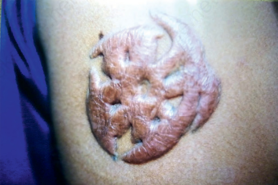
圖 35.52:在刺青旁緣發生的顯著蟹足腫 (keloid)。承蒙菲律賓馬尼拉 Dr J. Dayrit 提供。
Fig. 35.52 Prominent keloid developing at the side of a tattoo. By courtesy of Dr J. Dayrit, Manila, The Philippines.
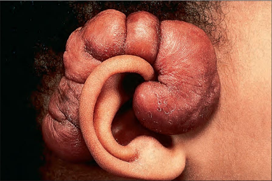
圖 35.53:蟹足腫 (keloid):病灶常繼發於創傷之後,並且是穿洞 (piercing) 的常見併發症。承蒙英國倫敦 Institute of Dermatology 提供。
Fig. 35.53 Keloid: lesions commonly follow trauma and are a frequent complication of piercing. By courtesy of the Institute of Dermatology, London, UK.
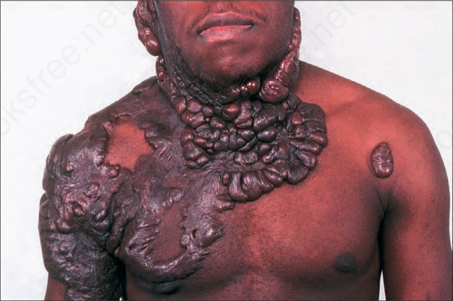
圖 35.54:蟹足腫 (keloid):廣泛的 keloid 形成可造成嚴重的外觀毀損 (disfiguring)。承蒙英國倫敦 Institute of Dermatology 提供。
Fig. 35.54 Keloid: extensive keloid formation can be very disfiguring. By courtesy of the Institute of Dermatology, London, UK.
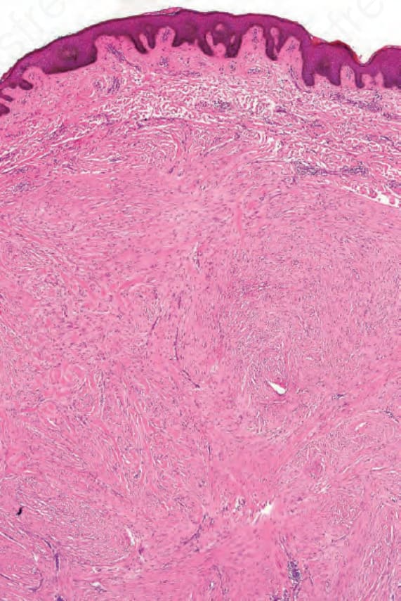
圖 35.55:蟹足腫 (keloid):此病灶藉由出現寬大的嗜伊紅性、玻璃樣化膠原蛋白束 (broad bundles of eosinophilic, hyalinized collagen) 而與肥厚性疤痕 (hypertrophic scar) 區別。
Fig. 35.55 Keloid: this lesion is distinguished from a hypertrophic scar by the presence of broad bundles of eosinophilic, hyalinized collagen.
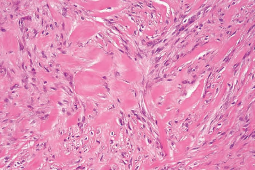
圖 35.56:蟹足腫 (keloid):高倍視野:注意腫脹的玻璃樣化膠原蛋白束 (swollen hyalinized collagen bundles) 與溫和的梭形細胞 (bland spindled cells) 混雜在一起。
Fig. 35.56 Keloid: high-power view: note the swollen hyalinized collagen bundles admixed with bland spindled cells.