上皮樣肉瘤 (Epithelioid Sarcoma)
臨床特徵 (Clinical Features)
-
上皮樣肉瘤 (epithelioid sarcoma) 是一種相對罕見的腫瘤,最常發生於年輕成人(尤其是男性)的遠端肢體(特別是手與腕部)。發生於兒童並不常見。
-
整體的年齡範圍與解剖分布都很廣。罕見病例可呈現於頭部,甚至發生於口腔與腮腺 (parotid gland)(Figs 35.676 與 35.677)。
-
它主要是一種真皮或皮下腫瘤,表現為緩慢生長、隆起、常具觸痛的結節,直徑小於 5 cm。潰瘍 (ulceration) 是常見特徵。
-
由於此腫瘤具有沿血管、神經與筋膜 (fascia) 廣泛擴散的特殊傾向,在距主要腫瘤一段距離處出現衛星結節 (satellite nodules) 是常見現象。
-
此腫瘤可能模擬其他疾病,包括穿透性環狀肉芽腫 (perforating granuloma annulare) 與 Dupuytren 病 (Dupuytren disease)。
-
由 SMARCB1 (INI1) 編碼、位於 22q11.23 的核內表現缺失具特徵性,且可能對其致病機轉至關重要。其他異常已見於 8q,亦曾記載第 21 對染色體單體 (monosomy 21)。整體而言,存在複雜的基因體 (complex genomic) 病例,且 CDKN2A 的缺失常見,這與具 SMARCB1 缺失的橫紋肌樣腫瘤 (rhabdoid tumor) 之簡單基因體輪廓 (simple genomic profile) 形成對比。第 8 與第 22 對染色體的異常也被發現參與近端型上皮樣肉瘤 (proximal-type epithelioid sarcoma)。
-
惰性 (indolent) 且反覆的局部區域性復發 (locoregional recurrence) 常見。轉移至淋巴結(這在其他肉瘤是少見的特徵)相當常見,其次為轉移至肺部。雖然整體 5 年存活率約為 70%,但 20 年存活率不超過 20–25%。預後改善似乎與較小的腫瘤體積有關。
-
一群發生於骨盆-會陰 (pelvi-perineal) 部位(包括外陰)的上皮樣肉瘤具有非常侵襲性的臨床病程,比一般變異型更為嚴重,被描述為近端型上皮樣肉瘤 (proximal-type epithelioid sarcoma)。類似病例偶爾也發生於其他部位。較差生物學行為的獨立指標為早期轉移與大腫瘤體積。
致病機轉與組織學特徵 (Pathogenesis and Histologic Features)
-
至今所研究的病例中,最一致的細胞遺傳學異常為第 22q 對染色體的雜合性缺失 (loss of heterozygosity)。INI1 (BAF47) 的缺失(見上文)。
-
在一例轉移性上皮樣肉瘤中曾描述 NRAS 致癌基因突變 (oncogene mutation)。
-
顯微鏡下表現具特徵性(Figs 35.678–35.680)。腫瘤由多個由多角形 (polygonal)、上皮樣 (epithelioid) 或紡錘狀 (spindle-shaped) 細胞構成的結節組成,這些細胞具嗜伊紅性細胞質 (eosinophilic cytoplasm) 並顯示不等程度的多形性 (pleomorphism)。有絲分裂 (mitoses) 通常稀少。偶可見巨細胞型 (giant cell forms)。在這些結節的中央,約 50% 的病例以局灶性壞死 (focal necrosis) 為顯著特徵,造成類似肉芽腫過程 (granulomatous process) 的外觀(Fig. 35.681)。其他病例顯示模糊的纖維素樣 (fibrinoid) 或黏液樣 (myxoid) 變性型態。後者在罕見病例中可能占主導。在結節周邊,腫瘤細胞傾向較呈紡錘狀,在罕見病例此特徵可能很顯著(見下文)。常存在血管與神經周圍侵犯 (vascular and perineural invasion)(Fig. 35.682)。
-
在少數病例,紡錘狀細胞占主導,排列成束,壞死極少或缺如。此變異型稱為纖維瘤樣 (fibroma-like)。在少數病例可見血管樣 (angiomatoid) 型態。異位骨形成 (heterotopic bone formation) 極為例外。
-
免疫組織化學上,超過 90% 的病例對 vimentin、cytokeratin 與 EMA 呈陽性(Figs 35.683 與 35.684),高達 60% 對 CD34 呈陽性。SMA 也常呈局灶陽性。vimentin、CD34 與 keratin 陽性的組合在上皮樣肉瘤的診斷上非常有用。近端型上皮樣肉瘤的免疫組織化學輪廓與典型上皮樣肉瘤相似。兩種型態在超過 90% 的病例皆顯示核內 INI1 表現缺失,這在合理的鑑別診斷範圍內是相對特異的發現。GLUT-1 在上皮樣肉瘤的診斷上並非有用的標記。據報導 CA125 在上皮樣肉瘤常呈陽性,而在可模擬此腫瘤的反應性與腫瘤性病灶呈陰性。近端型上皮樣肉瘤以瀰漫性生長型態 (diffuse growth pattern) 為特徵(Figs 35.685 與 35.686)。腫瘤細胞主要為上皮樣,有絲分裂活性常很活躍 (brisk)(Fig. 35.687)。常存在局灶性或廣泛的橫紋肌樣變化 (rhabdoid change)(Figs 35.688–35.690)。壞死似乎較一般變異型少見。
-
超微結構研究顯示,上皮樣腫瘤細胞含有發育良好的橋粒樣連接 (desmosome-like junctions) 與中間絲 (intermediate filaments) 聚集物,常位於核旁 (paranuclear) 位置。在一例近端型上皮樣肉瘤中,免疫電子顯微鏡 (immunoelectron microscopy) 顯示 keratin 絲但無 vimentin,提示其與上皮細胞的關係較與間葉細胞 (mesenchymal cells) 更為密切。
鑑別診斷 (Differential Diagnosis)
-
在適當的臨床情境下,其特殊的組織學特徵通常可避免診斷混淆。對此病灶缺乏認識可能導致誤診為壞死性轉移癌 (necrotic metastatic carcinoma) 或肉芽腫性發炎病灶 (granulomatous inflammatory lesion)。
-
與上皮樣血管內皮瘤 (epithelioid hemangioendothelioma) 或血管肉瘤 (angiosarcoma) 的區分可能困難,因為上皮樣肉瘤常有假血管裂隙 (pseudovascular clefts) 與局灶性細胞質空泡化 (cytoplasmic vacuolation)。然而,前者的細胞傾向至少局灶性地呈索狀 (cords) 生長,且常較大;它們對內皮標記 (endothelial markers) 染色陽性,且常為 keratin 陽性。
-
惡性橫紋肌樣腫瘤 (malignant rhabdoid tumor) 顯示許多具細胞質內包涵體 (intracytoplasmic inclusions) 的細胞,雖然免疫組織化學上腫瘤細胞對上皮標記呈陽性,但它們通常也對其他標記呈陽性,提示分歧分化 (divergent differentiation)。此外,橫紋肌樣腫瘤具特徵性的泡狀核 (vesicular nuclei) 與巨核仁 (macronucleoli)。
-
深部環狀肉芽腫 (deep granuloma annulare) 與類風濕結節 (rheumatoid nodule) 可模擬上皮樣肉瘤,特別是在低倍鏡檢查下。然而,前述這些病灶既無細胞學異型性 (cytologic atypia) 亦無有絲分裂活性,缺乏壞死,且存在伴有纖維素 (fibrin) 或黏蛋白 (mucin) 沉積的壞死膠原變性 (necrobiosis),組織球 (histiocytes) 對 CD68 呈陽性、對 keratin 與 CD34 呈陰性。
-
似乎與預後相關的因素包括年齡、腫瘤大小、組織學特徵、分期 (stage)、腫瘤分級 (tumor grade) 與分子改變。兒童與青少年的預後優於成人,而呈現於四肢的腫瘤其行為優於發生於頭頸部者。與較佳預後相關的組織學特徵:分化不良腫瘤所占比例(超過 20%)、直徑小於 5 cm(極小的滑膜肉瘤 (synovial sarcoma) 具極佳預後)、在 1.7 mm² 內少於 5 個有絲分裂,以及缺乏壞死。NY-ESO-1 表現有助於辨識適合免疫療法標靶治療 (immunotherapy targeted therapy) 的病例。
-
大多數滑膜肉瘤 (synovial sarcoma) 病例(包括單相 (monophasic) 與雙相 (biphasic) 變異型)顯示一種平衡型 t(X;18)(p11;q11),將 SSX1、SSX2 或極罕見的 SSX4(三者一同位於 X 染色體上)與 SS18(先前稱為 SYT)融合。亦曾記載一種導致 SS18L1-SSX1 融合的 t(X;20)(p11;q13)。SSX1 的參與在雙相腫瘤較常見,而在單相型則三者中任一者皆可參與。不同融合型別的預後價值仍有爭議,但可能很小。
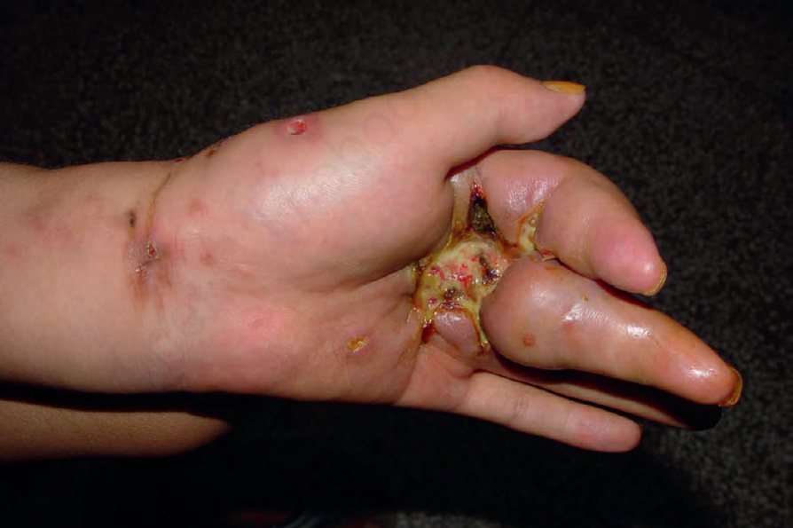
圖 35-676:上皮樣肉瘤 (epithelioid sarcoma):手部是常見的受侵犯部位。承蒙中國西安 Dr. Yi-Guo Feng 提供。
Fig. 35.676 Epithelioid sarcoma: the hand is a commonly affected site. By courtesy of Dr. Yi-Guo Feng, Xian, China.
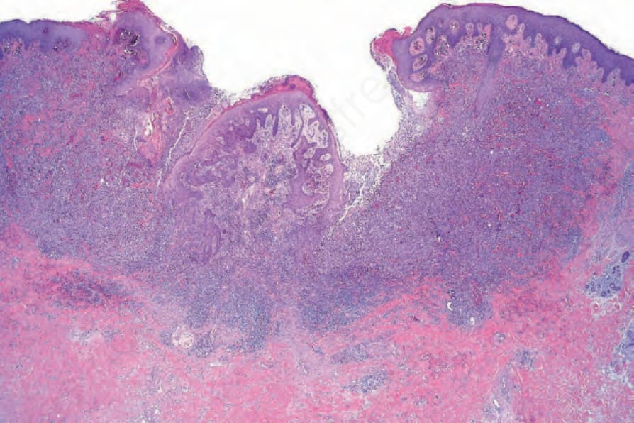
圖 35-678:上皮樣肉瘤 (epithelioid sarcoma):上層真皮被一個潰瘍性腫瘤瀰漫性浸潤。
Fig. 35.678 Epithelioid sarcoma: the upper dermis is diffusely infiltrated by an ulcerated tumor.
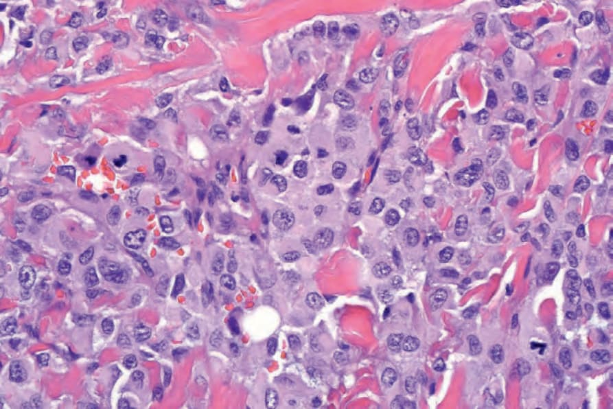
圖 35-679:上皮樣肉瘤 (epithelioid sarcoma):在此視野中,腫瘤細胞為上皮樣 (epithelioid),具豐富的嗜伊紅性細胞質與大的泡狀核 (vesicular nuclei)。
Fig. 35.679 Epithelioid sarcoma: in this field the tumor cells are epithelioid with abundant eosinophilic cytoplasm and large vesicular nuclei.
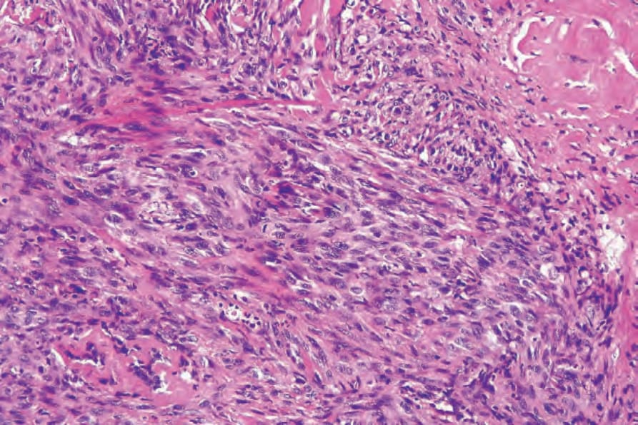
圖 35-680:上皮樣肉瘤 (epithelioid sarcoma):在其他處,腫瘤細胞具紡錘狀 (spindled) 型態。
Fig. 35.680 Epithelioid sarcoma: elsewhere the tumor cells have a spindled morphology.
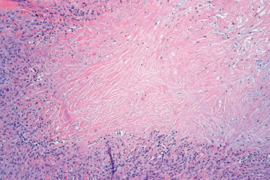
圖 35-681:上皮樣肉瘤 (epithelioid sarcoma):低倍鏡檢查下所見的地圖狀壞死 (geographical necrosis) 可能造成與肉芽腫過程 (granulomatous process) 的診斷混淆。
Fig. 35.681 Epithelioid sarcoma: geographical necrosis seen at low-power examination may result in diagnostic confusion with a granulomatous process.
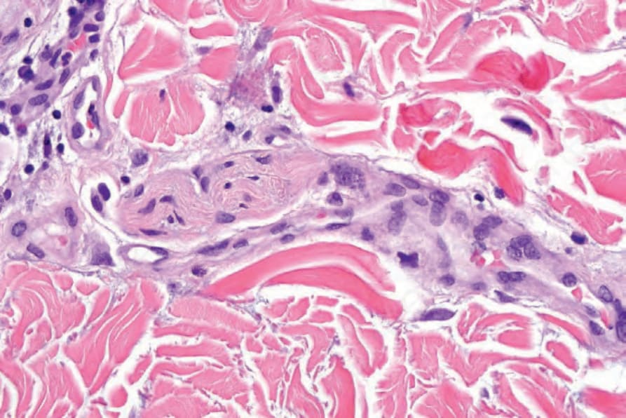
圖 35-682:上皮樣肉瘤 (epithelioid sarcoma):此腫瘤常沿神經幹 (nerve trunks) 延伸,部分解釋了其高復發率。
Fig. 35.682 Epithelioid sarcoma: the tumor commonly extends along nerve trunks, in part accounting for its high recurrence rate.
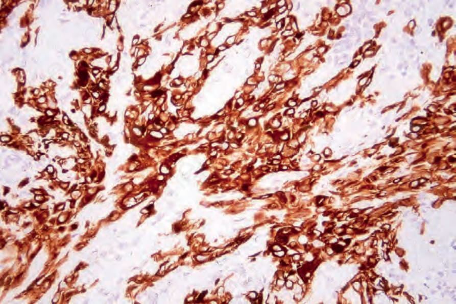
圖 35-683:上皮樣肉瘤 (epithelioid sarcoma):腫瘤細胞具特徵性地表現 keratin,如此視野所示。
Fig. 35.683 Epithelioid sarcoma: the tumor cells characteristically express keratin, as shown in this field.
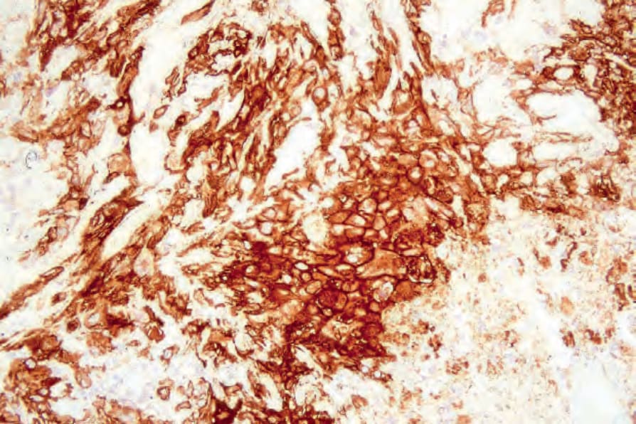
圖 35-684:上皮樣肉瘤 (epithelioid sarcoma):上皮膜抗原 (epithelial membrane antigen, EMA) 陽性通常明顯可見。
Fig. 35.684 Epithelioid sarcoma: epithelial membrane antigen positivity is usually evident.
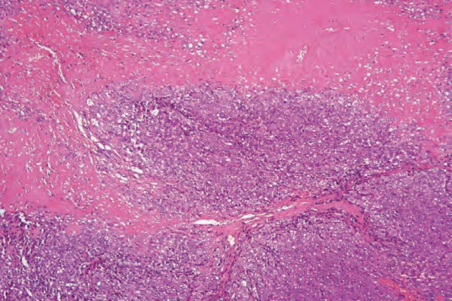
圖 35-685:近端型上皮樣肉瘤 (proximal epithelioid sarcoma):此腫瘤以瀰漫性細胞浸潤 (diffuse cellular infiltrate) 伴廣泛壞死 (necrosis) 為特徵。
Fig. 35.685 Proximal epithelioid sarcoma: the tumor is characterized by a diffuse cellular infiltrate with widespread necrosis.
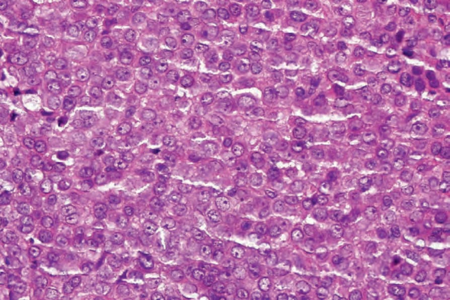
圖 35-686:近端型上皮樣肉瘤 (proximal epithelioid sarcoma):腫瘤細胞為上皮樣 (epithelioid),具嗜伊紅性細胞質與含明顯核仁 (nucleoli) 的圓形泡狀核 (round vesicular nuclei)。
Fig. 35.686 Proximal epithelioid sarcoma: the tumor cells are epithelioid with eosinophilic cytoplasm and round vesicular nuclei containing conspicuous nucleoli.
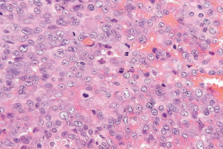
圖 35-687:近端型上皮樣肉瘤 (proximal epithelioid sarcoma):在此例中,有顯著的有絲分裂活性 (mitotic activity)。
Fig. 35.687 Proximal epithelioid sarcoma: in this example, there is marked mitotic activity.
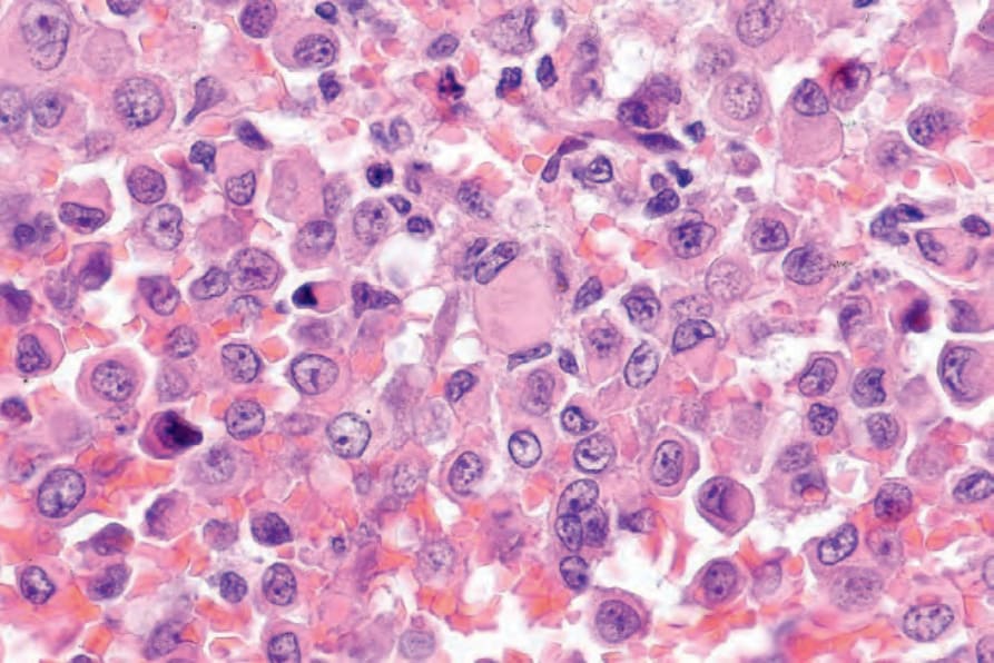
圖 35-688:近端型上皮樣肉瘤 (proximal epithelioid sarcoma):橫紋肌樣包涵體 (rhabdoid inclusions)(如視野中央所見)常存在。
Fig. 35.688 Proximal epithelioid sarcoma: rhabdoid inclusions, as seen in the center of the field, are often present.
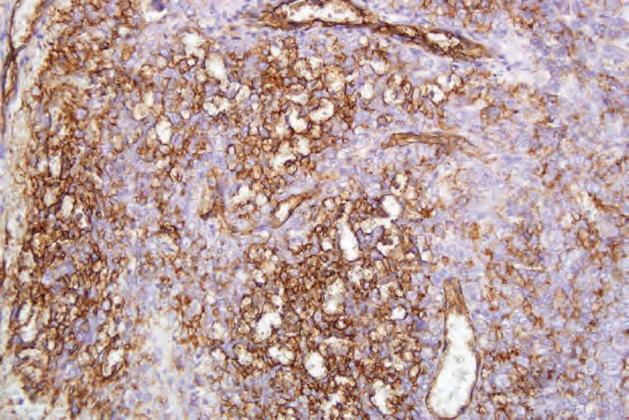
圖 35-689:近端型上皮樣肉瘤 (proximal epithelioid sarcoma):腫瘤細胞對 keratin (AE1/AE3) 呈陽性。
Fig. 35.689 Proximal epithelioid sarcoma: the tumor cells are positive for keratin (AE1/AE3).
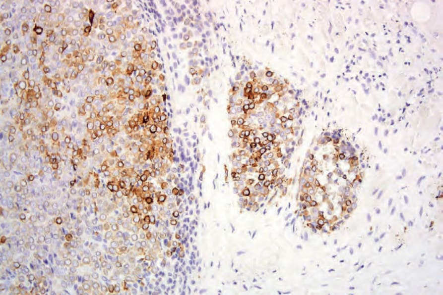
圖 35-690:近端型上皮樣肉瘤 (proximal epithelioid sarcoma):在此例中亦表現 CD34。
Fig. 35.690 Proximal epithelioid sarcoma: CD34 is also expressed in this example.
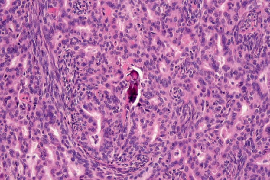
圖 35-691:滑膜肉瘤 (synovial sarcoma):此視野顯示由紡錘狀細胞與腺體腔隙 (glandular spaces) 構成的特徵性雙相 (biphasic) 細胞群。
Fig. 35.691 Synovial sarcoma: this field shows the characteristic biphasic population of spindle cells and glandular spaces.