淺表肢端纖維黏液瘤 (Superficial Acral Fibromyxoma)
臨床特徵 (Clinical Features)
- 淺表肢端纖維黏液瘤 (superficial acral fibromyxoma),又稱指趾纖維黏液瘤 (digital fibromyxoma),是一種具特徵性、相對少見的良性腫瘤,好發於手指與腳趾,其次為手掌。相同的病灶曾以 cellular digital fibroma 之名被描述。
- 累及甲區 (nail region) 非常常見。發生於足跟 (heel) 的病灶罕見。
- 患者通常為年輕至中年成人,並好發於男性。腫瘤體積小、生長緩慢且無症狀。切除後局部復發 (local recurrence) 罕見。
致病機轉與組織學特徵 (Pathogenesis and Histologic Features)
- 近期已記錄到 RB1 基因缺失 (RB1 gene deletions),提示其可能與其他 RB1 缺失型腫瘤有關,例如 spindle cell/pleomorphic lipoma、mammary-type myofibroblastoma 與 cellular angiofibroma。
- 組織學顯示一個界線相當清楚的真皮及/或皮下腫瘤,由溫和的紡錘形或星狀細胞 (bland, spindle-shaped or stellate cells) 組成,於黏液樣或膠原樣間質 (myxoid or collagenous stroma) 中呈局部車輻狀 (storiform) 或束狀 (fascicular) 排列 (Fig. 35.660)。病灶內含有散在的小血管腔 (vascular channels)。某些腫瘤的細胞密度高於其他腫瘤,這些腫瘤中的黏液樣變化 (myxoid change) 往往非常局部 (Fig. 35.661)。脂肪細胞成分 (adipocytic component) 屬例外罕見。
- 有絲分裂象 (mitotic figures) 罕見,細胞學異型性 (cytologic atypia) 輕微或缺如。常可見肥大細胞 (mast cells)。
- 免疫組化方面,腫瘤細胞通常對 CD34 呈陽性,並可能對 EMA、SMA、CD99、CD10 與 nestin 呈局部陽性。Desmin 陽性僅見於一例報告。RB1 表現喪失 (loss of expression of RB1)。超微結構上,腫瘤細胞顯示胞質內中間絲 (cytoplasmic intermediate filaments) 與粗面內質網 (rough endoplasmic reticulum),提示纖維母細胞分化 (fibroblastic differentiation)。
鑑別診斷 (Differential Diagnosis)
- 鑑別診斷包括 neurofibroma、onychomatricoma、dermatofibrosarcoma protuberans、minute synovial sarcoma 與 low-grade fibromyxoid sarcoma。
- Neurofibroma 在肢端部位罕見,且雖然腫瘤細胞可能對 CD34 呈局部陽性,但它們同時對 S100 protein 呈陽性。
- Onychomatricoma 中的間質成分可能與淺表肢端纖維黏液瘤所見者相同,含有 CD34 陽性細胞。鑑別依據在於 onychomatricoma 中存在特徵性的上皮變化。
- 局部車輻狀 (storiform) 排列可能模擬 dermatofibrosarcoma protuberans。然而,後者在遠端肢體 (distal extremities) 極為罕見,會瀰漫性浸潤皮下組織,且對 apolipoprotein D 呈陽性。
- 手足的 minute synovial sarcoma 可能與淺表肢端纖維黏液瘤外觀相似,具有黏液樣間質與溫和的紡錘形細胞。然而,前者有局部的鈣化區域,腫瘤細胞至少對 keratin 呈局部陽性,且細胞遺傳學分析顯示 t(X;18) 易位 (translocation)。MUC4 陰性可排除 low-grade fibromyxoid sarcoma。
含鐵血黃素纖維脂肪瘤性腫瘤/含鐵血黃素纖維組織細胞性脂肪瘤性病變 (Hemosiderotic fibrolipomatous tumor/hemosiderotic fibrohistiocytic lipomatous lesion)
臨床特徵 (Clinical Features)
- 含鐵血黃素纖維脂肪瘤性腫瘤 (hemosiderotic fibrolipomatous tumor),又稱含鐵血黃素纖維組織細胞性脂肪瘤性病變 (hemosiderotic fibrohistiocytic lipomatous lesion),幾乎僅發生於足部,尤其是踝部 (ankle),並好發於女性。多數患者為成人,但兒童偶可受累。它生長緩慢且無症狀。單純切除 (simple excision) 為首選治療。可能發生局部復發。
致病機轉與組織學特徵 (Pathogenesis and Histologic Features)
- 過去認為 hemosiderotic fibrohistiocytic lipomatous lesion 是創傷 (trauma) 的結果。亦有人提出該病灶可能因鬱滯 (stasis) 而發展而來。然而,現已明確它代表一種來源未明 (unknown histogenesis) 的腫瘤性過程。已有報告描述持續存在的 t(1;10) 並伴有 TGFBR3 與 MGEA5 的重排 (rearrangements)。相同的易位也見於一部分 myxoinflammatory fibroblastic sarcoma。有人提出此腫瘤與 pleomorphic hyalinizing angiectatic tumor 及 myxoinflammatory fibroblastic sarcoma 之間存在組織發生上的關聯。此點已於後者條目下討論。根據多項研究所得的結論為:雖然 hemosiderotic fibrohistiocytic lipomatous tumor 與 pleomorphic hyalinizing angiectatic tumor 之間存在致病機轉上的關聯,但這些腫瘤不太可能與純粹的 myxoinflammatory fibroblastic sarcoma 病例有關。
- 組織學顯示一個界線相當清楚的腫塊,由豐富的成熟脂肪組織 (mature adipose tissue) 與局部成束、胞體飽滿的紡錘形細胞 (plump spindle-shaped cells) 混合組成,這些細胞具有泡狀核 (vesicular nuclei) 與不明顯的小核仁。細胞學異型性輕微,有絲分裂象非常罕見。一個顯著特徵是存在明顯的含鐵血黃素沉積 (hemosiderin deposition),尤其見於紡錘細胞區域 (Figs 35.662 and 35.663)。腫瘤的局部區域可能顯示類似 myxoinflammatory fibroblastic sarcoma 的特徵。早期病灶顯示與 pleomorphic hyalinizing angiectatic tumor 所見者相同的組織學特徵。
- 免疫組化方面,紡錘細胞對 vimentin、calponin 與 CD34 呈陽性,並對 KP1 呈局部陽性。
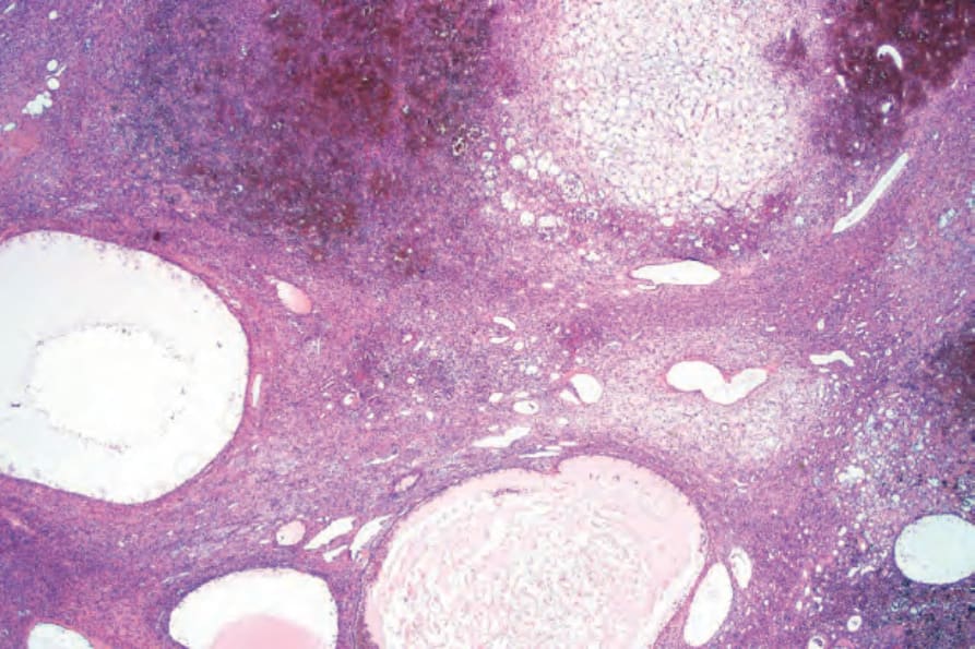
圖 35-656:磷酸鹽尿性間葉腫瘤 (phosphaturic mesenchymal tumor):掃描視野顯示囊腔、血管、局部出血與黏液樣間質。
Fig. 35.656 Phosphaturic mesenchymal tumor: scanning view showing cysts, blood vessels, focal hemorrhage and a myxoid stroma.
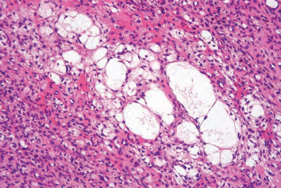
圖 35-657:磷酸鹽尿性間葉腫瘤 (phosphaturic mesenchymal tumor):腫瘤細胞具有嗜伊紅性胞質 (eosinophilic cytoplasm) 與深染的紡錘形核 (hyperchromatic spindled nuclei)。可見微囊 (microcysts)。
Fig. 35.657 Phosphaturic mesenchymal tumor: the tumor cells have eosinophilic cytoplasm and hyperchromatic spindled nuclei. Microcysts are present.
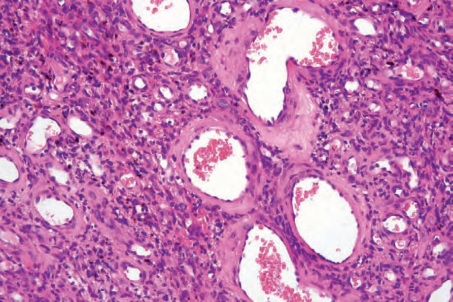
圖 35-658:磷酸鹽尿性間葉腫瘤 (phosphaturic mesenchymal tumor):高倍視野顯示腫瘤細胞與血管。
Fig. 35.658 Phosphaturic mesenchymal tumor: higher-power view showing tumor cells and blood vessels.
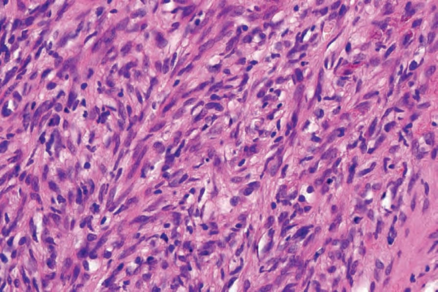
圖 35-659:磷酸鹽尿性間葉腫瘤 (phosphaturic mesenchymal tumor):紡錘細胞的高倍視野,顯示模糊的束狀生長模式 (vaguely fascicular growth pattern)。
Fig. 35.659 Phosphaturic mesenchymal tumor: high-power view of spindled cells showing a vaguely fascicular growth pattern.
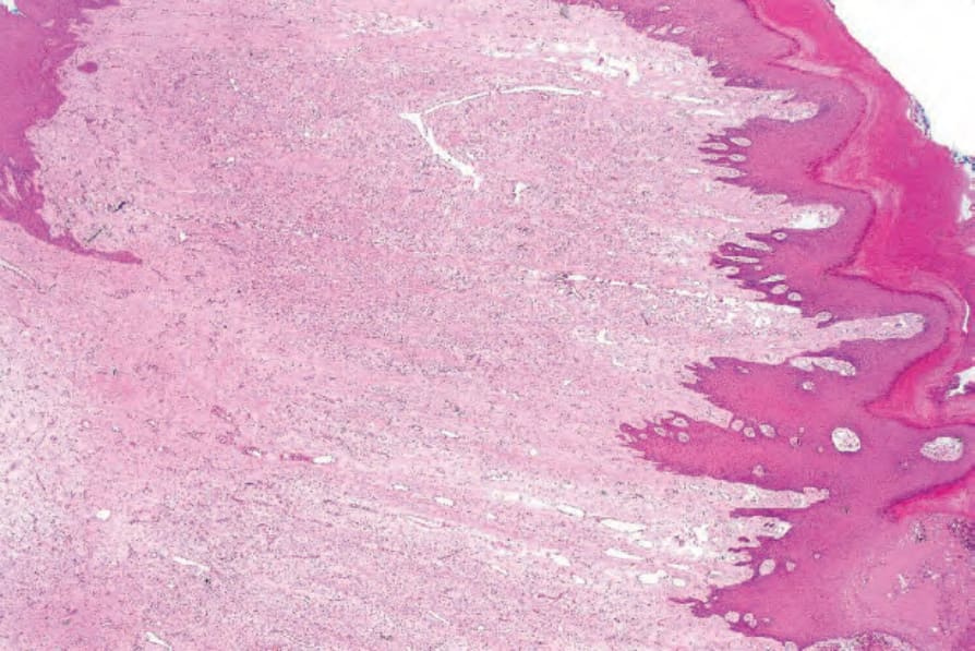
圖 35-660:淺表肢端纖維黏液瘤 (superficial acral fibromyxoma):血管性紡錘細胞腫瘤的低倍視野,具有黏液樣間質 (myxoid stroma)。
Fig. 35.660 Superficial acral fibromyxoma: low-power view of a vascular spindle cell tumor with a myxoid stroma.
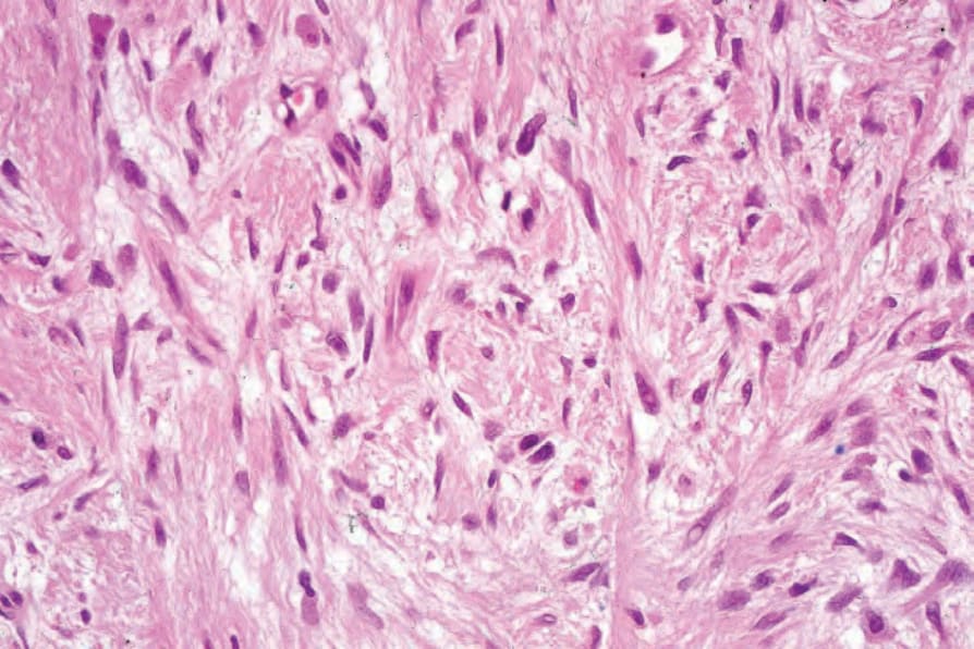
圖 35-661:淺表肢端纖維黏液瘤 (superficial acral fibromyxoma):黏液樣間質中溫和紡錘細胞 (bland spindled cells) 的高倍視野。
Fig. 35.661 Superficial acral fibromyxoma: high-power view of bland spindled cells in a myxoid stroma.
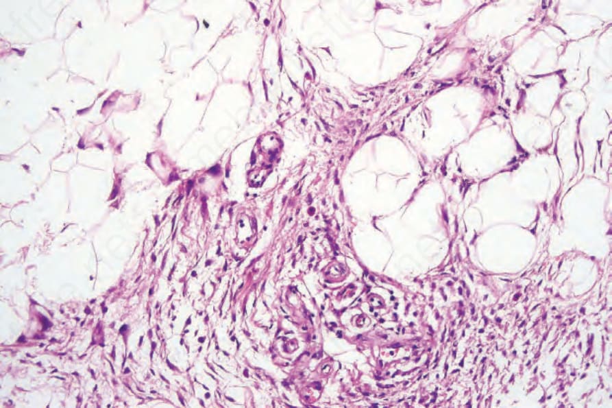
圖 35-662:含鐵血黃素纖維組織細胞性脂肪瘤性病變 (hemosiderotic fibrohistiocytic lipomatous lesion):可見成熟脂肪細胞 (mature adipocytes) 的小葉,內含散在的紡錘細胞與組織細胞 (histiocytes) 區域。
Fig. 35.662 Hemosiderotic fibrohistiocytic lipomatous lesion: there are lobules of mature adipocytes containing scattered areas of spindled cells and histiocytes.
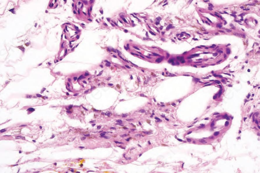
圖 35-663:含鐵血黃素纖維組織細胞性脂肪瘤性病變 (hemosiderotic fibrohistiocytic lipomatous lesions):可見溫和的紡錘形細胞 (bland spindle-shaped cells) 與明顯的含鐵血黃素 (hemosiderin)。
Fig. 35.663 Hemosiderotic fibrohistiocytic lipomatous lesions: there are bland spindle-shaped cells and prominent hemosiderin.