臨床特徵 (Clinical Features)
- 淺表性血管黏液瘤 (superficial angiomyxoma) 為一群相對少見的病灶,好發於成人的頭部、頸部或軀幹,表現為緩慢生長、單發、無症狀的結節或息肉,大小介於 1 至 5 cm。較大的病灶罕見發生。
- 腫瘤在生殖器區域也似乎相對常見,而在口腔與咽部則極為罕見。足底病灶屬例外。已記載三例與毛母質瘤 (pilomatricoma) 相關的病例。局部復發常見,可發生於高達 25% 的病例。
- Carney complex 中所描述的 myxoma 與 superficial angiomyxoma 非常相似,甚至可能完全相同。此 complex 於 1985 年被描述,為一種體染色體顯性 (autosomal dominant) 疾病,與 PRKAR1A 的去活化突變 (inactivating mutations) 相關,PRKAR1A 編碼 protein kinase A 的一個調節次單元 (regulatory subunit),其組成包括 myxoma、斑點狀色素沉著 (spotty pigmentation;臉部特別是嘴唇上的 lentigines) 與內分泌過度活性 (Cushing syndrome、pituitary adenoma 與 testicular tumors)。
- 此 complex 的其他特徵包括 blue nevi 與惡性黑色素性神經鞘瘤 (malignant melanotic schwannoma,先前稱為 psammomatous melanotic schwannoma)。這些 myxoma 可出現於皮膚、乳房與心臟。其在皮膚中的辨識相當重要,因為它們可能是此症候群的首發表現。它們通常為多發、出現於年輕成人,且具有廣泛的解剖分布,特別偏好眼瞼 (eyelids)、耳部 (ears) 與乳頭 (nipples)。
組織學特徵 (Histologic Features)
- 組織學上,病灶位於真皮與皮下,由多個界線不清的黏液樣小葉 (myxoid lobules) 組成,其中含有溫和的紡錘狀 (spindle-shaped) 或星狀 (stellate) 細胞與豐富的小血管 (Figs 35.647 與 35.648)。
- 通常也存在稀疏的發炎細胞浸潤,含有 lymphocytes 與 neutrophils。約 30% 的病例中——無論在原發病灶或其復發病灶——存在上皮成分 (epithelial component)。後者由上皮索 (epithelial strands)、角質囊腫 (keratin cysts) 或基底樣細胞 (basaloid cells) 巢所組成。它可能類似毛囊性腫瘤,如 trichofolliculoma。在單一例外陰部腫瘤中,於病灶內發現壞死性血管炎 (necrotizing vasculitis)。
- 免疫組織化學 (Immunohistochemistry) 顯示腫瘤細胞對 vimentin 呈陽性,並對 CD34 呈不定的局部陽性,對 actin 則較少呈陽性。
鑑別診斷 (Differential Diagnosis)
- Nerve sheath myxoma 由分離、界線分明的結節組成,其中含有 S100 protein 陽性的細胞。
- Low-grade myxofibrosarcoma 同樣為多小葉腫瘤,但位置較深,並含有具有有絲分裂活性 (mitotic activity) 的多形性細胞 (pleomorphic cells)。
- Superficial angiomyxoma 幾乎不會發生於手指,這一點連同更顯著的血管增生 (vascular proliferation)、較高的細胞密度 (cellularity) 與局部發炎細胞浸潤,使其得以與 myxoid cyst 區別。
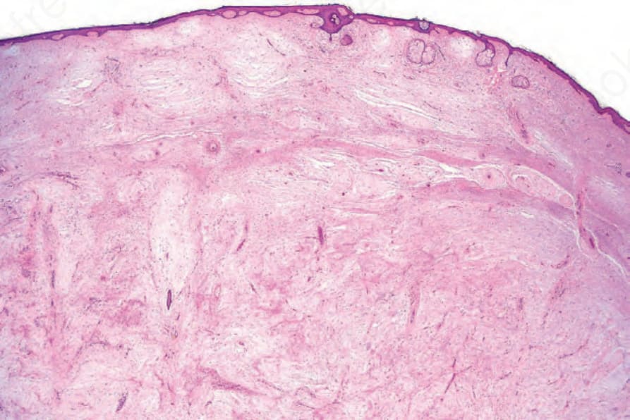
圖 35-647:淺表性血管黏液瘤 (superficial angiomyxoma):真皮內有大量黏液樣變化 (myxoid change),並伴隨眾多小血管。
Fig. 35.647 Superficial angiomyxoma: there is massive myxoid change in the dermis associated with numerous small vessels.
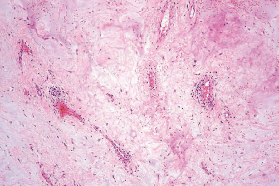
圖 35-648:淺表性血管黏液瘤 (superficial angiomyxoma):黏液樣沉積物 (myxoid deposits) 中含有星狀細胞 (stellate cells) 與薄壁血管 (thin-walled vessels)。
Fig. 35.648 Superficial angiomyxoma: the myxoid deposits contain stellate cells and thinwalled vessels.