Chondroid lipoma
Chondroid lipoma
臨床特徵 (Clinical Features)
軟骨樣脂肪瘤 (chondroid lipoma) 是一種獨特的腫瘤,主要發生於成年女性,好發於近端肢體與肢帶 (limb girdles)。發生於兒童的腫瘤極為罕見。病灶通常較小,主要發生於較深層的軟組織,較少見於皮下組織 (subcutis)。罕見病例可表現於口腔、鼻咽 (nasopharynx) 與骨盆。曾有一例表現於腓神經 (peroneal nerve) 內的案例被描述。其臨床特徵並不具特異性。腫瘤為良性,單純切除後無復發傾向。
致病機轉與組織學特徵 (Pathogenesis and Histologic Features)
此腫瘤之特徵為 t(11;16)(q13;p13),融合 C11orf95 與 MLK2。
此類腫瘤主要見於男性,主要發生於頸部後方、肩部或上背部,特徵性地見於第六或第七十年(60–70 歲)。多發性病灶罕見,部分為家族性。罕見病例可發生於其他部位,包括足、手、臀部、脅腹 (flank)、口腔(含舌)、喉 (larynx)、眼眶 (orbit)、肛周區軟組織、會陰 (perineum)、腹股溝 (groin)、主動脈瓣 (aortic valve)、縱膈 (mediastinum) 與乳房。最後一項可能代表軟組織之乳腺型肌纖維母細胞瘤 (mammary-type myofibroblastoma) 之案例,此病灶與 spindle cell lipoma 密切相關。有趣的是,女性的病灶常發生於非典型部位。它們較常見於肢體與顏面,不少為真皮內 (dermal),且傾向發生於較年輕者。它們通常呈現為界限清楚、緩慢生長、單發、皮下或(罕見,多達百分之十三的病例)真皮內病灶,直徑小於 5 cm。肌內 (intramuscular) 表現極為罕見。曾有一例報告發生於以化學治療處理過的嬰兒型纖維肉瘤 (infantile fibrosarcoma) 部位。Spindle cell lipoma 為一完全良性、
此病灶界限清楚、呈分葉狀,且常有包膜。它由成熟脂肪細胞 (mature adipocytes)、單空泡或多空泡之脂肪母細胞 (uni- or multivacuolated lipoblasts),以及具有顆粒狀嗜伊紅胞質之冬眠瘤樣細胞 (hibernoma-like cells) 混合組成,位於黏液樣與軟骨樣基質 (myxoid and chondroid matrix) 中,該基質可呈現玻璃樣變 (hyalinization)(圖 35.23
1708 Connective tissue tumors
不復發之病灶。Atypical spindle cell lipoma 一詞被提出用以指稱低度惡性脂肪腫瘤,其細胞數量不定,由具有低至中度細胞學異型性 (cytologic atypia) 之梭形細胞、脂肪細胞、脂肪母細胞 (lipoblasts) 與黏液樣基質 (myxoid stroma) 以不同比例組成,且局部復發潛能低(見下文)。
致病機轉與組織學特徵 (Pathogenesis and Histologic Features)
在 spindle cell lipoma 中發現了多種染色體異常,與 pleomorphic lipoma 所見者相同。第 13 與 16 號染色體的單體 (monosomy) 或部分缺失是最常見的變異,亦見於 pleomorphic lipoma,強烈提示此兩種病灶以形態學連續譜 (morphological continuum) 之形式存在。有趣的是,類似的 13q 缺失亦見於乳腺型肌纖維母細胞瘤 (mammary-type myofibroblastoma) 與細胞性血管纖維瘤 (cellular angiofibroma),提示這些腫瘤之間有密切關聯。13q 缺失似乎參與由氧化壓力誘發之 p38 mitogen-activated protein kinase 的活化。
皮下病灶傾向界限清楚,而真皮腫瘤則界限不清。位於顏面的病灶通常顯示對骨骼肌的浸潤。除了成熟之單空泡脂肪細胞外,可見不規則聚集之纖細梭形細胞,具有淡嗜伊紅胞質、均勻的細胞核與罕見的核分裂(圖 35.25)。亦可能存在玻璃樣膠原束 (hyaline bundles of collagen) 與偶見之巨細胞,但脂肪母細胞 (lipoblasts) 罕被辨識出。肥大細胞 (mast cells) 常很多(圖 35.26)。脂肪組織與梭形細胞之相對比例在個別病例間有差異(圖 35.27)。部分病例僅含少量脂肪細胞,罕見地完全不含脂肪細胞。血管分布亦有差異,有時可見局灶性類血管外皮瘤樣 (hemangiopericytoma-like) 區域。廣泛的黏液樣變化可導致顯著的退化性特徵,呈現一種假血管型態 (pseudovascular pattern),其中乳頭狀結構突入空腔內(圖 35.28)。然而,已有研究顯示,至少在某些呈現此種變化的案例中,這些空腔確實由內皮細胞 (endothelial cells) 襯覆,這些應命名為 angiomatous spindle cell lipoma。顯著的黏液樣變化在表現於口腔的病灶中更為常見。除了 spindle cell lipoma 之組織學特徵外,pleomorphic lipoma 的特徵為大量深染且不規則之多核巨細胞,其細胞核常排列成同心圓的花環型態 (floret giant cells)(圖 35.29–35.31)。花環樣細胞 (floret-like cells) 亦可見於因退化過程而脫垂的眼眶脂肪 (prolapsed orbital fat)。核分裂象可能罕見地明顯,偶爾存在多空泡脂肪母細胞 (multivacuolated lipoblasts)(圖 35.32)。顯示少量或無脂肪細胞的病灶可能造成診斷上的挑戰,這可以是具 pleomorphic lipoma 特徵之腫瘤的一項特點。
免疫組化與特殊染色 (Immunohistochemistry & Special Stains)
梭形細胞 CD34 陽性,但 S100 protein 通常陰性。然而,在某些病例中曾報告梭形細胞對後者(S100)呈陽性。Estrogen receptor 在某些病例中可能呈局灶陽性。成熟脂肪細胞 S100 陽性。RB1 (retinoblastoma protein) 為陰性。
超微結構研究顯示具有成熟脂肪細胞特徵之細胞,以及代表未分化間葉細胞 (undifferentiated mesenchymal cells) 之梭形細胞。
鑑別診斷 (Differential Diagnosis)
雖然臨床病史可能具有足夠特徵以協助診斷,但與脂肪肉瘤 (liposarcoma) 之區別在於缺乏脂肪細胞異型性 (adipocytic atypia) 或脂肪細胞大小的變異。在組織學上可相比擬、且侵犯較深層軟組織的病灶,應歸類為 atypical spindle cell lipomatous tumor(先前稱為 spindle cell liposarcoma),以反映其遠較高的復發傾向。
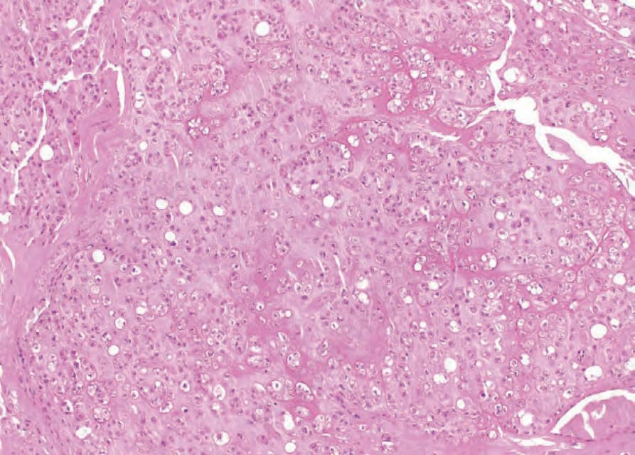
圖 35.23:軟骨樣脂肪瘤 (chondroid lipoma):黏液樣與玻璃樣變之基質 (myxoid and hyalinised stroma),與不同程度空泡化之細胞相關聯。
Fig. 35.23 Chondroid lipoma: myxoid and hyalinised stroma associated with variably vacuolated cells.
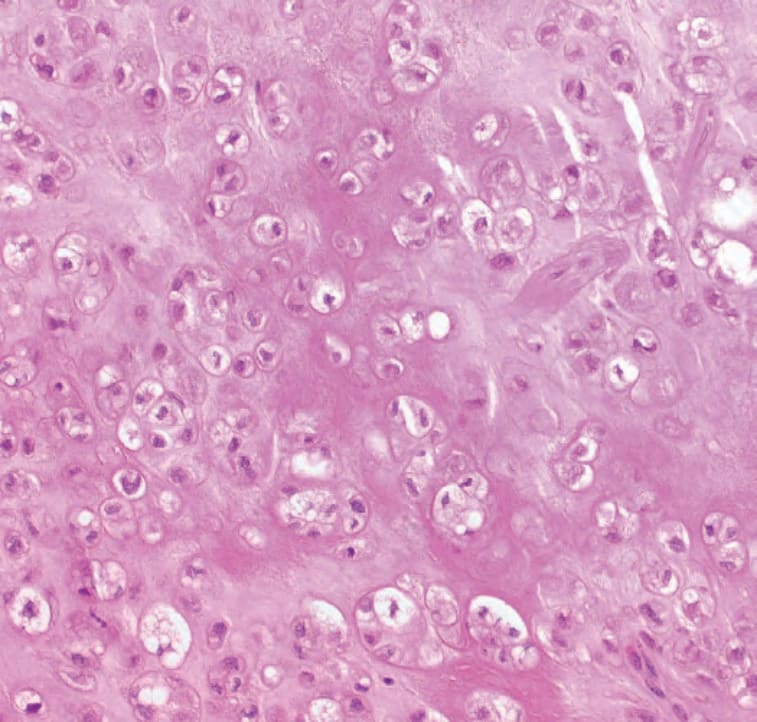
圖 35.24:軟骨樣脂肪瘤 (chondroid lipoma):高倍視野,凸顯廣泛範圍之脂肪母細胞分化 (lipoblastic differentiation)。
Fig. 35.24 Chondroid lipoma: high-power view highlighting wide range of lipoblastic differentiation.
圖 35.25:梭形細胞脂肪瘤 (spindle cell lipoma):腫瘤由成熟脂肪細胞與纖細梭形細胞混合組成,常與寬大之玻璃樣膠原束 (hyalinized collagen) 相關聯。
Fig. 35.25 Spindle cell lipoma: the tumor consists of an admixture of mature adipocytes and delicate spindle cells, often associated with broad bundles of hyalinized collagen.
圖 35.26:梭形細胞脂肪瘤 (spindle cell lipoma):高倍視野。注意顯著的肥大細胞 (mast cells)。
Fig. 35.26 Spindle cell lipoma: high-power view. Note the conspicuous mast cells.
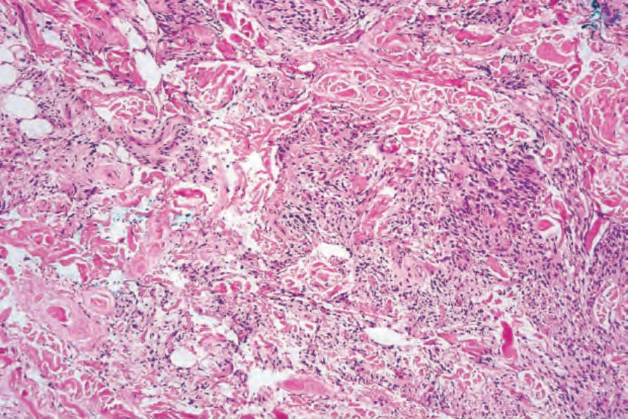
圖 35.27:梭形細胞脂肪瘤 (spindle cell lipoma):在某些案例中,梭形細胞佔優勢,以致病灶容易被誤認為另一種結締組織腫瘤 (connective tissue tumor)。
Fig. 35.27 Spindle cell lipoma: in some examples, the spindled cells predominate such that the lesion is easily mistaken for another connective tissue tumor.
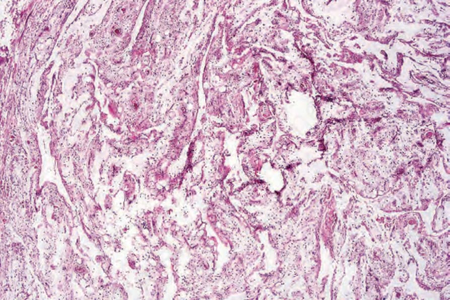
圖 35.28:梭形細胞脂肪瘤 (spindle cell lipoma):罕見地,大量黏液樣退化 (massive myxoid degeneration) 導致假血管空腔 (pseudovascular spaces) 之形成(所謂的 lymphangiomatous variant)。
Fig. 35.28 Spindle cell lipoma: rarely, massive myxoid degeneration results in the formation of pseudovascular spaces (the so-called lymphangiomatous variant).
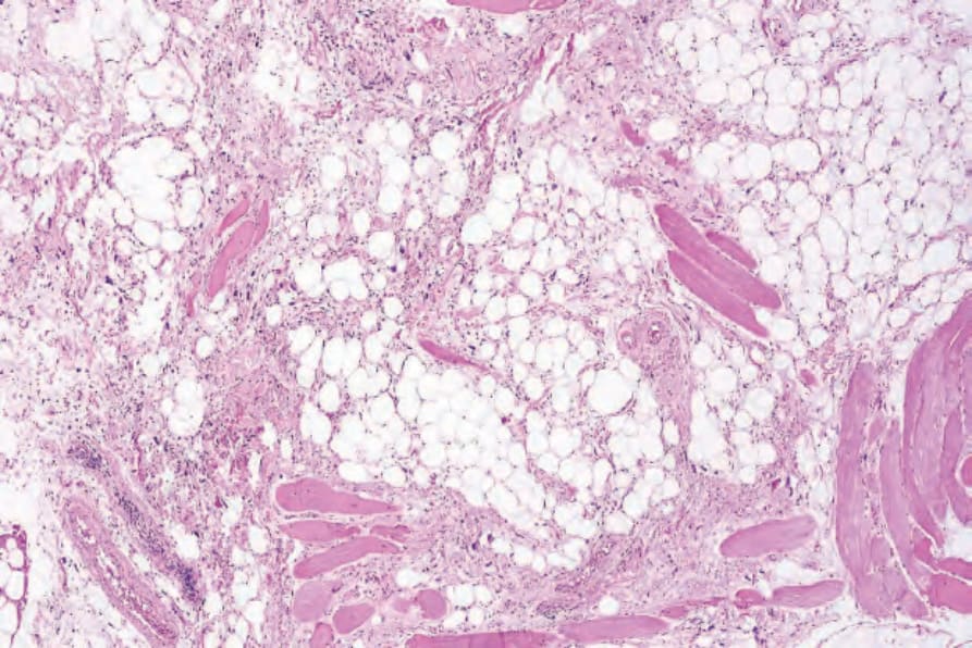
圖 35.29:多形性脂肪瘤 (pleomorphic lipoma):此視野顯示脂肪細胞、粗大膠原束與梭形細胞。
Fig. 35.29 Pleomorphic lipoma: this view shows adipocytes, thick collagen bundles, and spindled cells.
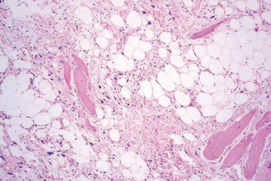
圖 35.30:多形性脂肪瘤 (pleomorphic lipoma):可見多個深染巨細胞 (hyperchromatic giant cells)。
Fig. 35.30 Pleomorphic lipoma: multiple hyperchromatic giant cells are seen.
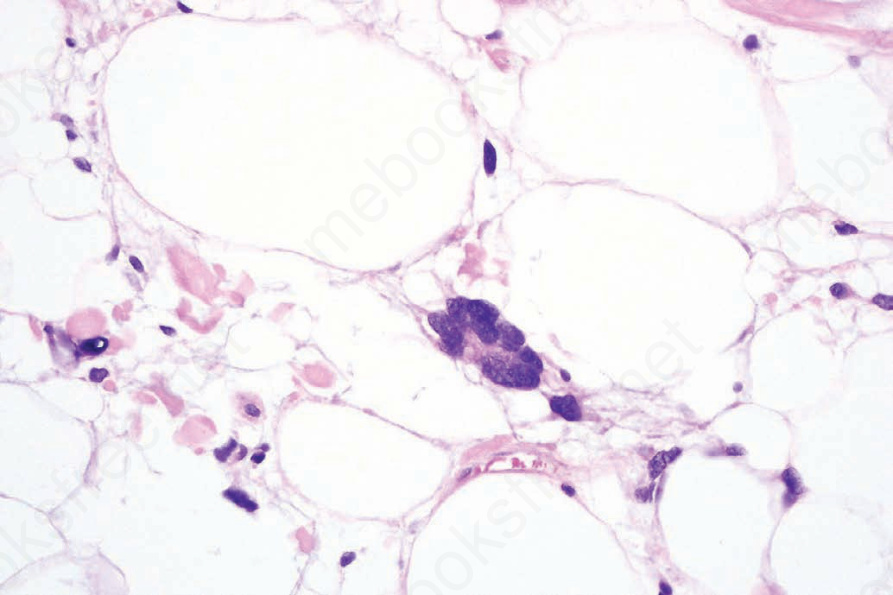
圖 35.32:多形性脂肪瘤 (pleomorphic lipoma):高倍視野。
Fig. 35.32 Pleomorphic lipoma: high-power view.