Chondroid lipoma
Chondroid lipoma
Clinical features Chondroid lipoma is a distinctive tumor that presents predominantly in adult females, with a predilection for the proximal extremities and limb girdles.1,2 Tumors in children are very rare.3 Lesions are usually small and arise mainly in deeper soft tissues and (less commonly) in the subcutis. Rare examples present in the oral cavity, nasopharynx, and pelvis.4–6 A case presenting within the peroneal nerve has been described.7 Clinical features are not distinctive. Tumors are benign and there is no tendency for recurrence after simple excision.
Pathogenesis and histologic features This tumor is characterized by t(11;16)(q13;p13) fusing C11orf95 and MLK2.8–11
Found predominantly in males, they arise mainly in the posterior portion of the neck, shoulder or upper back, and are characteristically seen in the sixth or seventh decade. Multiple lesions are rare and some are familial.10,11 Rare cases can occur at other locations including the foot, hand, buttock, flank, oral cavity (including the tongue), larynx, orbit, soft tissues of the perianal area, perineum, groin, aortic valve, mediastinum and breast.12–22 The last may represent an example of mammary-type myofibroblastoma of soft tissue, a lesion closely related to spindle cell lipoma.23 Interestingly, lesions in women commonly occur in atypical locations.24 They are more common on the extremities and face, are not infrequently dermal, and tend to occur at a younger age. They usually occur as a well-circumscribed, slowly growing, solitary, subcutaneous or (rarely, in up to 13% of cases) dermal lesion, less than 5 cm in diameter.25–27 Intramuscular presentation is very rare.28–31 A case has been reported at the site of an infantile fibrosarcoma treated with chemotherapy.32 Spindle cell lipoma is an entirely benign,
The lesion is well circumscribed, lobular and often encapsulated. It consists of an admixture of mature adipocytes, uni- or multivacuolated lipoblasts and hibernoma-like cells with granular eosinophilic cytoplasm in a myxoid and chondroid matrix, which can show hyalinization (Figs 35.23
1708 Connective tissue tumors
nonrecurring lesion. Atypical spindle cell lipoma is a term proposed to refer to low-grade malignant adipose tumors with variable cellularity consisting of spindle cells with low to moderate cytologic atypia, adipocytes, lipoblasts, and myxoid stroma in varying proportions and with low potential for local recurrence (see below).33–35
Pathogenesis and histologic features A number of chromosomal abnormalities have been found in spindle cell lipoma, identical to those found in pleomorphic lipoma. Monosomy or partial loss of chromosomes 13 and 16 are the most common alterations also seen in pleomorphic lipoma, strongly suggesting that these two lesions exist as a morphological continuum.36–39 Interestingly, similar loss of 13q has been found in mammary-type myofibroblastoma and cellular angiofibroma, suggesting a close link between these tumors.25,40–42 The 13q deletion seems to be involved in the activation of p38 mitogen-activated protein kinase induced by oxidative stress.43
Subcutaneous lesions tend to be well circumscribed while dermal tumors are ill defined.27 Lesions located on the face usually display infiltration of skeletal muscle.44 In addition to mature univacuolated adipocytes, irregular collections of slender spindled cells are seen with pale eosinophilic cytoplasm, uniform nuclei and rare mitoses (Fig. 35.25). Hyaline bundles of collagen and occasional giant cells may also be present, but lipoblasts are rarely identified. Mast cells are often numerous (Fig. 35.26). The relative proportions of adipose tissue to spindled cells vary between individual cases (Fig. 35.27). Some cases contain few or, rarely, no adipocytes at all.45 Vascularity also varies and focal hemangiopericytoma-like areas are sometimes seen. Extensive myxoid change can lead to striking degenerative features with a pseudovascular pattern in which papillary structures project into empty spaces (Fig. 35.28). However, it has been shown that at least in some examples showing this change, the spaces are truly lined by endothelial cells, and these should be named angiomatous spindle cell lipoma.46,47 Prominent myxoid change is more common in lesions presenting in the oral cavity. In addition to the histologic features of spindle cell lipoma, pleomorphic lipoma is characterized by numerous hyperchromatic and irregular multinucleated giant cells, with nuclei often arranged in a concentric floret pattern (floret giant cells) (Figs 35.29–35.31). Floret-like cells can also be seen in prolapsed orbital fat as a result of a degenerative process.48 Mitotic figures may rarely be evident and occasional multivacuolated lipoblasts are sometimes present (Fig. 35.32). Lesions that show few or no adipocytes may pose a diagnostic challenge and this can be a feature of tumors with features of pleomorphic lipoma.49,50
The spindled cells are positive for CD34 but usually negative for S100 protein. However, positivity in the spindle cells for the later has been
1709 Benign adipocytic tumors and tumorlike lesions
reported in some cases.51 Estrogen receptor may be focally positive in some cases. Mature adipocytes are positive for S100. RB1 (retinoblastoma protein) is negative.
Ultrastructural studies show cells with features of mature adipocytes and spindled cells representing undifferentiated mesenchymal cells.46
Differential diagnosis Although the clinical history may be sufficiently characteristic to aid the diagnosis, distinction from liposarcoma is made by the absence of either adipocytic atypia or variation in adipocyte size. Histologically comparable lesions involving deeper soft tissue should be classified as atypical spindle cell lipomatous tumor (previously termed spindle cell liposarcoma), reflecting their much greater tendency to recur.
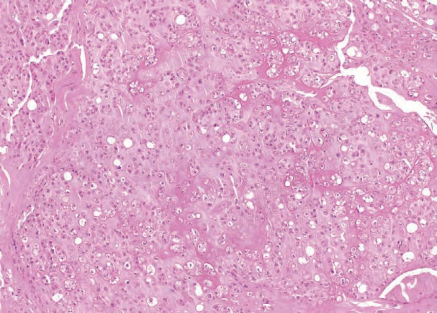
Fig. 35.23 Chondroid lipoma: myxoid and hyalinised stroma associated with variably vacuolated cells.
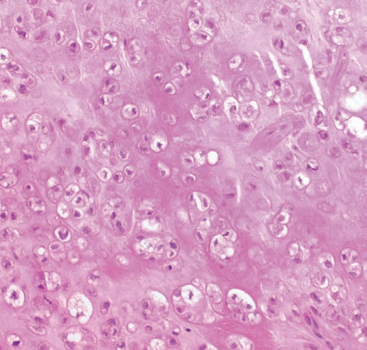
Fig. 35.24 Chondroid lipoma: high-power view highlighting wide range of lipoblastic differentiation.
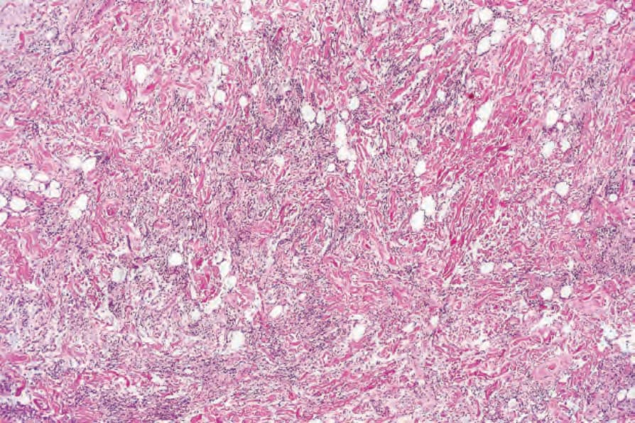
Fig. 35.25 Spindle cell lipoma: the tumor consists of an admixture of mature adipocytes and delicate spindle cells, often associated with broad bundles of hyalinized collagen.
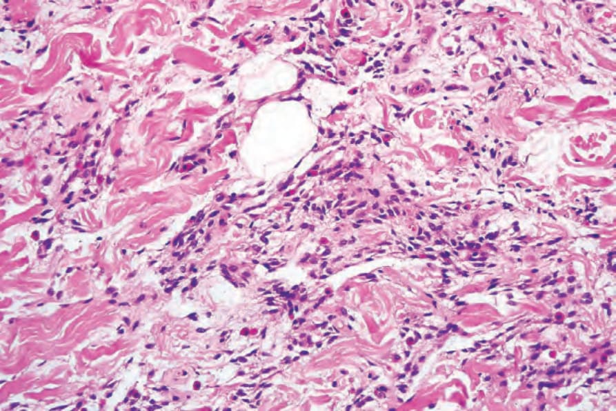
Fig. 35.26 Spindle cell lipoma: high-power view. Note the conspicuous mast cells.
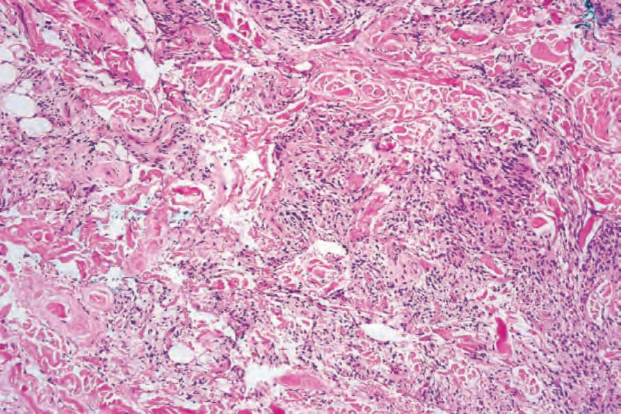
Fig. 35.27 Spindle cell lipoma: in some examples, the spindled cells predominate such that the lesion is easily mistaken for another connective tissue tumor.
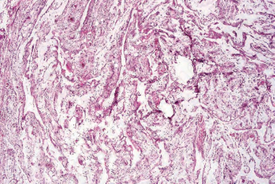
Fig. 35.28 Spindle cell lipoma: rarely, massive myxoid degeneration results in the formation of pseudovascular spaces (the so-called lymphangiomatous variant).
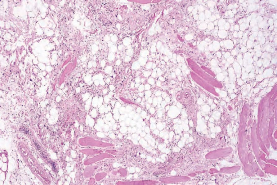
Fig. 35.29 Pleomorphic lipoma: this view shows adipocytes, thick collagen bundles, and spindled cells.
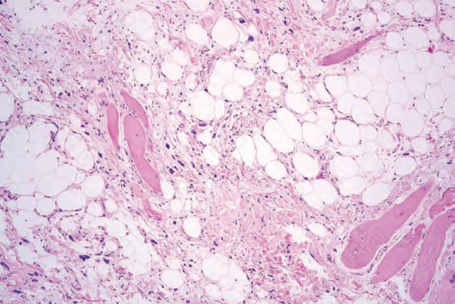
Fig. 35.30 Pleomorphic lipoma: multiple hyperchromatic giant cells are seen.
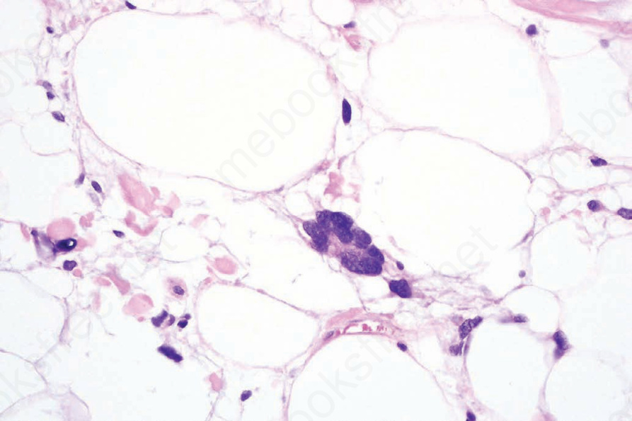
Fig. 35.32 Pleomorphic lipoma: high-power view.