疾病定義與分類
淋巴管瘤 (lymphangioma) 主要有四種形式:
- 海綿狀淋巴管瘤 (cavernous lymphangioma)
- 囊狀水瘤 (cystic hygroma)
- 局限性淋巴管瘤 (lymphangioma circumscriptum)
- 後天性進行性淋巴管瘤 (acquired progressive lymphangioma)(良性淋巴管內皮瘤,benign lymphangioendothelioma)。真正的微血管型淋巴管瘤 (capillary lymphangioma) 是否存在,極具爭議。
臨床特徵 (Clinical Features)
海綿狀淋巴管瘤 (Cavernous Lymphangioma)
Cavernous lymphangioma 為一種先天性或嬰兒期病灶,男女發生率相等,最常出現於頭頸部(尤其是舌部)及四肢。它表現為一個大型、瀰漫性、質地相當柔軟麵糰狀 (doughy) 的腫塊,在單純切除後極易局部復發。少數病例首次發病於成人。與 lymphangioma circumscriptum 並存者極為罕見。
囊狀水瘤 (Cystic Hygroma)
Cystic hygroma 同樣是一種嬰兒期病灶,表現為大型囊性腫塊,最常見於頸部、腋窩或腹股溝區。亦曾有陰囊病灶的報告。也可發生於腹腔內及胸腔內。除非廣泛切除,否則同樣易於局部復發,但此傾向遠不如海綿狀腫瘤明顯。與葡萄酒色斑 (port-wine stain) 並存者極為例外。已有發生於成人的病例報告。Cystic hygroma 曾與第 21、13、18 號染色體三體 (trisomy 21, 13, and 18),以及 Turner 與 Noonan 症候群相關聯。
局限性淋巴管瘤 (Lymphangioma Circumscriptum)
Lymphangioma circumscriptum 同樣最常見於嬰兒期,但可發生於任何年齡,男女分布相等。雖然可發生於任何皮膚部位,但通常侵犯四肢近端及肢帶 (limb girdles)。病灶表現為局部聚集的眾多小水疱或泡狀突起 (blebs),有時可融合形成較大的腫塊,內含透明液體或血液(圖 35.595)。偶見單發性病灶。除非被病人搔抓刺激,否則通常無症狀。
後天性進行性淋巴管瘤(良性淋巴管內皮瘤)(Acquired Progressive Lymphangioma [Benign Lymphangioendothelioma])
Acquired progressive lymphangioma 是一種罕見腫瘤,最初被描述為較常見於兒童。然而較新近的文獻顯示其在成人中較為常見。男女發生率相等,尤其侵犯四肢,特別是上肢,但其解剖分布範圍甚廣。它表現為一個單發、邊界清楚的紅斑性斑點 (macule) 或斑塊 (plaque),並逐漸增大。單純切除通常可治癒,僅有例外的局部復發。偶見部分自發性消退者極為罕見。曾有一例記錄於放射治療後發生,另有一例發生於股動脈攝影 (femoral arteriography) 之後,並有一例報告發生於 HIV 陽性病人。
致病機轉與組織病理特徵 (Pathogenesis and Histologic Features)
近期已顯示,許多以孤立形式出現,或作為複雜血管畸形 (complex vascular malformations) 一部分而出現的淋巴管腫瘤,帶有 PIK3CA 的體細胞突變 (somatic mutations)。
Cavernous lymphangioma 典型為位於真皮或皮下脂肪中、邊界不清的病灶,由眾多擴張的淋巴管道組成,內皮無異型性 (without endothelial atypia)(圖 35.596)。周圍間質可不明顯,或由明顯的外膜型網狀纖維 (adventitial-type reticulin fibers) 組成,並伴有慢性發炎細胞浸潤。
Cystic hygroma 在組織學上幾乎與海綿狀病灶無法區別,差別僅在於其薄壁的淋巴間隙呈現粗大囊狀擴張 (gross cystic dilatation)。除淋巴球浸潤外,散在的淋巴濾泡 (lymphoid follicles) 亦屬常見。
在這兩種病灶中,血管腔內常含有蛋白性、淡染、嗜伊紅性的淋巴液 (proteinaceous, pale, eosinophilic lymph),且管壁可能含有不完整的一層平滑肌。
Lymphangioma circumscriptum 通常位於淺層真皮。它由多條擴張的淋巴管道組成,這些管道往往管壁相當厚,且常顯得延伸至其上方的表皮(圖 35.597)。後者(表皮)常呈棘層肥厚 (acanthotic),有時可見間質淋巴球浸潤。較深的真皮中可見海綿狀間隙,偶爾可見一條肌性淋巴管道(常被視為供應血管,feeding vessel)(圖 35.598)。
雖然這些腫瘤大多數很可能代表發育性畸形 (developmental malformations),但有一小部分為後天性,通常發生於區域淋巴結廓清術 (block dissection of regional lymph nodes) 或放射治療之後。同樣地,少數病例與潛在的海綿狀或囊狀淋巴管瘤相關。外陰部病灶為特發性,或曾與 Crohn disease 及放射治療相關。已描述罕見的外陰部病灶與惡性腫瘤及化膿性汗腺炎 (hidradenitis suppurativa) 相關。切除後復發相當常見。
在 progressive lymphangioma 中,淺層真皮的侵犯通常很明顯,但延伸至深層真皮及淺層皮下組織者亦不少見(圖 35.599)。可見由單層扁平、變薄的內皮細胞 (flat attenuated endothelial cells) 所襯覆的水平、不規則、薄壁血管道,分隔解剖膠原束 (dissecting the collagen bundles)(圖 35.600)。這些管道看似空虛,但偶見蛋白性物質或紅血球。有時可見局灶性乳頭狀突起 (focal papillary projections)。某些血管間隙可能有一層平滑肌。間質發炎並非其特徵。
鑑別診斷 (Differential Diagnosis)
Progressive lymphangioma 可能類似低度惡性血管肉瘤 (low-grade angiosarcoma) 及斑片期 Kaposi 肉瘤 (patch-stage Kaposi sarcoma)。前者至少有局灶性細胞異型性 (cytologic atypia) 與多層化 (multilayering),且臨床情境不同。後者通常有多發性病灶,且組織學上有含鐵血黃素沉積 (hemosiderin deposition),伴外滲的紅血球及鄰近的發炎細胞(包括漿細胞,plasma cells)。與淋巴管瘤病 (lymphangiomatosis) 的區別主要藉由病灶的臨床範圍來判定。
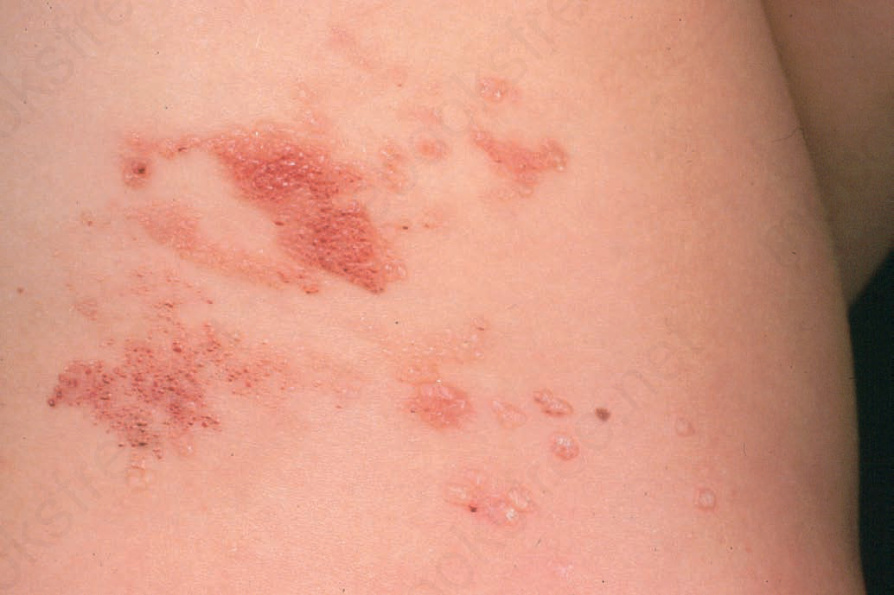
圖 35-595:局限性淋巴管瘤 (lymphangioma circumscriptum):病灶表現為數量不一的淺表充液泡狀突起 (fluid-filled blebs)。取自已故 N.P. Smith 醫師之收藏,英國倫敦皮膚科研究所 (the Institute of Dermatology, London, UK)。
Fig. 35.595 Lymphangioma circumscriptum: the lesion presents as variable numbers of superficial fluid-filled blebs. From the collection of the late N.P. Smith, MD, the Institute of Dermatology, London, UK.
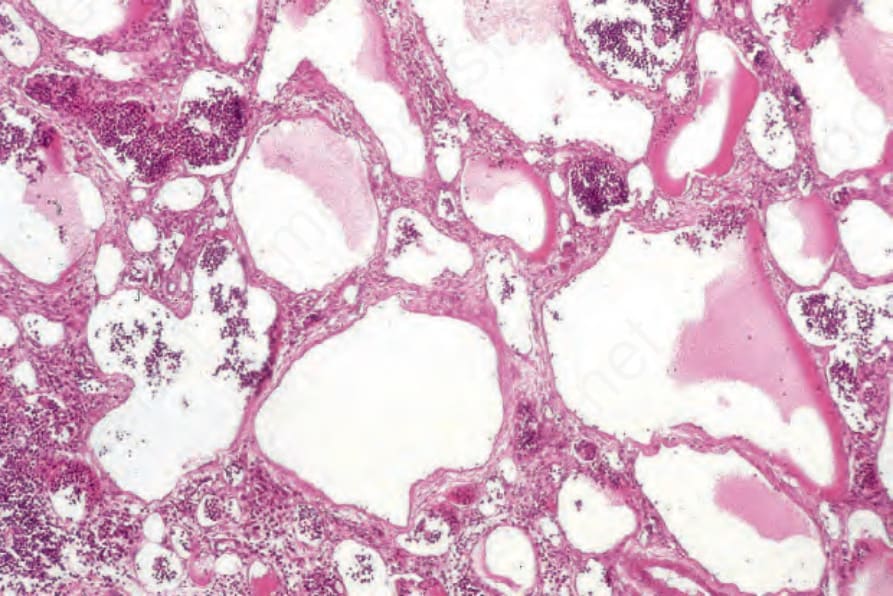
圖 35-596:海綿狀淋巴管瘤 (cavernous lymphangioma):廣泛擴張、充滿淋巴液的管道為其特徵。
Fig. 35.596 Cavernous lymphangioma: widely dilated lymph-filled channels are characteristic.
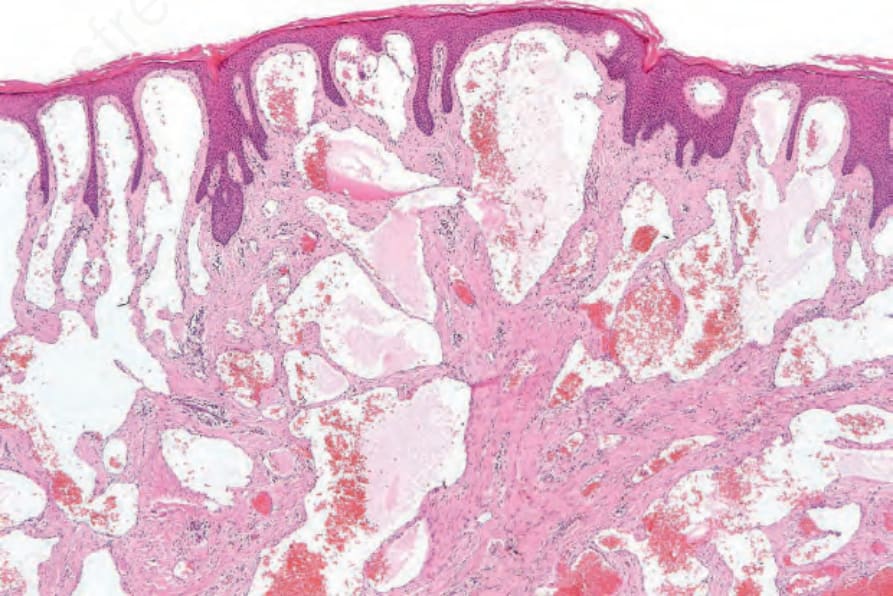
圖 35-597:局限性淋巴管瘤 (lymphangioma circumscriptum):網狀真皮 (reticular dermis) 與乳頭真皮 (papillary dermis) 中皆可見薄壁淋巴管道。
Fig. 35.597 Lymphangioma circumscriptum: thin-walled lymphatic channels are present in both the reticular and papillary dermis.
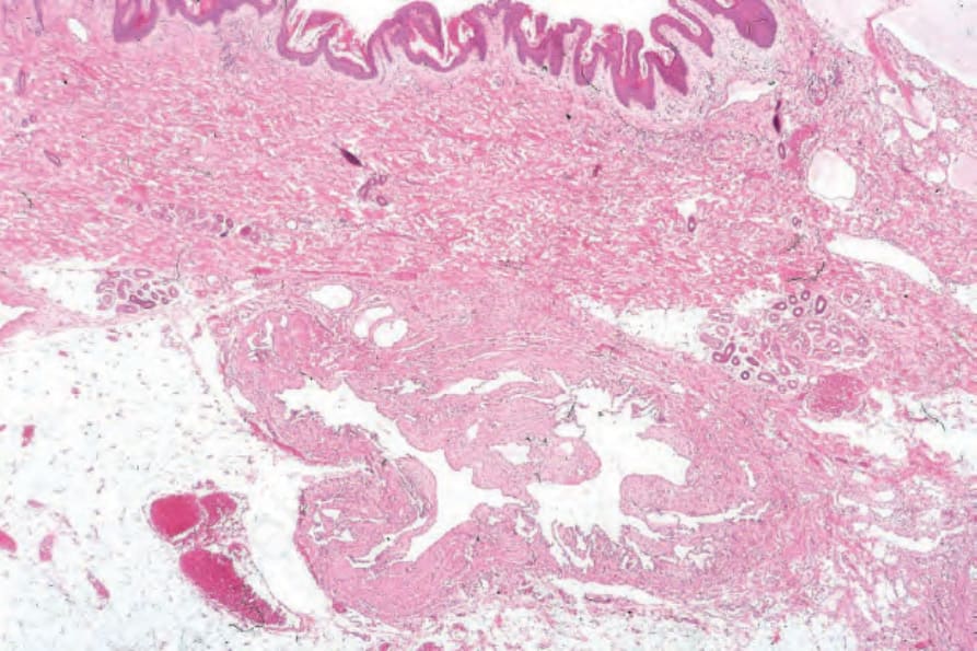
圖 35-598:局限性淋巴管瘤 (lymphangioma circumscriptum):皮下脂肪內可見一條大型肌性「供應」淋巴主幹 (muscular ‘feeder’ lymphatic trunk)。若手術時未予結紮,復發風險甚高。
Fig. 35.598 Lymphangioma circumscriptum: within the subcutaneous fat is a large muscular ‘feeder’ lymphatic trunk. If this is not ligated at surgery, there is a high risk of recurrence.
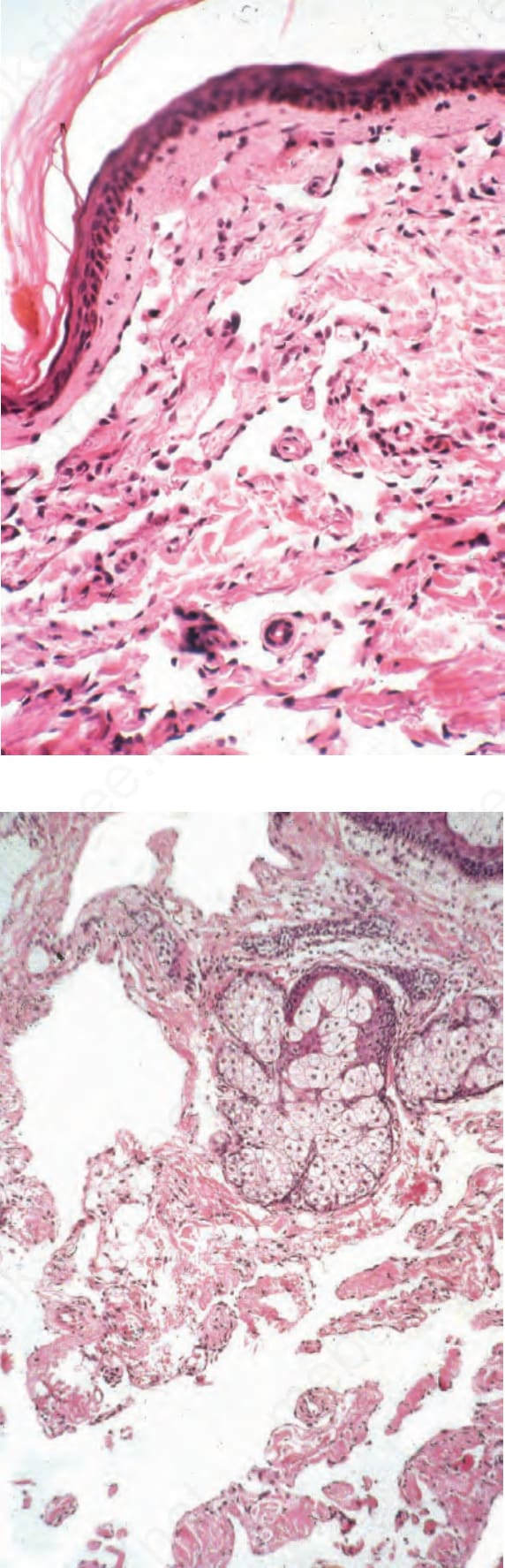
圖 35-599:進行性淋巴管瘤 (progressive lymphangioma):儘管構造上與血管肉瘤 (angiosarcoma) 相似,但完全沒有內皮多層化 (endothelial multilayering) 或核異型性 (nuclear atypia)。
Fig. 35.599 Progressive lymphangioma: despite the architectural resemblance to angiosarcoma, there is a complete absence of endothelial multilayering or nuclear atypia.
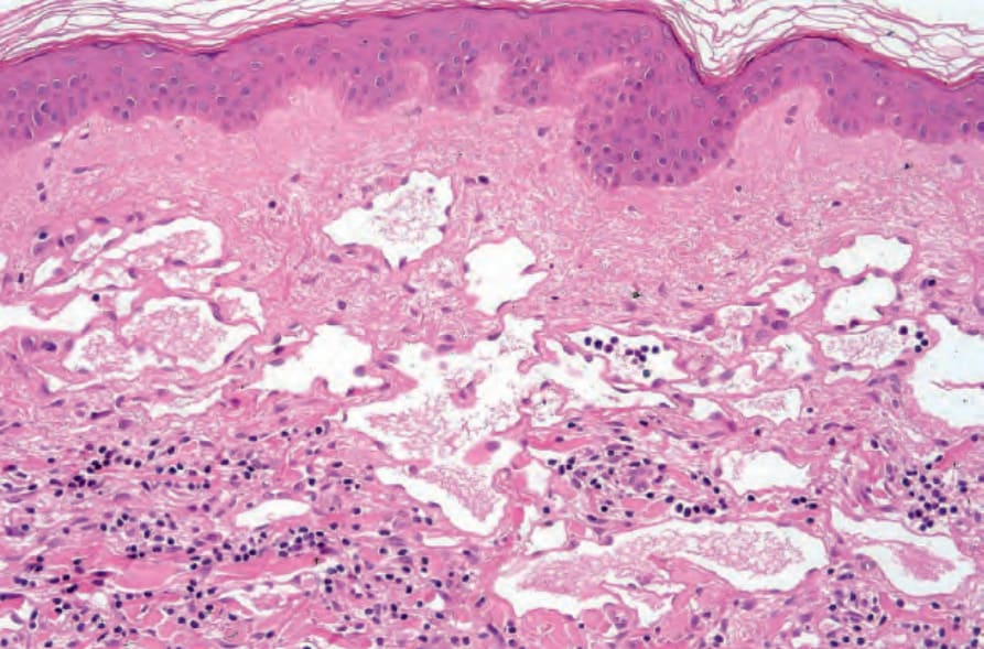
圖 35-600:進行性淋巴管瘤 (progressive lymphangioma):部分病例顯示真皮結構被擴張的淋巴間隙分隔解剖 (dissection) 的情形更為明顯。
Fig. 35.600 Progressive lymphangioma: some cases show more dissection of dermal structures by the dilated lymphatic spaces.