臨床特徵 (Clinical Features)
- 上皮樣血管肉瘤 (epithelioid angiosarcoma) 代表上皮樣血管腫瘤譜系中惡性的一端。此名詞保留用於幾乎完全由上皮樣細胞 (epithelioid cells) 組成的腫瘤,因為傳統血管肉瘤 (conventional angiosarcomas) 也相當常見呈局灶性上皮樣型態。
- 我們將此名詞保留用於具上皮樣型態、且發生於前述皮膚血管肉瘤 (cutaneous angiosarcoma) 傳統情境之外的腫瘤。雖然此腫瘤侵犯皮膚屬罕見,但其發生頻率似乎比過去所認為的更高。在它被劃分為一個獨特疾病實體之前,許多病例很可能被誤診為黑色素細胞性 (melanocytic) 或上皮性 (epithelial) 腫瘤。
- 病灶有廣泛的解剖分布,通常出現於成人,無性別偏好 (Fig. 35.588)。腫瘤為單發,較少見為多發。
- 發生於皮膚、軟組織及其他器官的腫瘤預後極差。原發性皮膚病灶可見早期轉移與高達 55% 的高死亡率。
- 已有文獻記載源自心臟、縱膈、骨及血管內上皮樣血管肉瘤的罕見皮膚轉移。
組織病理特徵 (Histopathology)
- 病灶具浸潤性,由成片的大型卵圓形或圓形細胞組成,胞質豐富、呈嗜伊紅性 (eosinophilic) 或雙嗜性 (amphophilic),細胞核呈泡狀 (vesicular),並具明顯的嗜伊紅性核仁 (Figs 35.589–35.591)。
- 雖然存在細胞學異型性 (cytologic atypia),腫瘤細胞相對單一型 (monomorphic)。有絲分裂 (mitosis)、壞死 (necrosis) 與出血 (hemorrhage) 為常見表現。局灶可見少數細胞具胞質內腔隙 (intracytoplasmic lumina),內含偶見的紅血球 (Fig. 35.592)。血管形成 (blood vessel formation) 也可為一項特徵。
- Reticulin 染色有助於凸顯其血管形成性 (vasoformative) 結構。
免疫組化與特殊染色 (Immunohistochemistry & Special Stains)
- 免疫組化上,腫瘤細胞一致對 ERG、CD31、FLI1 或 von Willebrand factor 呈陽性 (Fig. 35.593)。
- Cytokeratin 在高達 50–60% 的病例中亦呈陽性,EMA 在約 25% 的病例中呈局灶性陽性 (Fig. 35.594)。INI 在腫瘤細胞中常呈局灶性或瀰漫性陽性。
- 可見對 Melan-A 與 smooth muscle actin 的罕見局灶性陽性。一例於乳癌放射治療情境下發生的純上皮樣血管肉瘤對 CD30 呈陽性。
- 在考慮與癌 (carcinoma,特別是轉移性) 或上皮樣肉瘤 (epithelioid sarcoma) 的鑑別診斷時,這些重要發現必須謹記在心。
鑑別診斷 (Differential Diagnosis)
- 鑑別診斷包括轉移性癌 (metastatic carcinoma)、melanoma、epithelioid sarcoma 及上皮樣惡性許旺氏瘤 (epithelioid malignant schwannoma),這些病灶皆對內皮標記 (endothelial markers) 呈陰性,且缺乏局灶性血管形成與胞質內腔隙 (intracytoplasmic lumina)。
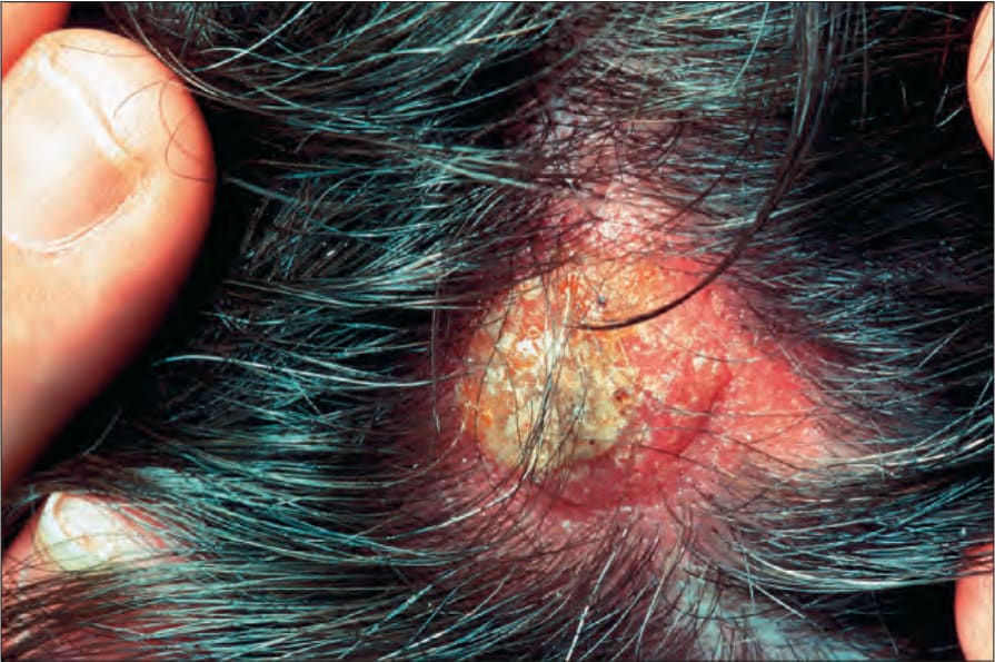
圖 35-588:上皮樣血管肉瘤 (epithelioid angiosarcoma):於頭皮 (scalp) 的表現並不少見。與較典型的血管肉瘤相比,此病灶的血管性較不明顯。By courtesy of the Institute of Dermatology, London, UK.
Fig. 35.588 Epithelioid angiosarcoma: presentation on the scalp is not uncommon. The lesion is less obviously vascular when compared with more typical angiosarcoma. By courtesy of the Institute of Dermatology, London, UK.
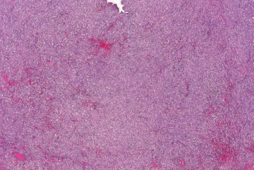
圖 35-589:上皮樣血管肉瘤 (epithelioid angiosarcoma):可見瀰漫性上皮樣細胞浸潤,伴多處出血 (hemorrhage) 病灶。
Fig. 35.589 Epithelioid angiosarcoma: there is a diffuse epithelioid cell infiltrate with multiple foci of hemorrhage.
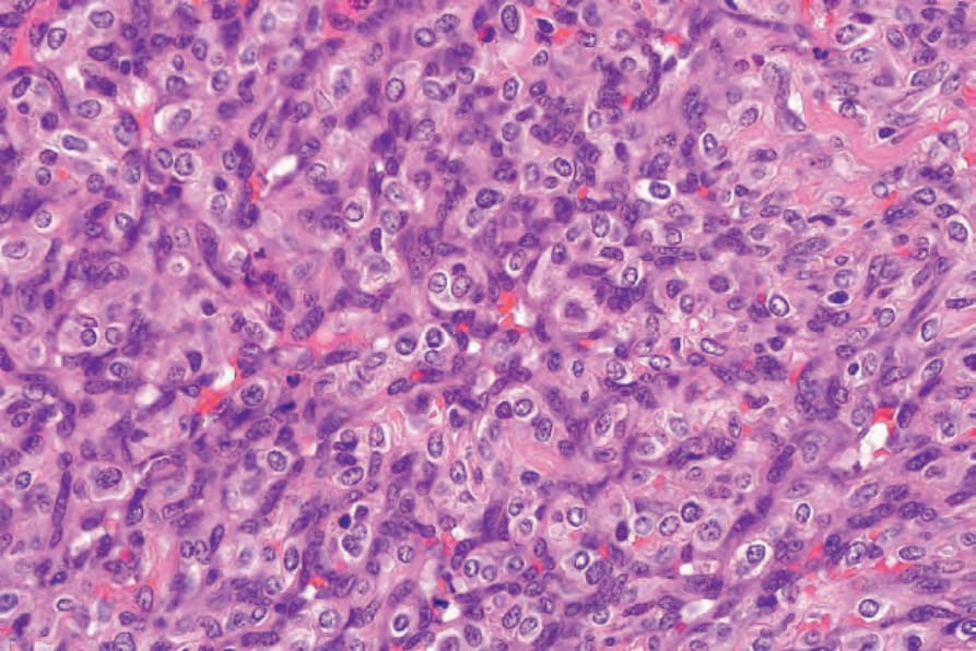
圖 35-590:上皮樣血管肉瘤 (epithelioid angiosarcoma):腫瘤細胞具豐富的嗜伊紅性胞質 (eosinophilic cytoplasm) 與多形性泡狀核 (pleomorphic vesicular nuclei)。
Fig. 35.590 Epithelioid angiosarcoma: the tumor cells have abundant eosinophilic cytoplasm and pleomorphic vesicular nuclei.
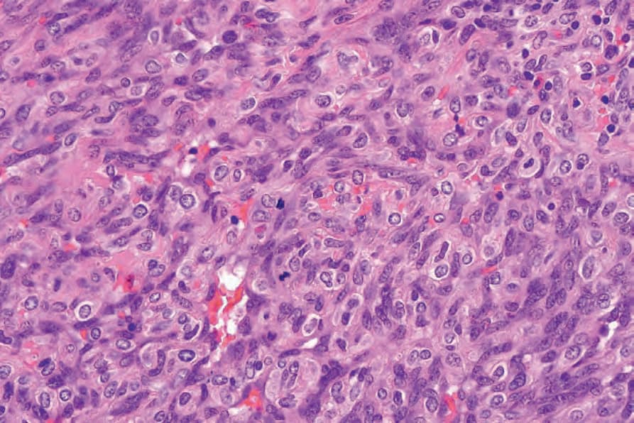
圖 35-591:上皮樣血管肉瘤 (epithelioid angiosarcoma):注意有絲分裂 (mitoses)。
Fig. 35.591 Epithelioid angiosarcoma: note the mitoses.
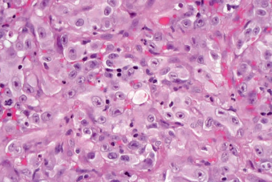
圖 35-592:上皮樣血管肉瘤 (epithelioid angiosarcoma):視野中央可見胞質內腔隙 (intracytoplasmic lumina)。
Fig. 35.592 Epithelioid angiosarcoma: intracytoplasmic lumina are present in the center of the field.
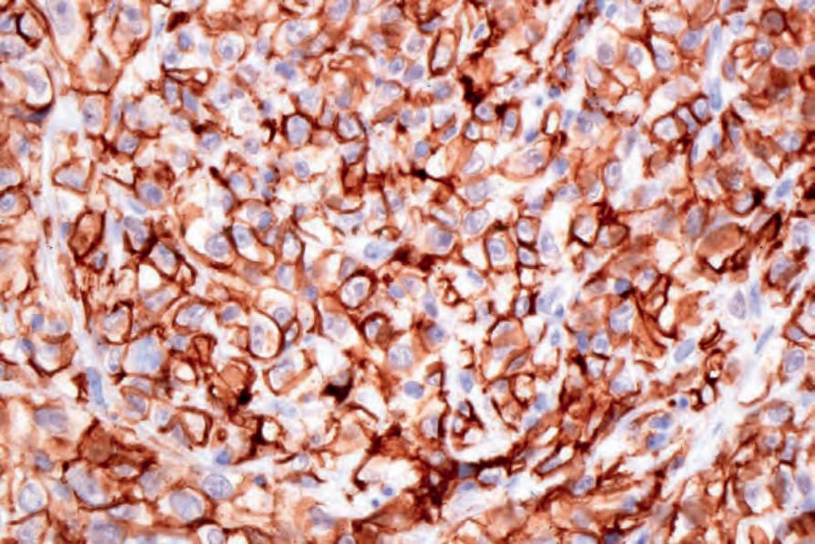
圖 35-593:上皮樣血管肉瘤 (epithelioid angiosarcoma):腫瘤細胞表現 CD31。
Fig. 35.593 Epithelioid angiosarcoma: the tumor cells express CD31.
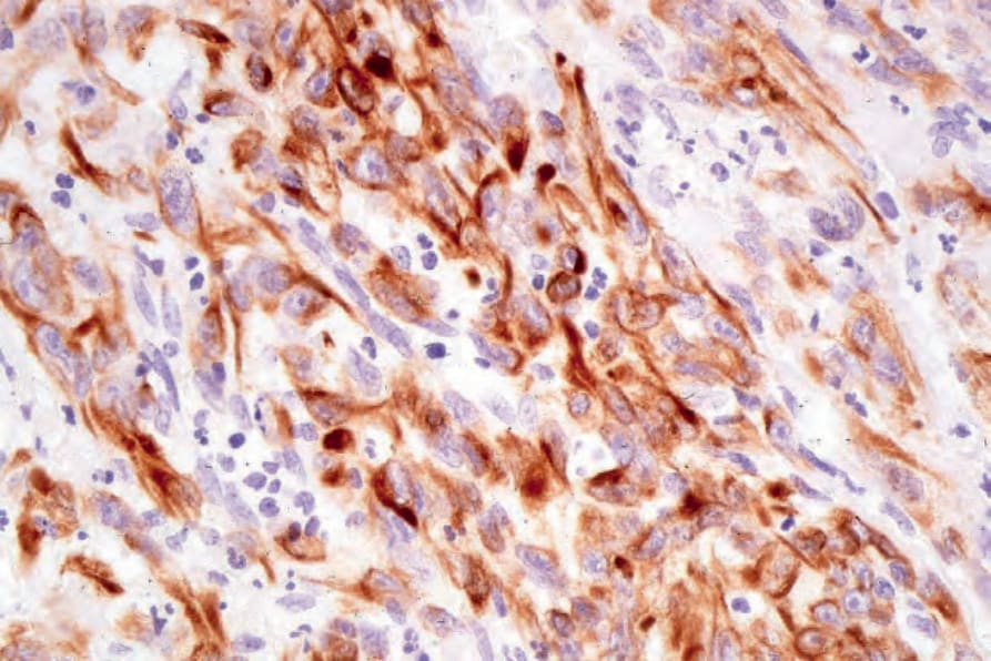
圖 35-594:上皮樣血管肉瘤 (epithelioid angiosarcoma):此例亦表現 keratin (MNF-116)。
Fig. 35.594 Epithelioid angiosarcoma: this example also expressed keratin (MNF -116).