Epithelioid angiosarcoma
Epithelioid angiosarcoma
Clinical features Epithelioid angiosarcoma represents the malignant end of the spectrum of epithelioid vascular neoplasms.1–5 The term is reserved for tumors composed almost exclusively of epithelioid cells, as conventional angiosarcomas are quite often focally epithelioid. We reserve this term to tumors with epithelioid morphology occurring outside the conventional settings of cutaneous angiosarcoma described earlier. Although involvement of the skin by this tumor is rare, it appears to occur more often than was previously thought. It is likely that before it was delineated as a distinctive entity, cases were misdiagnosed as melanocytic or epithelial neoplasms. Lesions have a wide anatomical distribution and usually present in adults, with no sex predilection (Fig. 35.588). Tumors are single or less commonly multiple.4 Tumors in the skin, soft tissues and other organs have a dismal prognosis. Early metastasis and high mortality of up to 55% is seen in primary cutaneous
Exceptional cutaneous metastasis from cardiac, mediastinal, bone and intravascular epithelioid angiosarcomas has been documented.4,13,14
Histologic features Lesions are infiltrative and composed of sheets of large oval or round cells with abundant eosinophilic or amphophilic cytoplasm and vesicular nuclei with prominent eosinophilic nucleoli (Figs 35.589–35.591). Although cytologic atypia is present, the tumor cells are relatively monomorphic. Mitosis, necrosis and hemorrhage are common findings. Focally, a few cells show intracytoplasmic lumina containing occasional red blood cells (Fig. 35.592). Blood vessel formation can also be a feature.
A reticulin stain is useful to highlight the vasoformative architecture. Immunohistochemically, the tumor cells are consistently positive for ERG, CD31, FLI1 or von Willebrand factor (Fig. 35.593). Cytokeratin is also positive in up to 50–60% of cases and EMA is focally positive in about 25% of cases (Fig. 35.594).1,4 INI is often focally or diffusely positive in tumor
1863 Malignant vascular tumors
cells.4 Exceptional focal positivity for Melan-A and smooth muscle actin can be seen.4 A case of pure epithelioid angiosarcoma arising in the setting of radiotherapy for breast cancer was positive for CD30.15 These important findings need to be borne in mind when considering the differential diagnosis from a carcinoma (especially metastatic) or epithelioid sarcoma.
Differential diagnosis The differential diagnosis includes metastatic carcinoma, melanoma, epithelioid sarcoma and epithelioid malignant schwannoma, all of which are negative for endothelial markers and lack focal blood vessel formation and intracytoplasmic lumina.
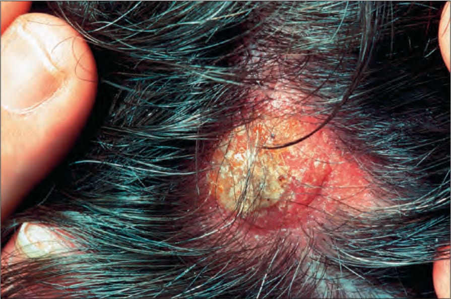
Fig. 35.588 Epithelioid angiosarcoma: presentation on the scalp is not uncommon. The lesion is less obviously vascular when compared with more typical angiosarcoma. By courtesy of the Institute of Dermatology, London, UK.
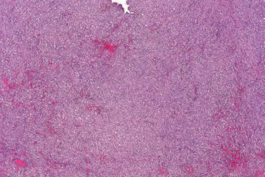
Fig. 35.589 Epithelioid angiosarcoma: there is a diffuse epithelioid cell infiltrate with multiple foci of hemorrhage.
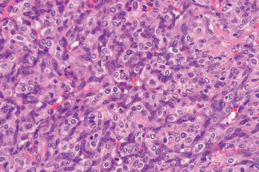
Fig. 35.590 Epithelioid angiosarcoma: the tumor cells have abundant eosinophilic cytoplasm and pleomorphic vesicular nuclei.
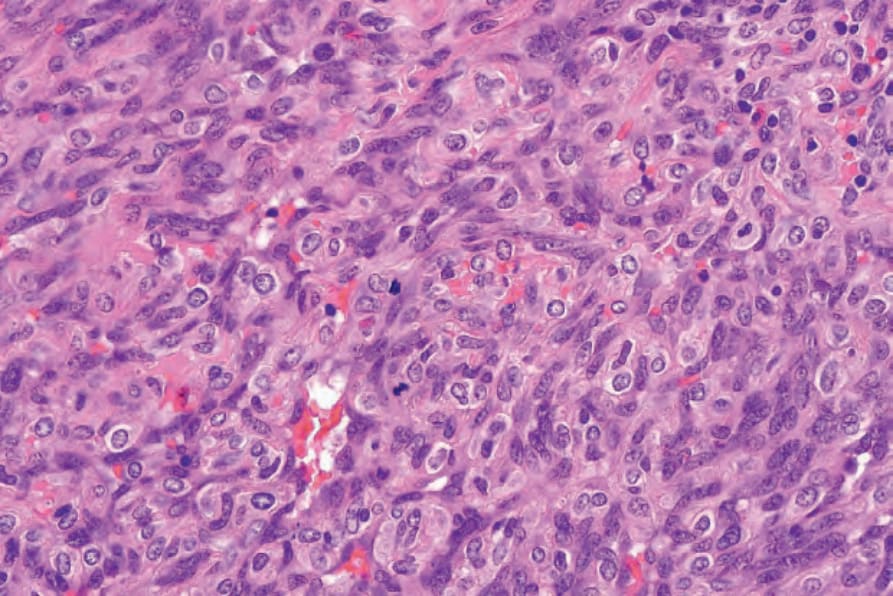
Fig. 35.591 Epithelioid angiosarcoma: note the mitoses.
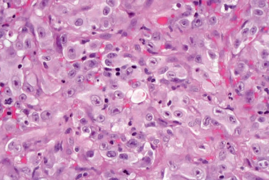
Fig. 35.592 Epithelioid angiosarcoma: intracytoplasmic lumina are present in the center of the field.
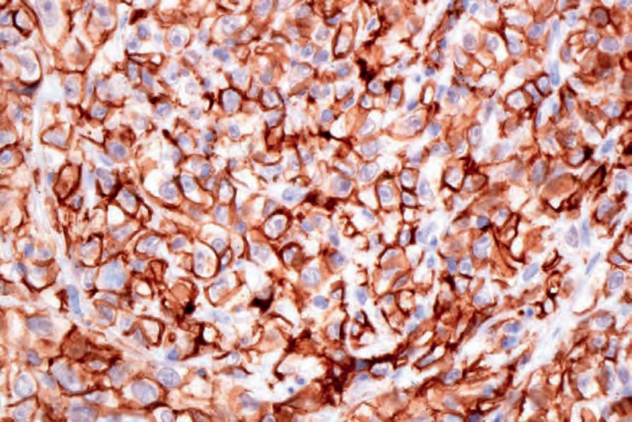
Fig. 35.593 Epithelioid angiosarcoma: the tumor cells express CD31.
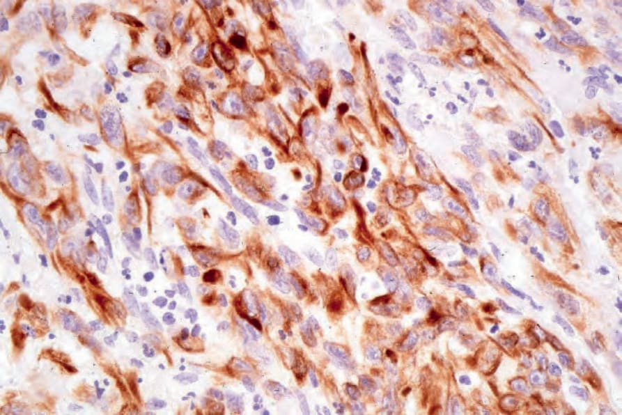
Fig. 35.594 Epithelioid angiosarcoma: this example also expressed keratin (MNF -116).