血管肉瘤 (Angiosarcoma)
血管肉瘤 (angiosarcoma)
angiosarcoma 一詞與 hemangiosarcoma 及 lymphangiosarcoma 同義。
臨床特徵 (Clinical Features)
皮膚 angiosarcoma 主要發生於以下三種臨床情境之一:
- 頭頸部的特發性血管肉瘤 (idiopathic angiosarcoma of the head and neck),
- 淋巴水腫相關血管肉瘤 (lymphedema-associated angiosarcoma),
- 放射治療後血管肉瘤 (postirradiation angiosarcoma)。
發生於四肢的偶發性 (sporadic) 病例(與淋巴水腫無關)可見於任何年齡。極罕見病例曾被報告與 vinyl chloride 暴露、xeroderma pigmentosum、epidermolysis bullosa、stasis ulceration(瘀血性潰瘍)、痛風石 (gouty tophus)、病態肥胖 (morbid obesity) 之併發症,以及與關節成形術 (arthroplasty) 有關。有人提出,在 xeroderma pigmentosum 情境下發生的 angiosarcoma 其侵襲性可能不如其他 angiosarcoma。一名 Klippel-Trenaunay-Weber syndrome 患者在同一受侵犯的肢體上發生 angiosarcoma 及 malignant peripheral nerve sheath tumor。亦曾記錄發生於畸胎瘤 (teratoma) 內的 angiosarcoma 及一例先天性病例。發生於其他器官的 angiosarcoma 可能轉移至皮膚。罕見病例曾被報告與腎臟移植患者的慢性免疫抑制以及與 HIV 有關。兒童的 angiosarcoma 屬例外,主要傾向發生於軟組織與內臟器官,尤其是頭頸部與縱膈 (mediastinum)。除前述者之外,兒童的相關狀況尚包括先天性淋巴水腫 (congenital lymphedema)、Aicardi syndrome 與先天性血管瘤 (congenital hemangioma)。此腫瘤偶爾可能發生於血管內、血管瘤 (hemangioma) 內、神經內,以及良性或惡性神經鞘瘤 (nerve sheath tumors) 內。曾有一例與肝臟瀰漫性 angiosarcoma 相關之多發性皮膚及內臟血管畸形 (vascular malformations) 的報告。
1858 結締組織腫瘤 (Connective tissue tumors)
頭頸部的特發性血管肉瘤 (Idiopathic angiosarcoma of the head and neck)
頭頸部的特發性血管肉瘤 (idiopathic angiosarcoma of the head and neck) 主要是成年晚期的腫瘤,男女發生率相等,且好發於頭皮 (scalp) 與面部中央。曾記錄侷限於眼瞼 (eyelid) 的侵犯。它呈現為單發或多發、隆起的紅色或紫色斑塊 (plaques)、丘疹 (papules) 或結節 (nodules),其生長速度可有變異。高惡性度 (high-grade) 病灶傾向潰瘍並易出血 (Figs 35.569–35.574)。此腫瘤實際範圍通常遠較臨床上所見更為廣泛。自發性消退 (spontaneous regression) 極為例外地發生。罕見病例可能模擬其他疾病,包括 rosacea(酒糟)與 rhinophyma。血小板減少 (thrombocytopenia) 可能罕見發生,可能是腫瘤內血小板消耗與破壞的結果。alopecia(脫髮)是不常見的表現。
淋巴水腫相關血管肉瘤 (Lymphedema-associated angiosarcoma)
淋巴水腫相關血管肉瘤 (lymphedema-associated angiosarcoma)(傳統上稱為 lymphangiosarcoma)典型發生於多年前曾接受乳房切除術 (mastectomy) 合併腋下淋巴結廓清 (axillary lymph node dissection) 或放射治療的老年女性的手臂上(Stewart-Treves syndrome)(Fig. 35.575)。它也可能發生於其他形式的醫源性淋巴水腫 (iatrogenic lymphedema)、先天性淋巴水腫,極罕見地發生於淋巴管瘤畸形 (lymphangiomatous malformation) 內,與象皮病 (elephantiasis)、filariasis(絲蟲病)相關,甚至發生於繼發於病態肥胖之淋巴水腫區域。曾有一例發生於下肢具瘀血變化之 lipodermatosclerosis 區域的報告。病灶典型呈現為許多紫色結節或水疱 (vesicles),常分布於廣大區域。
放射治療後血管肉瘤 (Postirradiation angiosarcoma)
放射治療後血管肉瘤 (postirradiation angiosarcoma) 是三種變異型中最罕見者,可在針對良性(hemangiomas、tinea capitis)或惡性狀況進行放射治療後多年才發生。多數病例與乳癌及婦科癌症的放射治療有關。在皮膚的乳房放射治療後 angiosarcoma 中,通常沒有相關的淋巴水腫,
1859 惡性血管腫瘤 (Malignant vascular tumors)
且潛伏期較 Stewart-Treves syndrome 為短。某些乳房 postirradiation angiosarcoma 病例可能與慢性淋巴水腫有關,這可能促成疾病的發生。曾有一例於轉移性黑色素瘤 (metastatic melanoma) 治療後發生的報告。
所有 postirradiation angiosarcomas 均顯示 MYC 的高度擴增 (high-level amplification),反映染色體 8q24 的增益,這被視為腫瘤發展中早期且必要的改變。在約百分之二十五的這些病例中,存在 FLT4 的共擴增 (co-amplification),FLT4 編碼 VEGFR3。值得注意的是,這些改變在與放射治療相關的非典型血管增生 (atypical vascular proliferations) 中並未發現。其他類型的皮膚 angiosarcomas 也顯示 MYC 擴增。
所有形式的 angiosarcoma 均預後極差,伴隨反覆局部復發、快速播散,多達百分之八十的病例死亡,且常於相當短的時間內發生。一項針對頭皮及面部 angiosarcoma 的回溯性研究發現,5 年存活率改善至百分之四十三,歸因於合併多模式治療 (combined modality therapy)。另一項納入所有偶發性皮膚 angiosarcomas(包括來自頭皮與面部者及具純上皮樣 (pure epithelioid) 形態者)的研究發現,不良預後與壞死 (necrosis)、上皮樣形態 (epithelioid morphology) 及高齡(超過 70 歲)有關。局部復發與腫瘤深度有關。此研究證實了先前一項研究的發現,其中不良預後與腫瘤大小、侵犯深度及有絲分裂率 (mitotic rate) 相關。我們將發生於 angiosarcoma 三種常見臨床情境之外的純皮膚上皮樣血管肉瘤 (pure cutaneous epithelioid angiosarcomas) 視為一個獨特的腫瘤類別,預後極差(見下文)。較年輕的患者似乎預後較佳,且放射治療似乎能改善存活。有人提出,CD8 陽性的腫瘤浸潤性(淋巴球)數目增加與較佳預後相關。
在轉移部位中,以淋巴結與肺臟最為常見。值得注意的是,曾分別報告以 liposomal doxorubicin、paclitaxel,或後者與 sorefenib 之合併使用,治療一例抗放射線、一例無法手術及一例轉移性的 angiosarcoma 後達到完全緩解 (complete remission)。
致病機轉與組織學特徵 (Pathogenesis and histologic features)
對少數淺層及深層 angiosarcomas 進行的細胞遺傳學分析顯示複雜的染色體異常,主要涉及第 5、7、8、13、15、20、22 號染色體及 Y 染色體。曾記錄 KDR 及其他可能適合治療性標靶之基因的活化突變 (activating mutations)。近期描述了 PTPRB、PLCG1 及 ERK/MAPK 路徑的突變。涉及 ERK/MAPK 路徑的遺傳異常包括 KRAS、HRAS、NRAS、BRAF、MAPK1 及 NF1 的突變,或 MAPK1/CRKL、CRAF 或 BRAF 的擴增。在較少數病例中也發現 TP53 突變及 CDKN2A 的缺失。
顯微鏡下,除了共存的淋巴水腫之外,所有臨床變異型大致無法區分,因此一併討論。其外觀為一界限不清的浸潤性真皮內團塊,由眾多口徑不一、互相吻合 (anastomosing) 的血管腔道組成 (Figs 35.576–35.579)。內皮 (endothelium) 可為單層或多層,典型為飽滿 (plump)、多形性 (pleomorphic) 且具有絲分裂活性(異常有絲分裂相當常見),並可在血管腔內形成乳頭 (papillae) 或實心巢 (solid nests)。此血管增生傾向於在真皮內分枝蔓延,「分割 (dissecting)」膠原束 (collagen bundles) (Figs 35.580 與 35.581)。局灶性上皮樣變化 (focal epithelioid change) 並不少見,在某些情況下可十分顯著。在某些病例中,腫瘤(局灶或瀰漫地)呈現實心、未分化、梭形細胞 (spindled cell) 外觀,不易辨識其為血管來源 (Figs 35.582–35.584)。
有若干病例似乎呈現真正的淋巴管分化 (true lymphatic differentiation),主要是位於頭皮與面部者。這些腫瘤的特徵為互相連通的不規則腔道,腔內缺乏紅血球,由非典型的鞋釘樣內皮細胞 (hobnail endothelial cells) 襯覆,具有間質淋巴聚集 (stromal lymphoid aggregates),並對淋巴管標記物呈陽性染色,包括 D2-40、prox-1 及 VEGFR-3。一小部分 angiosarcomas 模擬 Kaposi sarcoma,此特徵也提示淋巴管分化。在某些病例中與後者的區別非常困難,因為在許多具淋巴管分化的腫瘤(特別是 Kaposi sarcoma)中所見的岬狀徵象 (promontory sign),
1860 結締組織腫瘤 (Connective tissue tumors)
1861 惡性血管腫瘤 (Malignant vascular tumors)
也可能見於 angiosarcoma。然而,在後者中存在細胞學非典型性 (cytologic atypia)、有絲分裂活性及多層化 (multilayering)。
辨識此腫瘤血管本質的一個有用方法是 reticulin staining(網狀纖維染色),它會顯示在分化較佳的區域中,腫瘤細胞位於血管周圍的網狀纖維鞘 (perivascular reticulin sheath) 之內;單一細胞並未被網狀纖維框架所包圍 (Fig. 35.585)。散布於整個腫瘤的慢性發炎細胞常為一顯著特徵。在例外的病例中,此浸潤模擬淋巴瘤 (lymphoma) 並遮蔽真正的腫瘤。罕見的 angiosarcoma 病例主要由具顆粒狀細胞質 (granular cytoplasm) 或印戒樣 (signet ring) 外觀的細胞組成。亦曾報告一種由模擬組織球 (histiocytes) 的泡沫狀細胞 (foamy cells) 組成的變異型。
在 postirradiation 腫瘤中,可能出現微血管小葉 (capillary lobules),雖然這傳統上被視為良性增生的特徵,但在此情境下應警覺 angiosarcoma 的存在。
免疫組化與輔助檢查 (Immunohistochemistry & Ancillary Studies)
在分化不良的病例中,評估一組內皮標記物是有用的,因為個別抗體在不同腫瘤中傾向呈不同程度的陽性。這些包括 von Willebrand factor(factor VIII-related antigen)、CD31、CD34、FLI1 及 ERG (Fig. 35.586)。ERG 是一種 ETS family transcription factor,現被視為最敏感且最具特異性的血管標記物。FLI1 是相當敏感但較不具特異性的內皮細胞分化標記物。CD34 呈不同程度陽性,且在頭頸部的 angiosarcomas 中傾向呈陰性。Claudin-5 曾被提議為 angiosarcoma 的良好標記物,但它在其他血管腫瘤以及雙相型滑膜肉瘤 (byphasic synovial sarcoma) 與癌 (carcinomas) 中也呈陽性。CD30 在多達三分之一的 angiosarcomas 中表現,這在分化不良的腫瘤中可能造成混淆。
缺乏上皮樣形態的腫瘤通常不對 keratin 與 epithelial membrane antigen 呈陽性。重要的是要記住,沒有任何抗體是完全特異性的,組織球被 CD31 染色可能是出血性非典型纖維黃色瘤 (hemorrhagic atypical fibroxanthomas) 中一個令人混淆的特徵,並可能導致誤診為 angiosarcoma。組織球的 CD31 染色為細胞質性且呈顆粒狀,而具內皮細胞分化的細胞染色不僅顯示細胞質染色,還顯示清晰的細胞質膜陽性 (crisp cytoplasmic membrane positivity)。
Angiosarcomas 通常對 HHV-8 呈陰性,但發生於 AIDS 患者的腫瘤除外。
在超微結構上,Weibel-Palade bodies 的存在可確認腫瘤的血管本質 (Fig. 35.587)。然而,後者這項技術如今已甚少使用。
鑑別診斷 (Differential Diagnosis)
內皮細胞非典型性 (endothelial cell atypia)、多層化及有絲分裂活性的存在,使其能輕易與良性血管瘤 (benign hemangioma)(或淋巴管瘤 lymphangioma)及 Masson tumor 區分。偶有病例可能需與梭形細胞黑色素瘤 (spindle cell melanoma) 或癌區分,在這些情況下免疫組化最有幫助。
上皮樣血管肉瘤 (epithelioid angiosarcoma)。偶有病例曾與異物 (foreign body)、放射治療、動靜脈廔管 (arteriovenous fistula)、血管 dacron 移植物及骨科關節置換物 (orthopedic joint prostheses) 有關。一例發生於造口周圍 (peristomal) 部位,一例發生於卵巢畸胎瘤 (ovarian teratoma) 內。
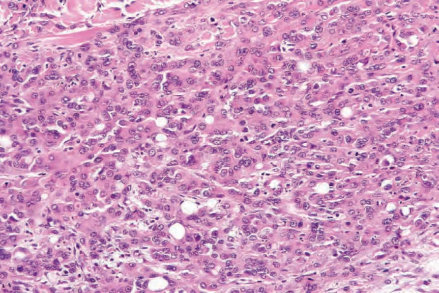
圖 35-566:上皮樣血管內皮細胞瘤 (epithelioid hemangioendothelioma):此例細胞密度高出許多。細胞質內腔 (intracytoplasmic lumina) 仍十分明顯。
Fig. 35.566 Epithelioid hemangioendothelioma: this example is much more cellular. Intracytoplasmic lumina are still conspicuous.
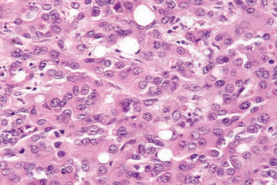
圖 35-567:上皮樣血管內皮細胞瘤 (epithelioid hemangioendothelioma):高倍視野。
Fig. 35.567 Epithelioid hemangioendothelioma: high-power view.
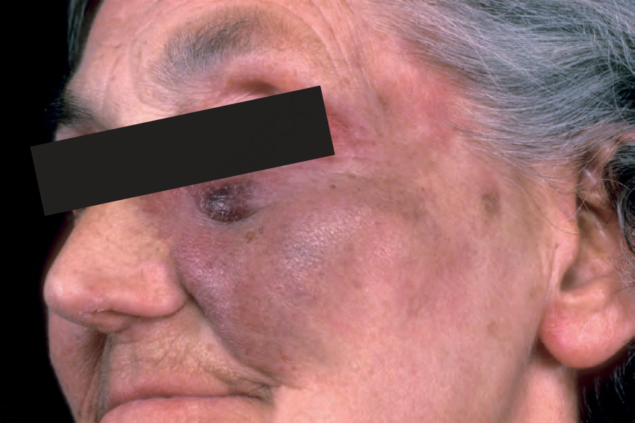
圖 35-569:血管肉瘤 (angiosarcoma):面部有紫色瘀傷樣 (bruise-like) 變色,並伴有一個眶下 (infraorbital) 結節。來自已故 N.P. Smith 醫師的收藏,英國倫敦皮膚科研究所 (the Institute of Dermatology, London, UK)。
Fig. 35.569 Angiosarcoma: there is a purplish bruise-like discoloration of the face with an infraorbital nodule. From the collection of the late N.P. Smith, MD, the Institute of Dermatology, London, UK.
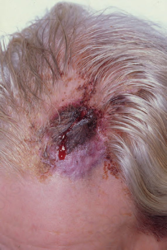
圖 35-570:血管肉瘤 (angiosarcoma):額部頭皮上潰瘍出血的斑塊。來自已故 N.P. Smith 醫師的收藏,英國倫敦皮膚科研究所 (the Institute of Dermatology, London, UK)。
Fig. 35.570 Angiosarcoma: ulcerated and hemorrhagic plaque on the frontal scalp. From the collection of the late N.P. Smith, MD, the Institute of Dermatology, London, UK.
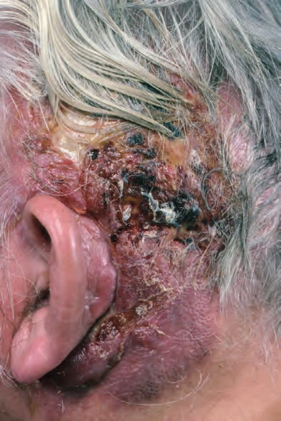
圖 35-571:血管肉瘤 (angiosarcoma):注意此瀰漫性結痂 (crusted) 且潰瘍的病灶。面部與頭皮為好發部位。來自已故 N.P. Smith 醫師的收藏,英國倫敦皮膚科研究所 (the Institute of Dermatology, London, UK)。
Fig. 35.571 Angiosarcoma: note this diffuse crusted and ulcerated lesion. The face and scalp are sites of predilection. From the collection of the late N.P. Smith, MD, the Institute of Dermatology, London, UK.
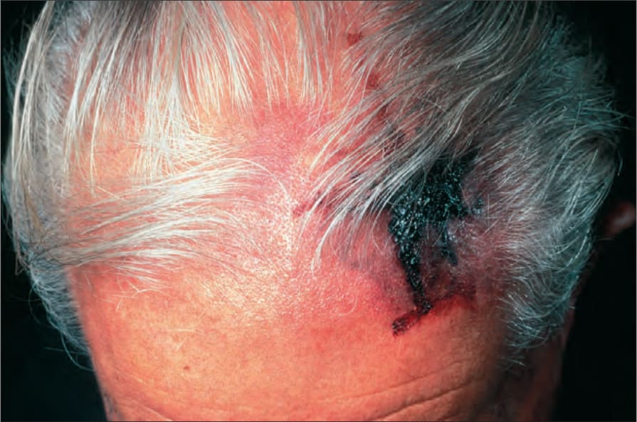
圖 35-572:血管肉瘤 (angiosarcoma):患者常以瘀傷樣 (bruise-like) 病灶表現。承蒙英國倫敦皮膚科研究所 (the Institute of Dermatology, London, UK) 提供。
Fig. 35.572 Angiosarcoma: patients often present with a bruise-like lesion. By courtesy of the Institute of Dermatology, London, UK.
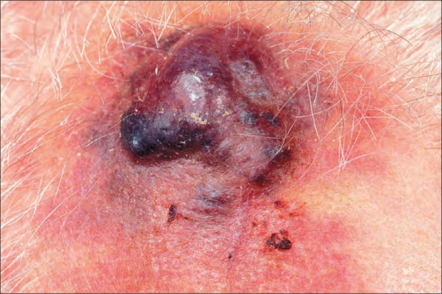
圖 35-573:血管肉瘤 (angiosarcoma):頭皮,特別是禿頭者,是常受侵犯的部位。承蒙英國倫敦皮膚科研究所 (the Institute of Dermatology, London, UK) 提供。
Fig. 35.573 Angiosarcoma: the scalp, particularly in bald individuals, is a commonly affected site. By courtesy of the Institute of Dermatology, London, UK.
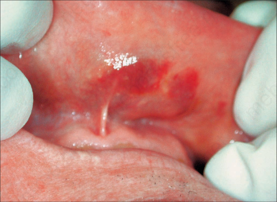
圖 35-574:血管肉瘤 (angiosarcoma):一名僅有極輕微皮膚侵犯的患者出現口腔病灶,凸顯此病程的多灶性 (multifocality)。承蒙英國倫敦皮膚科研究所 (the Institute of Dermatology, London, UK) 提供。
Fig. 35.574 Angiosarcoma: oral lesions in a patient with minimal cutaneous involvement highlighting the multifocality of the process. By courtesy of the Institute of Dermatology, London, UK.
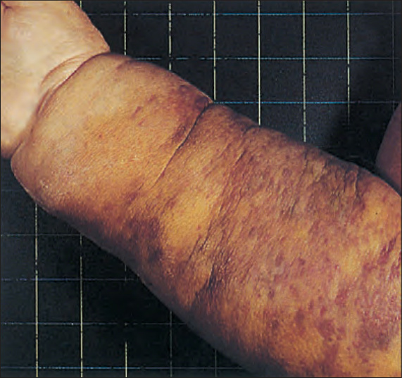
圖 35-575:淋巴水腫相關血管肉瘤 (lymphedema-associated angiosarcoma)(Stewart-Treves tumor):此老年女性患者在根除性乳房切除術 (radical mastectomy) 後併發極為顯著的淋巴水腫。手臂受腫瘤瀰漫性侵犯。
Fig. 35.575 Lymphedema-associated angiosarcoma (Stewart- Treves tumor): very marked lymphedema has complicated radical mastectomy in this elderly female patient. There is diffuse involvement of the arm by tumor.
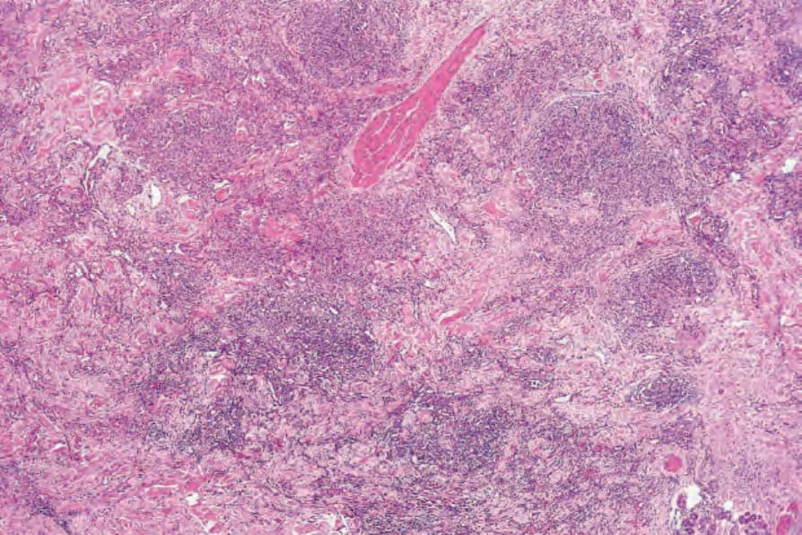
圖 35-576:血管肉瘤 (angiosarcoma):此低倍視野顯示血管腫瘤對真皮的廣泛浸潤。
Fig. 35.576 Angiosarcoma: this low-power view shows extensive infiltration of the dermis by a vascular tumor.
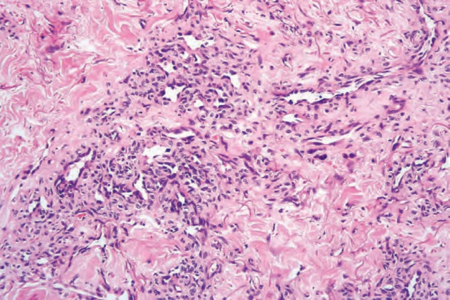
圖 35-577:血管肉瘤 (angiosarcoma):內皮細胞呈多形性 (pleomorphic) 且深染 (hyperchromatic)。
Fig. 35.577 Angiosarcoma: the endothelial cells are pleomorphic and hyperchromatic.
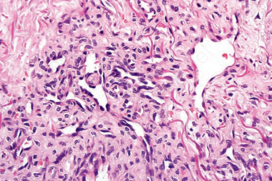
圖 35-578:血管肉瘤 (angiosarcoma):高倍視野。
Fig. 35.578 Angiosarcoma: high-power view.
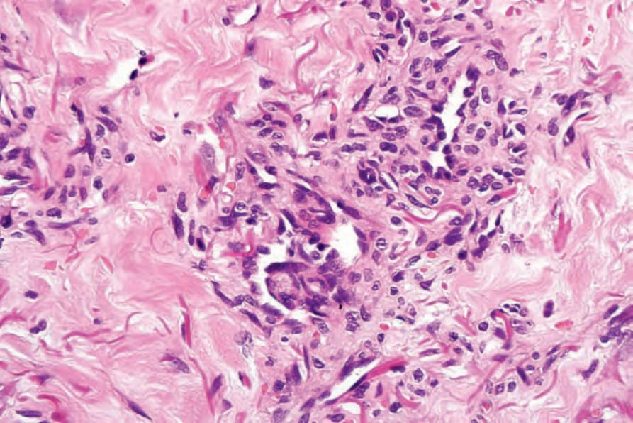
圖 35-579:血管肉瘤 (angiosarcoma):在此例中可見腔內乳頭 (intraluminal papillae)。
Fig. 35.579 Angiosarcoma: in this example, intraluminal papillae are present.
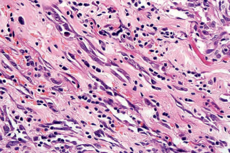
圖 35-580:血管肉瘤 (angiosarcoma):注意梭形細胞 (spindled cell) 族群,具有泡狀核 (vesicular nuclei) 及顯著核仁 (prominent nucleoli)。
Fig. 35.580 Angiosarcoma: note the spindled cell population with vesicular nuclei and prominent nucleoli.
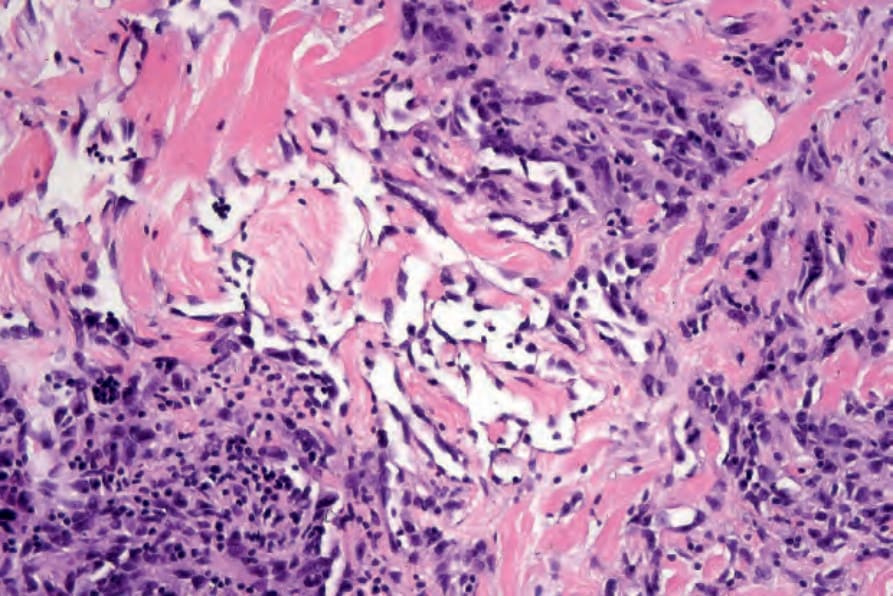
圖 35-581:血管肉瘤 (angiosarcoma):可見明顯的膠原分割 (dissection of collagen)。
Fig. 35.581 Angiosarcoma: there is conspicuous dissection of collagen.
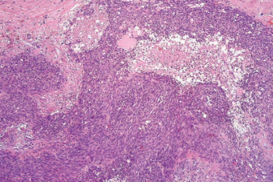
圖 35-582:血管肉瘤(梭形細胞變異型)(angiosarcoma, spindle cell variant):真皮被一梭形細胞腫瘤廣泛浸潤。
Fig. 35.582 Angiosarcoma (spindle cell variant): the dermis is extensively infiltrated by a spindled cell tumor.
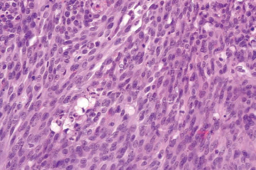
圖 35-583:血管肉瘤(梭形細胞變異型)(angiosarcoma, spindle cell variant):梭形細胞具有嗜伊紅性細胞質 (eosinophilic cytoplasm) 及多形性、泡狀核。可見細胞質內腔 (intracytoplasmic lumina)。
Fig. 35.583 Angiosarcoma (spindle cell variant): the spindle cells have eosinophilic cytoplasm and pleomorphic, vesicular nuclei. Intracytoplasmic lumina are apparent.
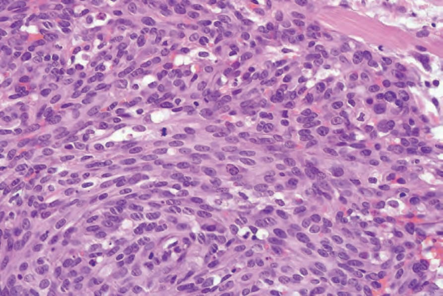
圖 35-584:血管肉瘤(梭形細胞變異型)(angiosarcoma, spindle cell variant):注意有絲分裂活性 (mitotic activity)。
Fig. 35.584 Angiosarcoma (spindle cell variant): note the mitotic activity.
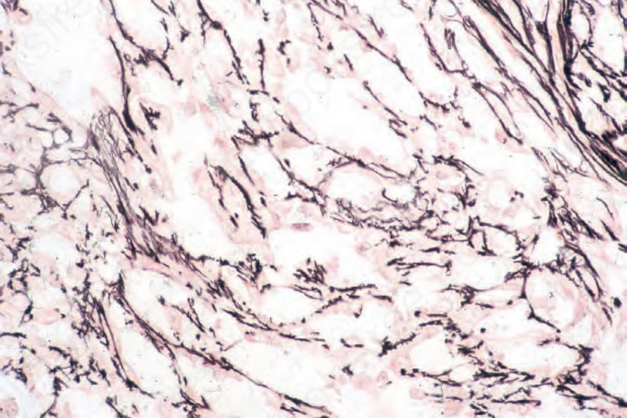
圖 35-585:血管肉瘤 (angiosarcoma):腫瘤細胞被包覆於網狀纖維鞘 (reticulin sheath) 之內。
Fig. 35.585 Angiosarcoma: the tumor cells are enclosed within a reticulin sheath.
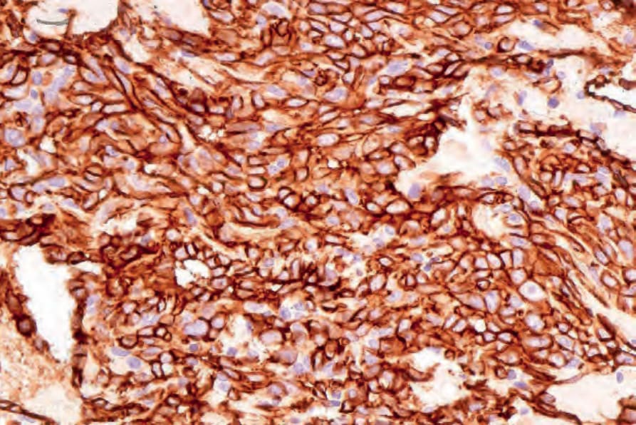
圖 35-586:血管肉瘤 (angiosarcoma):腫瘤細胞表現 CD31。
Fig. 35.586 Angiosarcoma: the tumor cells express CD31.
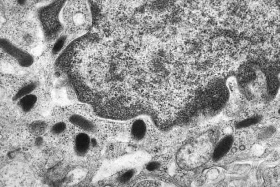
圖 35-587:血管肉瘤 (angiosarcoma):在梭形細胞變異型中,有時可藉由辨識 Weibel-Palade bodies 以超微結構方式確認診斷。
Fig. 35.587 Angiosarcoma: in spindle cell variants, the diagnosis is sometimes confirmed ultrastructurally by the identification of Weibel-Palade bodies.
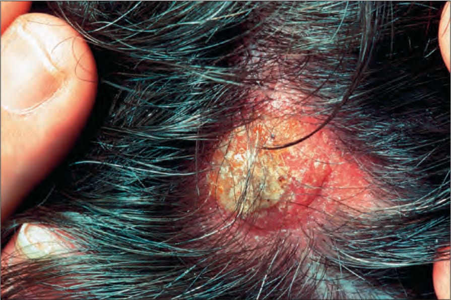
圖 35-588:上皮樣血管肉瘤 (epithelioid angiosarcoma):以頭皮為表現部位並不少見。與更典型的 angiosarcoma 相比,此病灶的血管性較不明顯。承蒙英國倫敦皮膚科研究所 (the Institute of Dermatology, London, UK) 提供。
Fig. 35.588 Epithelioid angiosarcoma: presentation on the scalp is not uncommon. The lesion is less obviously vascular when compared with more typical angiosarcoma. By courtesy of the Institute of Dermatology, London, UK.
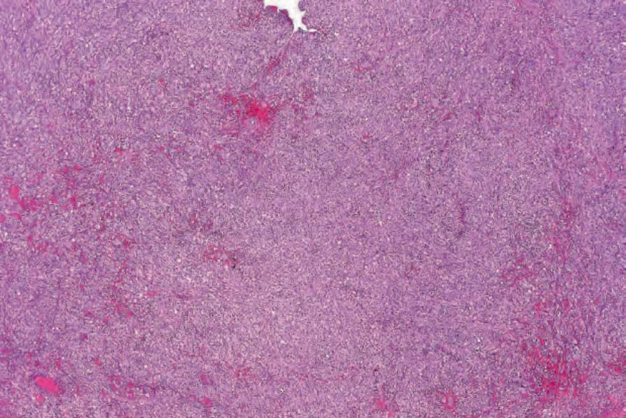
圖 35-589:上皮樣血管肉瘤 (epithelioid angiosarcoma):可見瀰漫性上皮樣細胞浸潤,並有多處出血灶 (foci of hemorrhage)。
Fig. 35.589 Epithelioid angiosarcoma: there is a diffuse epithelioid cell infiltrate with multiple foci of hemorrhage.