上皮樣血管內皮瘤 (Epithelioid Hemangioendothelioma)
臨床特徵 (Clinical Features)
- 上皮樣血管內皮瘤 (epithelioid hemangioendothelioma) 最初於 1982 年被描述為軟組織中一種獨特的低度惡性 (low-grade malignancy) 腫瘤。然而,鑑於其行為具有轉移擴散與致死的潛能(見下文),此腫瘤目前被歸類為完全惡性 (fully malignant)。
- 過去曾以不同名稱描述過侵犯其他器官(主要為肺、肝與骨)的相同病例。
- 大多數病例見於成人,僅罕見於兒童。
- 腫瘤可發生於任何器官,因為高達百分之五十的病例由血管發展而來,最常見為靜脈。
- 疼痛常為一項症狀。
- 多中心性 (multicentric) 病變常見,尤其在肺、肝與骨。
- 皮膚受侵犯相對罕見,可能與其下方的骨或軟組織病變相關,偶爾為多中心性。
- 皮膚 epithelioid hemangioendothelioma 罕見,解剖分布廣泛,未有獨特臨床特徵被描述。
- 皮膚病變可單獨出現,或與其他器官的病變相關,可能同時、之前或之後於皮膚表現出現。
- 口腔內 (intraoral) 病變極為罕見。
- 一名患有肝腫瘤的成人病患以 Kasabach-Merritt syndrome 表現;另一名病患的腫瘤則出現在針對先天性血管瘤 (congenital hemangioma) 接受放射治療之後。
- 轉移與致死率依所侵犯器官而異,但一般認為轉移率不超過百分之三十。
- 具孤立性皮膚病變的病例通常(但非總是)傾向於呈現惰性 (indolent) 行為。
致病機轉與組織學特徵 (Pathogenesis / Histologic Features)
- 細胞遺傳學 (cytogenetic) 研究顯示一獨特的 t(7;19)(q11;q13) 易位 (translocation),導致 SERPINE 1 與 FOSB 基因融合。
- 腫瘤不具血管形成性 (vasoformative),由界限不清的結節構成,這些結節由飽滿、明亮嗜酸性 (brightly eosinophilic) 的紡錘狀細胞 (spindle cells) 排列成束狀或片狀,細胞核呈囊泡狀 (vesicular),並混雜不等數量具橫紋肌樣外觀 (rhabdoid appearance) 的細胞(粉紅色細胞質與偏位細胞核)(Figs 35.558–35.560)。核異型性 (nuclear atypia) 一般輕微,有絲分裂 (mitoses) 稀少。
- t(1;3)(p36.3;q25) 易位導致 WWTR1-CAMTA1 基因融合,在幾乎所有 epithelioid hemangioendothelioma 病例中均可見。t(10;14)(p13;q24) 產生 YAP1-TFE3 融合也罕見地被辨識出。WWTR1-CAMTA1 與 YAP1-TFE3 基因重排有時可共存。
- 顯微鏡下,大多數腫瘤界限不清、呈浸潤性,由圓形、多角形或短紡錘狀的細胞構成,具粉紅色細胞質與囊泡狀細胞核。它們排列成短索狀 (cords) 或巢狀 (nests),被豐富的黏液樣 (myxoid) 或玻璃樣 (hyaline) 基質包圍(基質常呈某種程度的軟骨樣 (chondroid) 外觀,且富含硫酸化酸性黏多醣 (sulfated acid mucopolysaccharides))(Figs 35.561 and 35.562)。
- 內含偶見紅血球的胞質內腔隙 (intracytoplasmic lumina) 常很明顯,類似原始的血管腔道 (primitive vascular channels)(Figs 35.563–35.565)。
- 然而,形成良好的血管 (well-formed vessels) 並非大多數病例的特徵,或僅少見。
- 鈣化 (calcification)、骨化 (ossification) 以及(較少見的)破骨細胞樣巨細胞 (osteoclast-like giant cells) 可存在。
- 罕見病例顯示顯著的細胞學異型性 (cytologic atypia) 與高有絲分裂率,呈現出與上皮樣血管肉瘤 (epithelioid angiosarcoma) 連續譜 (continuum) 的關係 (Figs 35.566–35.568)。
- 較大尺寸與有絲分裂活性增加的腫瘤與較高致死率相關。預後不良與腫瘤大於 3 cm 以及每 50 個高倍視野 (high-power fields) 多於三個有絲分裂象 (mitotic figures) 相關。
- 壞死 (necrosis)、腫瘤部位、細胞學異型性以及腫瘤細胞的紡錘化 (spindling) 似乎不影響預後。
免疫組化與特殊染色 (Immunohistochemistry & Special Stains)
- 免疫組化上,腫瘤細胞表達血管標記,包括 ERG、CD31、CD34、podoplanin 與 FLI-1。
- CD10 通常為陽性,角蛋白 (keratin) 表達見於高達百分之二十五的病例。
- Actin 陽性也可能存在。
- CD30 表達也曾被報導。
- EMA 通常為陰性。
- CAMTA1 免疫組化通常表現為陽性核染色 (positive nuclear staining),有助於與其他具上皮樣形態的腫瘤做鑑別診斷。
鑑別診斷 (Differential Diagnosis)
- 在上皮樣血管瘤 (epithelioid hemangioma) 中,有明顯的發炎,且以形成良好的血管為主。
- 轉移性腺癌 (metastatic adenocarcinoma) 通常顯示較多的多形性 (pleomorphism),且對上皮標記(包括 EMA)呈陽性,對血管標記呈陰性。黏液染色 (mucin stains) 在腫瘤細胞質中常為陽性。
- 上皮樣肉瘤 (epithelioid sarcoma) 一般顯示較片狀的生長型態(至少在部分區域),且僅偶見胞質空泡 (cytoplasmic vacuoles)。它對角蛋白與 EMA 均為陽性,常為 CD34 陽性,但對 ERG、CD31、von Willebrand factor、INI1 與 CAMTA1 呈陰性。
- 上皮樣血管肉瘤 (epithelioid angiosarcoma) 缺乏纖維黏液樣 (fibromyxoid) 或硬化性 (sclerotic) 基質,傾向由片狀的多形性上皮樣細胞構成,具胞質內腔隙,且少有形成血管腔道的傾向。此外,後者不與 CAMTA1-WWTR1 融合相關。
- 黏液樣軟骨肉瘤 (myxoid chondrosarcoma) 具小葉狀結構 (lobular architecture);腫瘤細胞為 S100 protein 陽性,且缺乏胞質內腔隙。
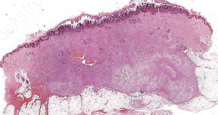
圖 35-558:類肌源性血管內皮瘤 (pseudomyogenic hemangioendothelioma):一浸潤性腫瘤明顯侵犯真皮與皮下組織。
Fig. 35.558 Pseudomyogenic hemangioendothelioma: prominent involvement of the dermis and subcutaneous tissue by an infiltrative tumor.
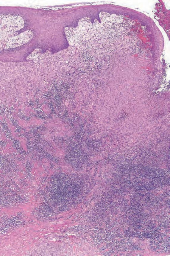
圖 35-561:上皮樣血管內皮瘤 (epithelioid hemangioendothelioma):此為一潰瘍性病變的邊緣。腫瘤位於淺層,邊緣被大量淋巴樣浸潤 (lymphoid infiltrate) 包圍。
Fig. 35.561 Epithelioid hemangioendothelioma: this is the edge of an ulcerated lesion. Tumor is present superficially and is bordered by a heavy lymphoid infiltrate.
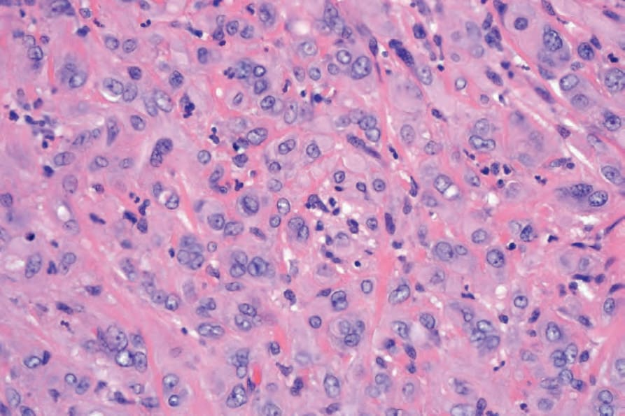
圖 35-562:上皮樣血管內皮瘤 (epithelioid hemangioendothelioma):腫瘤細胞具嗜酸性細胞質 (eosinophilic cytoplasm) 與大的囊泡狀細胞核 (vesicular nuclei)。
Fig. 35.562 Epithelioid hemangioendothelioma: the tumor cells have eosinophilic cytoplasm and large vesicular nuclei.
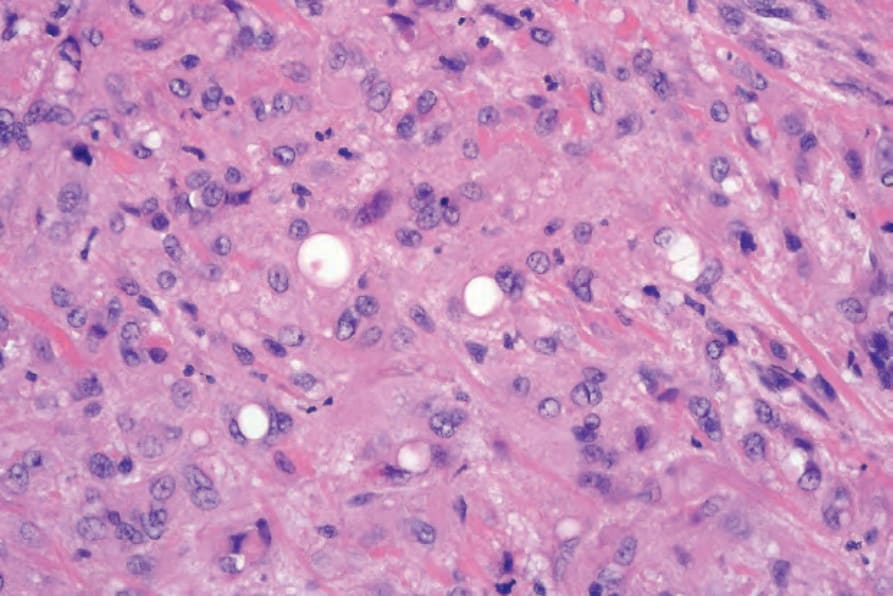
圖 35-563:上皮樣血管內皮瘤 (epithelioid hemangioendothelioma):胞質內腔隙 (intracytoplasmic lumina) 為一特徵性表現。
Fig. 35.563 Epithelioid hemangioendothelioma: intracytoplasmic lumina are a characteristic feature.
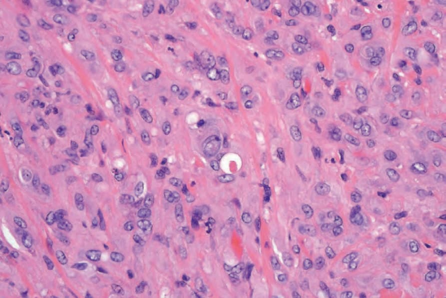
圖 35-564:上皮樣血管內皮瘤 (epithelioid hemangioendothelioma):仔細審視常可在胞質內腔隙 (intracytoplasmic lumina) 中發現紅血球 (erythrocytes)。
Fig. 35.564 Epithelioid hemangioendothelioma: careful scrutiny often reveals erythrocytes within the intracytoplasmic lumina.
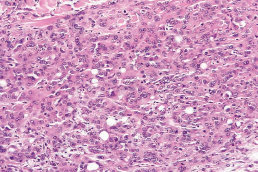
圖 35-566:上皮樣血管內皮瘤 (epithelioid hemangioendothelioma):此例細胞密度高得多。胞質內腔隙 (intracytoplasmic lumina) 仍很明顯。
Fig. 35.566 Epithelioid hemangioendothelioma: this example is much more cellular. Intracytoplasmic lumina are still conspicuous.
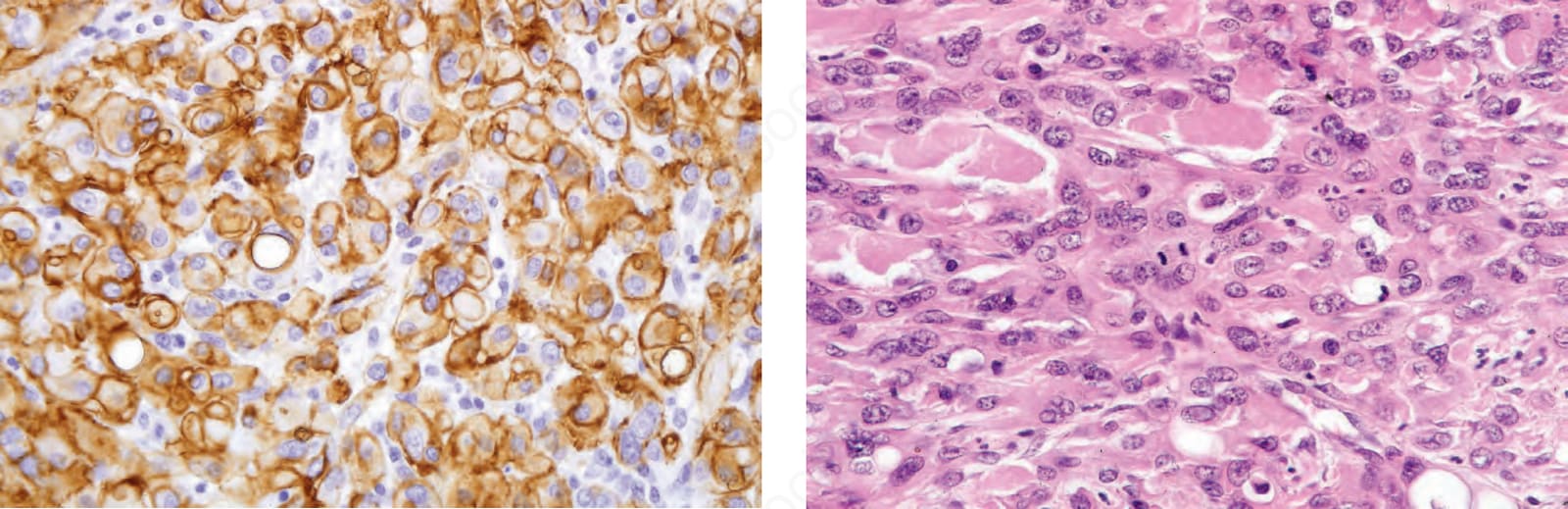
圖 35-568:上皮樣血管內皮瘤 (epithelioid hemangioendothelioma):注意有絲分裂象 (mitotic figures)。
Fig. 35.568 Epithelioid hemangioendothelioma: note the mitotic figures.