Kaposiform hemangioendothelioma
Kaposiform hemangioendothelioma
臨床特徵 (Clinical Features)
卡波西樣血管內皮瘤 (kaposiform hemangioendothelioma) 是一種相對少見的血管腫瘤,最初被描述為最常發生於嬰兒的腹膜後 (retroperitoneum) 或深部軟組織。腫瘤偶爾發生於膽總管 (choledocus)、腎臟、上頜竇 (maxillary sinus)、篩竇 (ethmoid sinus)、縱膈 (mediastinum)、喉部、內聽道 (internal auditory canal)、口咽 (oropharynx)、胸腺、脾臟與肺臟。侵犯皮膚與淺層軟組織的病灶也會發生,成人亦可受影響。曾有一例與創傷相關的病例記載。皮膚與軟組織腫瘤好發於四肢及頭頸部。多發性病灶 (multifocal lesions) 極為罕見。kaposiform hemangioendothelioma 的特徵為局部侵襲性、破壞性生長。一例曾報告同時侵犯皮膚與胸膜,且有一腫瘤因非免疫性胎兒水腫 (nonimmune fetal hydrops) 導致胎兒死亡。超過百分之五十的病例可見與 Kasabach-Merritt syndrome 的相關性,此為重要的致死原因。偶可見區域性結節周圍 (perinodal) 侵犯,但尚未有轉移性疾病的報告。罕見情況下會與淋巴管瘤病 (lymphangiomatosis) 相關。曾記載一例發生於患有類風濕性關節炎 (rheumatoid arthritis) 成人病人的病例、一例以胎兒水腫 (hydrops fetalis) 表現的病例,以及一例伴有大量胎兒乳糜性腹水 (chylous ascites) 的病例。亦曾描述一例極罕見、以水疱 (bullae) 表現並酷似先天性皮膚發育不全 (aplasia cutis congenita) 的病例。
分化朝向高內皮細胞 (high endothelial cells),後者在淋巴器官中正常負責淋巴球的選擇性歸巢 (selective homing)。對於 retiform hemangioendothelioma 也可提出類似的理論,其與 PILA 共有部分組織學特徵。腫瘤細胞對 VEGFR-3 的強表現使人提出這些腫瘤展現淋巴分化 (lymphatic differentiation) 的看法。然而,此標記作為淋巴分化指標的特異性仍有疑問。不過,D2-40 在襯覆血管腔的內皮細胞中呈陽性,進一步支持淋巴譜系 (lymphatic lineage)。
組織病理特徵 (Histopathology)
組織學顯示位於真皮且常延伸至皮下的腫瘤,由顯著擴張、薄壁的血管腔構成,類似海綿狀淋巴管瘤 (cavernous lymphangioma)。這些血管腔由良性的鞋釘狀內皮細胞 (hobnail endothelial cells) 襯覆,細胞核突出且細胞質極稀少。常出現顯著的血管內及血管外淋巴球性發炎細胞浸潤,且常見具膠原核心 (collagenous cores) 的血管內乳頭 (intravascular papillae)(Figs 35.525–35.527)。通常淋巴球看似與內皮細胞緊密貼附。腫瘤細胞對血管標記呈陽性染色,包括 ERG、CD31、CD34 與 von Willebrand factor。
有人提出 kaposiform hemangioendothelioma 與叢狀血管瘤 (tufted angioma) 之間有密切關係。此論點基於臨床與組織學的重疊,以及兩種增生皆可誘發 Kasabach-Merritt syndrome 的事實。此論點亦由兩種腫瘤共有相同免疫表型 (immunophenotype)、皆表現 PROX-1(一種淋巴內皮核轉錄因子)所佐證。在兩個 kaposiform hemangioendothelioma 的鼠類模型中,後者(PROX-1)的過度表現已被證實與促進侵襲有關。tufted angioma 很可能代表 kaposiform hemangioendothelioma 較侷限的變異型。
致病機轉與組織學特徵 (Pathogenesis and Histologic Features)
在單一病例中曾證實一平衡性轉位 (balanced translocation) t(13;16)(q14;p13.3)。
鑑別診斷 (Differential Diagnosis)
兒童的結節型卡波西氏肉瘤 (nodular Kaposi sarcoma) 通常侵犯淋巴結,具有顯著的發炎細胞浸潤,缺乏小葉狀 (lobular) 生長模式,並含有嗜伊紅小球 (eosinophilic globules)。

圖 35-525:乳頭狀淋巴管內血管內皮瘤(Dabska 腫瘤):腫瘤具擴張、薄壁的血管腔,酷似海綿狀淋巴管瘤。注意淋巴樣聚集 (Papillary intralymphatic angioendothelioma, Dabska tumor; cavernous lymphangioma; lymphoid aggregates)。
Fig. 35.525 Papillary intralymphatic angioendothelioma (Dabska tumor): tumors have dilated, thin-walled vascular spaces mimicking a cavernous lymphangioma. Note the lymphoid aggregates.
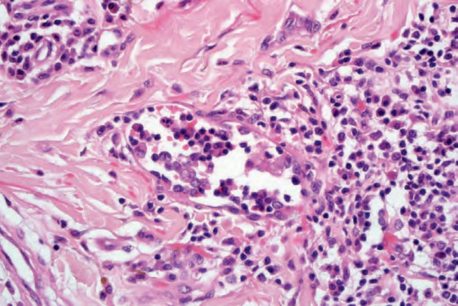
圖 35-526:乳頭狀淋巴管內血管內皮瘤(Dabska 腫瘤):高倍視野顯示鞋釘狀內皮細胞 (Papillary intralymphatic angioendothelioma, Dabska tumor; hobnail endothelial cells)。
Fig. 35.526 Papillary intralymphatic angioendothelioma (Dabska tumor): high-power view showing hobnail endothelial cells.
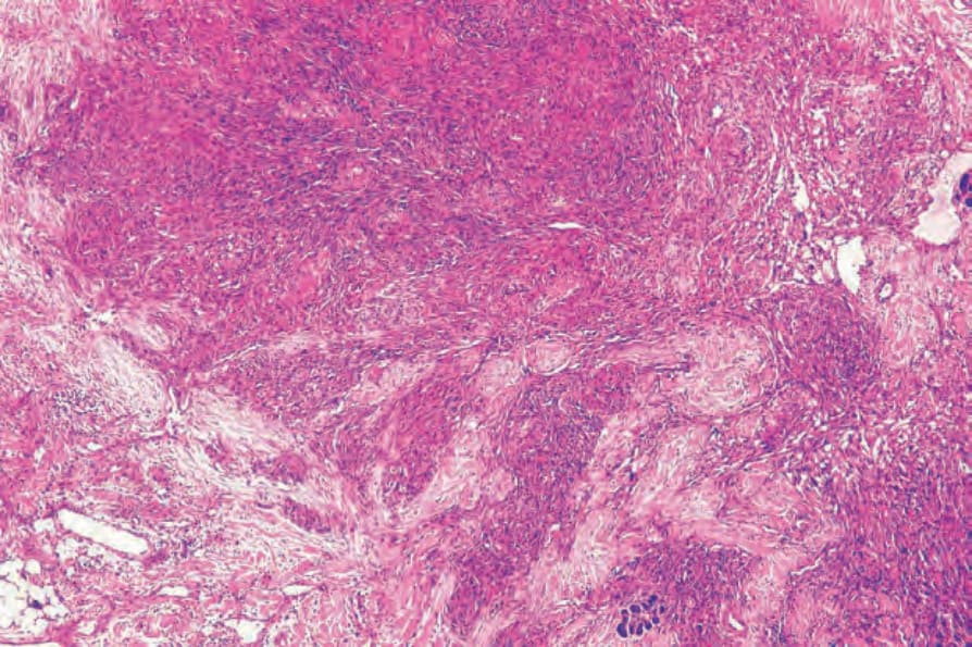
圖 35-528:卡波西樣血管內皮瘤:結節狀增生,含小葉狀、血管性及梭形細胞區域 (Kaposiform hemangioendothelioma; lobular, vascular and spindle cell areas)。
Fig. 35.528 Kaposiform hemangioendothelioma: nodular proliferation with lobular, vascular and spindle cell areas.
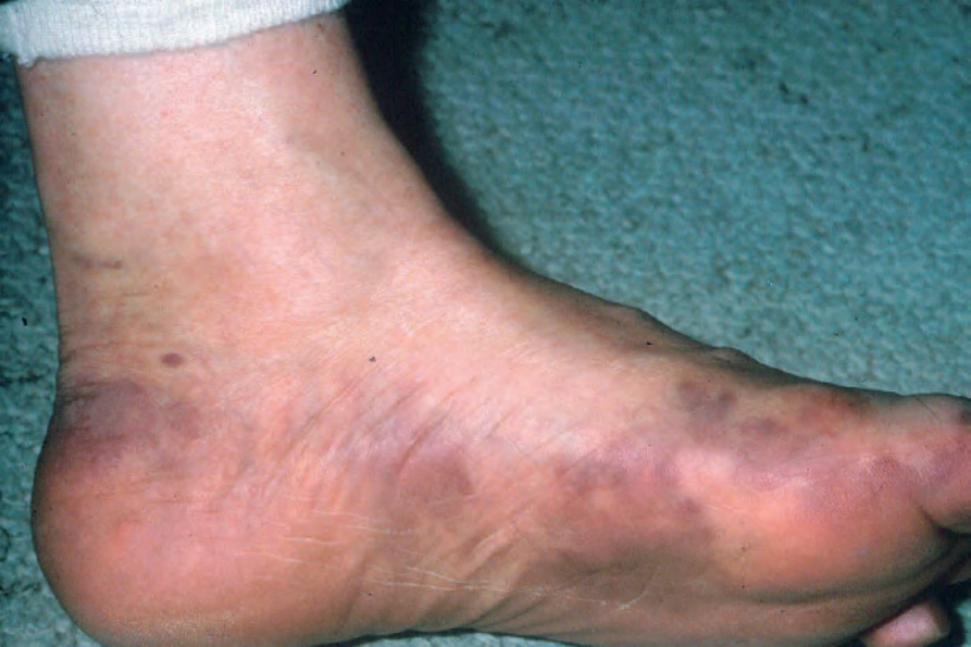
圖 35-530:經典型卡波西氏肉瘤:典型侵犯遠端肢體。取自已故 N.P. Smith 醫師之收藏,英國倫敦皮膚科學研究院 (Classic Kaposi sarcoma; distal extremities; From the collection of the late N.P. Smith, MD, the Institute of Dermatology, London, UK)。
Fig. 35.530 Classic Kaposi sarcoma: the distal extremities are typically involved. From the collection of the late N.P. Smith, MD, the Institute of Dermatology, London, UK.