Kaposiform hemangioendothelioma
Kaposiform hemangioendothelioma
Clinical features Kaposiform hemangioendothelioma is a relatively rare vascular tumor that was originally described as occurring most often in the retroperitoneum or deep soft tissues of infants.1–3 Tumors exceptionally occur in the choledocus, kidney, maxillary sinus, ethmoid sinus, mediastinum, larynx, internal auditory canal, oropharynx, thymus, spleen and lung.4–15 Lesions involving the skin and superficial soft tissues also occur and adults may also be affected.16,17 A case associated with trauma has been documented.18 Cutaneous and soft tissue tumors have predilection for the limbs and head and neck. Multifocal lesions are exceptional.19,20 Kaposiform hemangioendothelioma is characterized by locally aggressive and destructive growth. In one case, concurrent skin and concomitant pleural involvement was reported and a tumor led to fetal death due to nonimmune fetal hydrops.21,22 An association with Kasabach-Merritt syndrome is seen in more than 50% of cases and this is an important cause of mortality.1,3,17 Regional perinodal involvement is uncommonly seen but metastatic disease has not been reported.17 Rarely, there is association with lymphangiomatosis.3,23,24 A case developing in an adult patient with rheumatoid arthritis, one presenting as hydrops fetalis and one with massive fetal chylous ascites have been recorded.25–27 An exceptional case presenting with bullae and mimicking aplasia cutis congenita has been described.28
differentiate towards high endothelial cells, which are normally responsible for the selective homing of lymphocytes in lymphoid organs.12 A similar theory can be proposed for retiform hemangioendothelioma, which shares some of the histologic features of PILA. The strong expression of VEGFR-3 by tumor cells has led to suggestions that these tumors display lymphatic differentiation.2 The specificity of this marker as an indicator of lymphatic differentiation is, however, doubtful. However, D2-40 is positive in the endothelial cells lining the vascular channels giving further support to a lymphatic lineage.
Histology shows a dermal and often subcutaneous tumor composed of markedly dilated, thin-walled vascular channels resembling a cavernous lymphangioma. These vascular channels are lined by bland hobnail endothelial cells with protruding nuclei and very scanty cytoplasm. A prominent intra- and extravascular lymphocytic inflammatory cell infiltrate is often present, and intravascular papillae with collagenous cores are a frequent finding (Figs 35.525–35.527). Commonly, the lymphocytes appear to be in close apposition to the endothelial cells. Tumor cells stain for vascular markers including ERG, CD31, CD34 and von Willebrand factor.
It has been suggested that there is a close relationship between kaposiform hemangioendothelioma and tufted angioma.29–31 This is based on clinical and histologic overlap and the fact that both proliferations may induce Kasabach-Merritt syndrome. This is also substantiated by both tumors sharing an identical immunophenotype with expression of PROX-1, a lymphatic endothelial nuclear transcription factor.32 Overexpression of the latter has been shown to be associated with promotion of invasion in two murine models of kaposiform hemangioendothelioma.33 It is likely that tufted angioma represents a more localized variant of kaposiform hemangioendothelioma.34
Pathogenesis and histologic features A balanced translocation t(13;16)(q14;p13.3) has been demonstrated in a single case.35
1848 Connective tissue tumors
Differential diagnosis Nodular Kaposi sarcoma in children usually involves the lymph nodes, has a prominent inflammatory cell infiltrate, lacks a lobular growth pattern and contains eosinophilic globules.

Fig. 35.525 Papillary intralymphatic angioendothelioma (Dabska tumor): tumors have dilated, thin-walled vascular spaces mimicking a cavernous lymphangioma. Note the lymphoid aggregates.
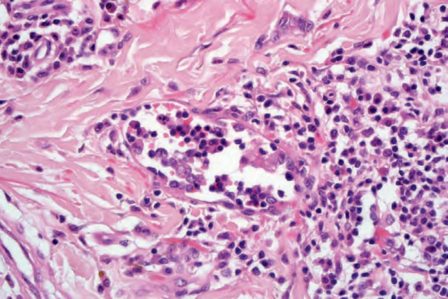
Fig. 35.526 Papillary intralymphatic angioendothelioma (Dabska tumor): high-power view showing hobnail endothelial cells.
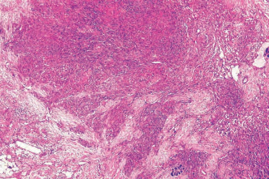
Fig. 35.528 Kaposiform hemangioendothelioma: nodular proliferation with lobular, vascular and spindle cell areas.
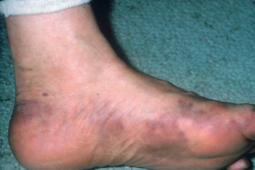
Fig. 35.530 Classic Kaposi sarcoma: the distal extremities are typically involved. From the collection of the late N.P. Smith, MD, the Institute of Dermatology, London, UK.