網狀血管內皮瘤 (Retiform Hemangioendothelioma)
臨床特徵 (Clinical Features)
- 網狀血管內皮瘤 (retiform hemangioendothelioma) 是低度惡性血管肉瘤 (low-grade angiosarcoma) 的一種變異型,與乳頭狀淋巴管內血管內皮瘤 (papillary intralymphatic angioendothelioma,Dabska tumor) 密切相關(且較其更為常見)(見下文)。
組織病理特徵 (Histopathology)
- 病灶界線不清,侵犯真皮 (dermis) 及/或皮下組織 (subcutis)。
- 一個顯著的特徵是腫瘤在組織學上類似正常的睪丸網 (rete testis)。此外觀來自長而呈樹枝狀分支 (long, arborizing, branching) 的血管,這些血管襯以單形性 (monomorphic)、外觀溫和 (bland) 的內皮細胞,具有明顯的頂端核 (prominent apical nuclei) 與稀少的細胞質 (scanty cytoplasm)(Fig. 35.521)。
- 這些細胞明顯突入血管腔內,呈典型的鞋釘樣外觀 (hobnail appearance)(Figs 35.522 與 35.523)。
- 一個常見但非恆定的特徵是在血管內與血管周邊、並與內皮細胞密切相關處,存在眾多淋巴球 (lymphocytes)(Fig. 35.524)。
- 局部可見具有膠原核心 (collagenous cores) 的血管內乳頭 (intravascular papillae)。
- 大多數腫瘤呈現由梭形細胞 (spindled cells) 及少數上皮樣細胞 (epithelioid cells) 構成的實性區 (solid areas)。在一例合併 Milroy disease 的患者所報告的病例中,實性區具有細胞學異型性 (cytologic atypia),提示該腫瘤可能代表一種複合性血管內皮瘤 (composite hemangioendothelioma) 的可能性。
免疫組化與特殊染色 (Immunohistochemistry & Special Stains)
- 免疫組化上,這些細胞對血管標記 (vascular markers) 包括 CD31 與 CD34 呈陽性染色。
- 對淋巴管標記 (lymphatic markers) 包括 D2-40 及特異性較低的 VEGFR-3 的染色,結果互相矛盾。
鑑別診斷 (Differential Diagnosis)
- 網狀血管內皮瘤與乳頭狀淋巴管內血管內皮瘤 (papillary intralymphatic angioendothelioma,PILA) 具有相似的臨床與組織學特徵,並有人提出前者為後者的成人型變異。然而,在 PILA 中並無網狀 (retiform) 結構,以海綿狀淋巴管瘤樣 (cavernous lymphangioma-like) 的血管腔為主,且具有膠原核心的血管內乳頭明顯可見。
- 標靶樣含鐵血黃素性血管瘤 (targetoid hemosiderotic hemangioma,hobnail hemangioma) 總是位置較表淺且較局限,且鞋釘樣內皮細胞 (hobnail endothelial cells) 僅局部出現。
- 血管肉瘤 (angiosarcoma) 通常出現於不同的臨床情境,且組織學上以至少局部的多形性 (pleomorphism)、有絲分裂 (mitosis)、缺乏鞋釘樣內皮細胞、以及多層化 (multilayering) 為特徵。
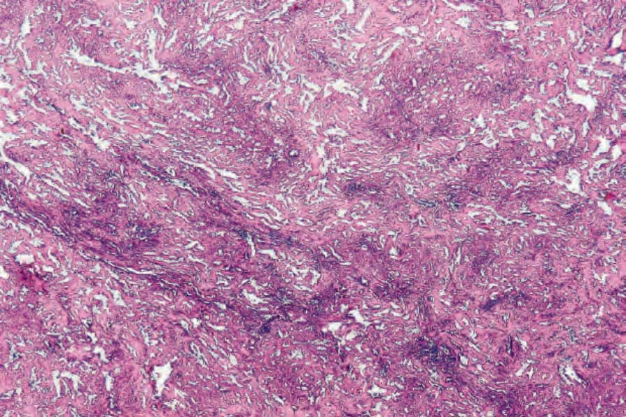
圖 35-521:網狀血管內皮瘤 (retiform hemangioendothelioma):低倍視野,顯示明顯的血管增生 (conspicuous vascularity)。
Fig. 35.521 Retiform hemangioendothelioma: low-power view showing the conspicuous vascularity.
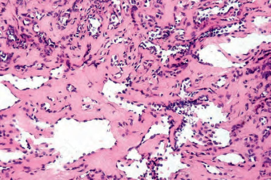
圖 35-522:網狀血管內皮瘤 (retiform hemangioendothelioma):突出的(鞋釘樣,hobnail)內皮細胞核是一項特徵性表現。
Fig. 35.522 Retiform hemangioendothelioma: protuberant (hobnail) endothelial cell nuclei are a characteristic feature.
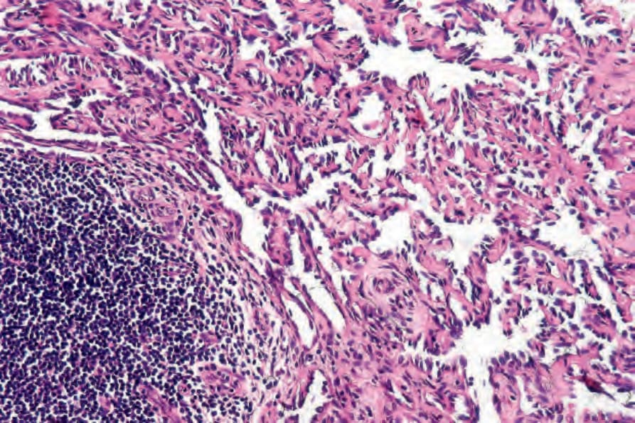
圖 35-524:網狀血管內皮瘤 (retiform hemangioendothelioma):常可見淋巴球聚集 (aggregates of lymphocytes)。
Fig. 35.524 Retiform hemangioendothelioma: aggregates of lymphocytes are frequently seen.