Retiform hemangioendothelioma
Retiform hemangioendothelioma
Clinical features Retiform hemangioendothelioma is a variant of low-grade angiosarcoma that is closely related to (and more common than) papillary intralymphatic angioendothelioma (Dabska tumor) (see below).1–4 It usually presents in
Histologic features Lesions are ill defined and involve the dermis and/or subcutis. A striking feature is the histologic resemblance of the tumor to normal rete testis. This appearance is conferred by the presence of long, arborizing, branching blood vessels, which are lined by monomorphic bland endothelial cells with prominent apical nuclei and scanty cytoplasm (Fig. 35.521). These cells protrude prominently into the vascular lumina, with a typical hobnail appearance (Figs 35.522 and 35.523). A common but not invariable feature is the presence of numerous lymphocytes both within and adjacent to the vessels and in close relation to the endothelial cells (Fig. 35.524). Focally, intravascular papillae with collagenous cores are present. Most tumors show solid areas
1846 Connective tissue tumors
composed of spindled and rare epithelioid cells. The case reported in the patient with Milroy disease had solid areas with cytologic atypia raising the possibility of the tumor representing a composite hemangioendothelioma.6
Immunohistochemically, the cells stain for vascular markers including CD31 and CD34. Staining for lymphatic markers including D2-40 and the less specific VEGFR-3 has yielded contradictory results.10,11
Differential diagnosis Retiform hemangioendothelioma has similar clinical and histologic features to papillary intralymphatic angioendothelioma (PILA) and it has been proposed that the former is an adult variant of the latter. However, in PILA, there is no retiform architecture, cavernous lymphangioma-like vascular spaces predominate and intravascular papillae with collagenous cores are prominent. Targetoid hemosiderotic hemangioma (hobnail hemangioma) is always more superficial and more localized, and hobnail endothelial cells are only focally present. Angiosarcoma usually presents in a different clinical setting and is characterized histologically by at least focal pleomorphism, mitosis, absence of hobnail endothelial cells and multilayering.
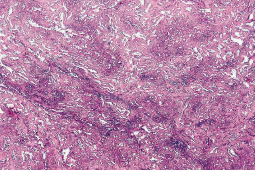
Fig. 35.521 Retiform hemangioendothelioma: low-power view showing the conspicuous vascularity.
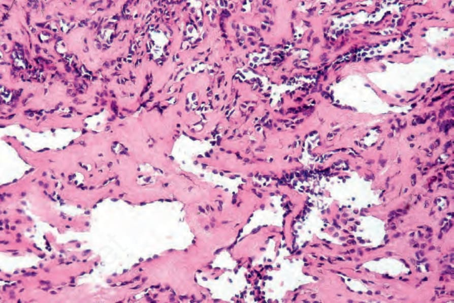
Fig. 35.522 Retiform hemangioendothelioma: protuberant (hobnail) endothelial cell nuclei are a characteristic feature.
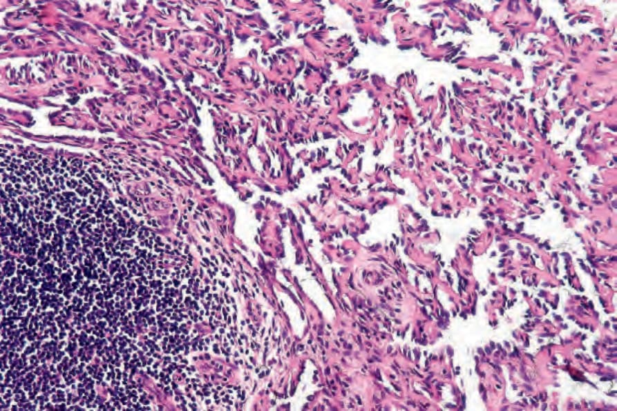
Fig. 35.524 Retiform hemangioendothelioma: aggregates of lymphocytes are frequently seen.