疾病定義與分類
- 合胞體性血管瘤 (symplastic hemangioma) 定義為在先前存在的血管增生(usually a vascular malformation,通常為一種血管畸形)內發生廣泛退化性變化 (extensive degenerative changes),其外觀酷似惡性腫瘤 (closely mimicking malignancy)。
- 文獻僅報告少數病例 (only a handful of cases)。
- 先前存在之 hemangioma 的亞型往往無法明確辨識。
臨床特徵 (Clinical Features)
- 通常出現於成人四肢 (the limbs of an adult),表現為一個長期存在、開始出現變化的病灶 (a long-standing lesion that starts changing)。
致病機轉與組織學特徵 (Pathogenesis and Histologic Features)
- 其組織學外觀可能反映 hemangioma 內、或更常見地在 vascular malformation 內所發生的退化性變化 (degenerative changes)(Figs 35.517 與 35.518)。
- 腫瘤常呈息肉狀 (polypoid)、位於真皮 (dermal) 且界限清楚 (well circumscribed)。
- 典型的組織學圖像由擴張且充血的薄壁至厚壁血管腔 (dilated and congested thin to thick-walled vascular spaces) 所構成,這些血管腔周圍環繞著數量不定的細胞性間質 (a variable cellular stroma),並常伴有黏液樣變化 (myxoid change) 與出血 (hemorrhage)。
- 間質細胞 (stromal cells) 與血管壁內的平滑肌細胞 (smooth muscle cells) 顯示程度不一的細胞學異型性 (cytologic atypia),包括核增大 (nuclear enlargement) 與深染 (hyperchromatism)(Fig. 35.519)。
- 細胞常呈奇異外觀 (bizarre appearance),且多核細胞 (multinucleated cells) 常見。
- 襯覆血管腔的內皮細胞 (endothelial cells) 可能呈飽滿狀 (plump),但不顯示細胞學異型性、多層化 (multilayering) 或有絲分裂活性 (mitotic activity),可藉此與 angiosarcoma 區分。
- 間質成分中可能找到有絲分裂相 (mitotic figures),但往往罕見 (Fig. 35.520)。極偶爾出現的非典型有絲分裂相 (atypical mitotic figures) 可為其特徵之一。
young adults as a slowly growing, asymptomatic tumor and shows a predilection for the distal extremities, especially the lower leg. Sex incidence is equal. Very rarely, cases occur in association with radiation therapy or chronic lymphedema. A patient with multiple lesions and one with Milroy disease have been documented.5,6 A case developing at the site of a cystic lymphangioma has been reported.7 Local, often repeated, recurrences are common, but so far only two cases have metastasized to regional lymph nodes and a further case metastasized to soft tissues close to the primary tumor.1,8,9 No distant spread or tumor-related death has been reported.
- 〔上述英文段落於原文中與本節主題不符、疑為錯置之殘留段落,依鐵則原樣保留、不刪除、不重新歸屬。〕
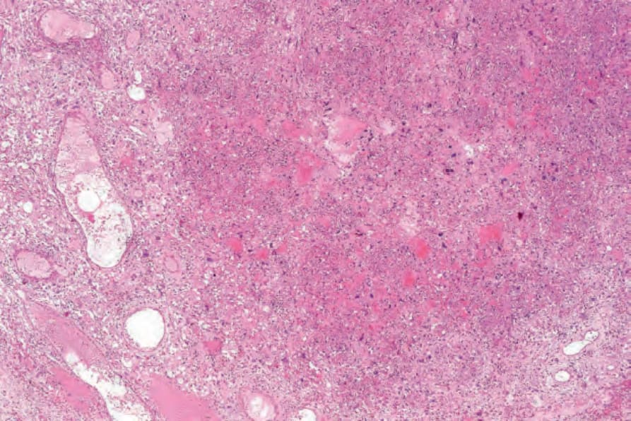
圖 35-517:合胞體性血管瘤 (symplastic hemangioma):低倍視野,顯示擴張的血管 (dilated vessels) 及含有明顯非典型細胞 (conspicuous atypical cells) 的細胞性間質。
Fig. 35.517 Symplastic hemangioma: low-power view showing dilated vessels, and a cellular stroma containing conspicuous atypical cells.
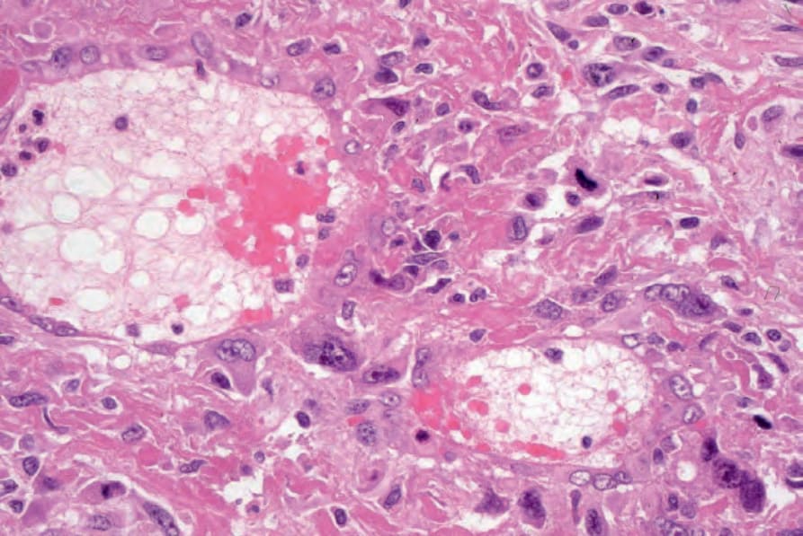
圖 35-518:合胞體性血管瘤 (symplastic hemangioma):較高倍視野,顯示擴張的血管 (dilated vessels) 與非典型間質細胞 (atypical stromal cells)。
Fig. 35.518 Symplastic hemangioma: higher-power view of dilated vessels and atypical stromal cells.
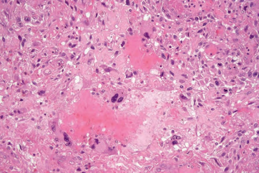
圖 35-519:合胞體性血管瘤 (symplastic hemangioma):可見明顯的核多形性 (nuclear pleomorphism) 與深染 (hyperchromatism)。
Fig. 35.519 Symplastic hemangioma: there is marked nuclear pleomorphism and hyperchromatism.
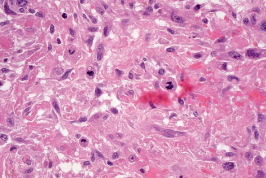
圖 35-520:合胞體性血管瘤 (symplastic hemangioma):注意其有絲分裂相 (mitotic figures)。
Fig. 35.520 Symplastic hemangioma: note the mitotic figures.