Symplastic hemangioma
Symplastic hemangioma
Symplastic hemangioma is defined as extensive degenerative changes in a pre-existing vascular proliferation, usually a vascular malformation, closely mimicking malignancy.1–4 Only a handful of cases have been reported. The variant of pre-existing hemangioma is often not clearly identifiable.
Clinical features It usually presents in the limbs of an adult, as a long-standing lesion that starts changing.
Pathogenesis and histologic features The histologic appearances are likely to reflect degenerative changes within a hemangioma or more often, a vascular malformation (Figs 35.517 and 35.518). Tumors are often polypoid, dermal and well circumscribed. The
1845 Vascular tumors of low-grade or borderline malignancy
typical histologic picture consists of dilated and congested thin to thick-walled vascular spaces surrounded by a variable cellular stroma with frequent myxoid change and hemorrhage. Stromal cells and smooth muscle cells within the vessel walls show variable cytologic atypia consisting of nuclear enlargement and hyperchromatism (Fig. 35.519). Often cells have a bizarre appearance, and multinucleated cells are common. The endothelial cells lining the vascular spaces may be plump but do not display cytologic atypia, multilayering or mitotic activity, allowing distinction from an angiosarcoma. Mitotic figures may be found in the stromal component but tend to be rare (Fig. 35.520). Very occasional atypical mitotic figures can be a feature.
young adults as a slowly growing, asymptomatic tumor and shows a predilection for the distal extremities, especially the lower leg. Sex incidence is equal. Very rarely, cases occur in association with radiation therapy or chronic lymphedema. A patient with multiple lesions and one with Milroy disease have been documented.5,6 A case developing at the site of a cystic lymphangioma has been reported.7 Local, often repeated, recurrences are common, but so far only two cases have metastasized to regional lymph nodes and a further case metastasized to soft tissues close to the primary tumor.1,8,9 No distant spread or tumor-related death has been reported.
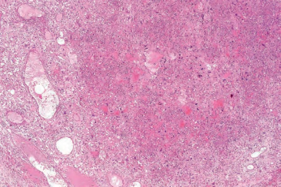
Fig. 35.517 Symplastic hemangioma: low-power view showing dilated vessels, and a cellular stroma containing conspicuous atypical cells.
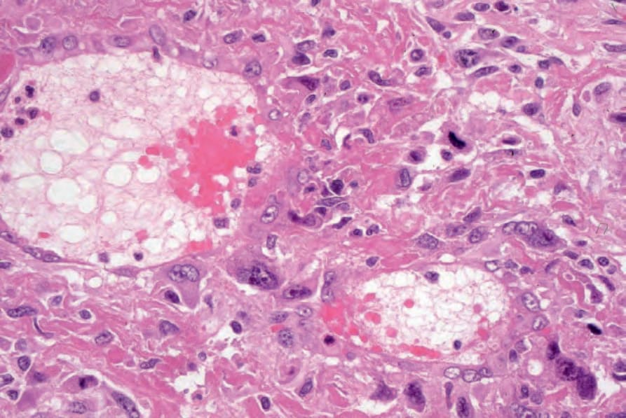
Fig. 35.518 Symplastic hemangioma: higher-power view of dilated vessels and atypical stromal cells.
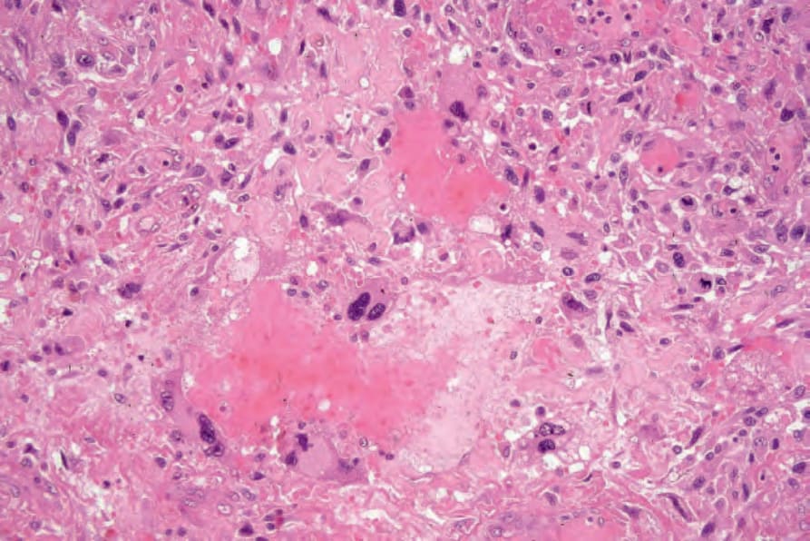
Fig. 35.519 Symplastic hemangioma: there is marked nuclear pleomorphism and hyperchromatism.
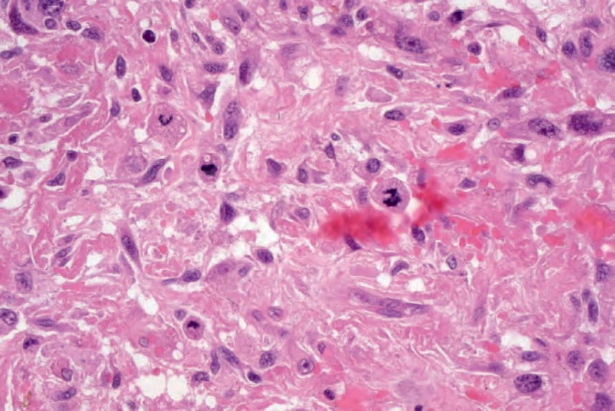
Fig. 35.520 Symplastic hemangioma: note the mitotic figures.