梭形細胞血管瘤 (Spindle Cell Hemangioma)
臨床特徵 (Clinical Features)
梭形細胞血管內皮瘤 (spindle cell hemangioendothelioma) 於 1986 年首次被描述為一種低度惡性血管肉瘤 (low-grade angiosarcoma) 的變異型。此一提議是基於該系列病例中一名病人發生了轉移的事實。然而,幾乎可以確定此轉移源自放射線誘發的肉瘤 (radiation-induced sarcoma),而非源自原始病灶。較近期的證據強烈支持此病灶很可能是一種血管畸形 (vascular malformation),或是疊加於畸形之上的良性過程(即 spindle cell hemangioma)。
它最常侵犯遠端肢體 (distal extremities) 的真皮 (dermis) 或皮下組織 (subcutis),表現為單發或(在 50% 的病例中)多發的紅藍色結節 (red–blue nodules),且相當常伴有疼痛 (Fig. 35.509)。罕見病灶發生於頭頸部,包括口腔。當為多發時,病灶於數年間緩慢發展,臨床病程惰性 (indolent)。多數病人處於前三十年(前三個十年)的年齡層,男女發生率相當。部分病例與早發性靜脈曲張 (early-onset varicose veins)、先天性淋巴水腫 (congenital lymphedema)、Klippel-Trenaunay syndrome 或 Maffucci syndrome 相關。
致病機轉與組織學特徵 (Pathogenesis and Histologic Features)
在散發性 (sporadic) 與 Maffucci syndrome 相關的病灶中皆已發現 IDH R132C 突變 (IDH R132C mutation),證實此腫瘤代表一種腫瘤性病變 (neoplasm)。
病灶界限不清,由薄壁、充血的海綿狀血管腔隙 (thin-walled, congested cavernous vascular spaces) 所構成,並與不同比例的溫和梭形至上皮樣細胞 (bland spindled to epithelioid cells) 相互交雜,後者具有泡狀核 (vesicular nuclei) (Figs 35.510–35.513)。常可見胞質內腔 (intracytoplasmic lumina),為有助於診斷的特徵 (Fig. 35.514)。血管腔隙襯覆單層溫和的內皮細胞 (bland endothelial cells),罕見情況下可顯現退行性核多形性 (degenerative nuclear pleomorphism)。血栓形成 (thrombosis) 與類似 Masson tumor 所見的乳頭狀突起 (papillary projections) 為常見特徵。平滑肌束 (bundles of smooth muscle) 相當常出現於血管周圍及梭形細胞區域。在許多病灶的周邊,可見厚壁、不規則的血管,類似局部動靜脈分流 (localized arteriovenous shunt)。罕見病例可與上皮樣血管內皮瘤 (epithelioid hemangioendothelioma) 相關聯。
免疫組化 (Immunohistochemistry)
免疫組化方面,血管標記 (vascular markers) 主要標記血管的內皮 (endothelium) 以及間質中較具上皮樣型態的細胞。與後者混雜的是 actin 陽性的周細胞 (actin-positive pericytes)。實質性區域的網狀纖維染色 (reticulin staining) 顯示具血管形成性的構造 (vasoformative architecture)。
鑑別診斷 (Differential Diagnosis)
在結節型 Kaposi sarcoma (nodular Kaposi sarcoma) 中,通常不會有海綿狀血管腔隙或空泡化的上皮樣細胞 (vacuolated epithelioid cells),且梭形細胞內常出現玻璃樣小球 (hyaline globules)。Kaposi sarcoma 中的梭形細胞一致地呈 CD34 陽性。
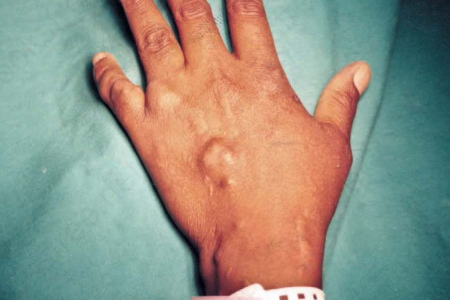
圖 35-509:梭形細胞血管瘤 (spindle cell hemangioma):於特徵性部位出現多發結節。
Fig. 35.509 Spindle cell hemangioma: multiple nodules are present at a characteristic site.
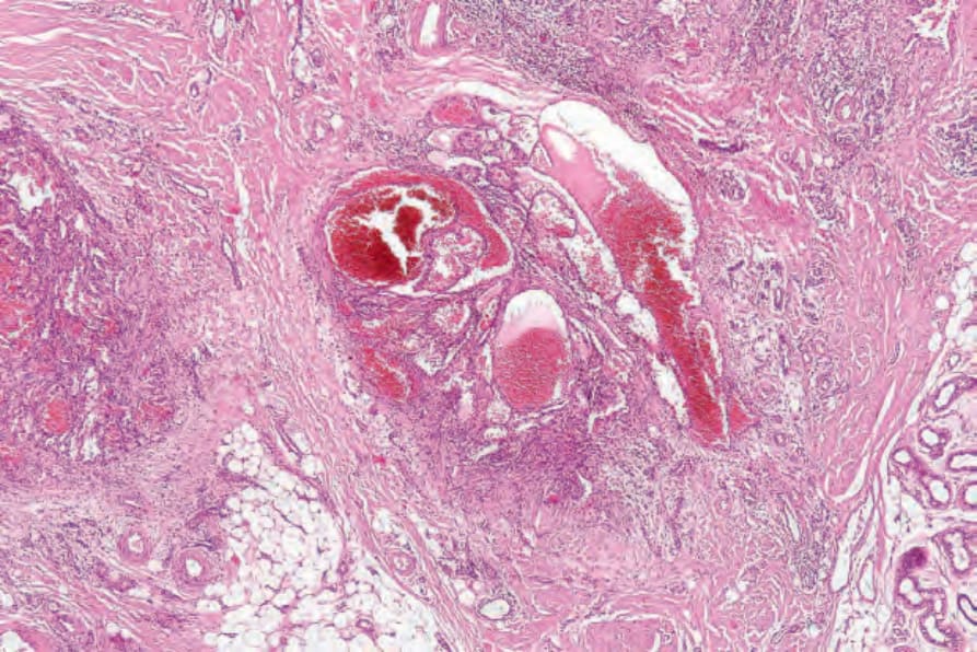
圖 35-510:梭形細胞血管瘤 (spindle cell hemangioma):低倍視野顯示明顯的擴張血管通道 (dilated vascular channels)。
Fig. 35.510 Spindle cell hemangioma: low-power view showing conspicuous dilated vascular channels
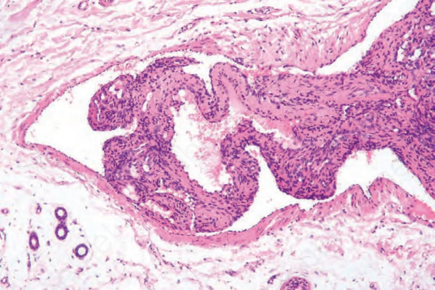
圖 35-511:梭形細胞血管瘤 (spindle cell hemangioma):存在血管內成分 (intravascular component)。
Fig. 35.511 Spindle cell hemangioma: there is an intravascular component.
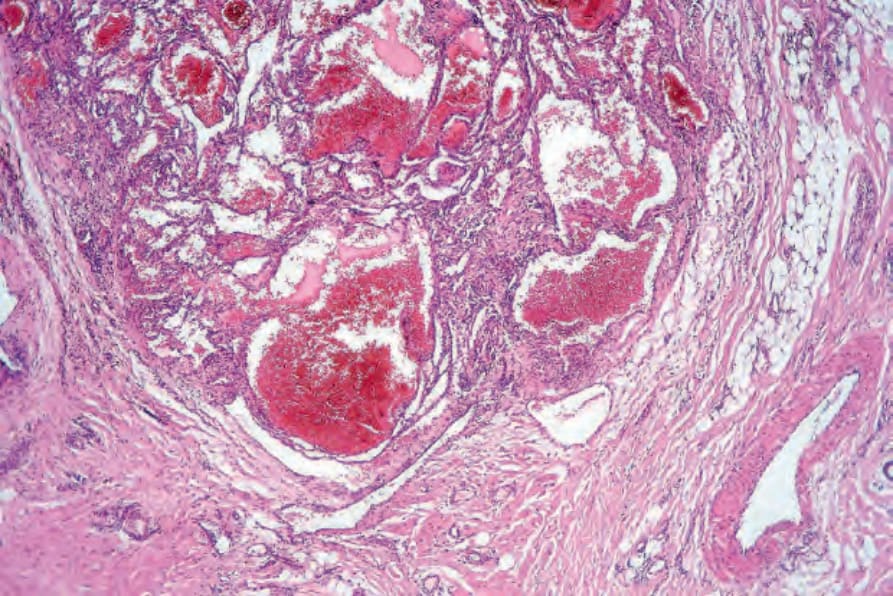
圖 35-512:梭形細胞血管瘤 (spindle cell hemangioma):腫瘤由梭形細胞 (spindle cells) 與常見的海綿狀血管通道 (cavernous vascular channels) 混合構成。
Fig. 35.512 Spindle cell hemangioma: the tumor is composed of an admixture of spindle cells and, often, cavernous vascular channels.
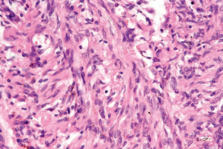
圖 35-513:梭形細胞血管瘤 (spindle cell hemangioma):梭形細胞溫和 (bland),具有相當規則的卵圓形或拉長的細胞核。
Fig. 35.513 Spindle cell hemangioma: the spindle cells are bland and have fairly regular oval or elongated nuclei.
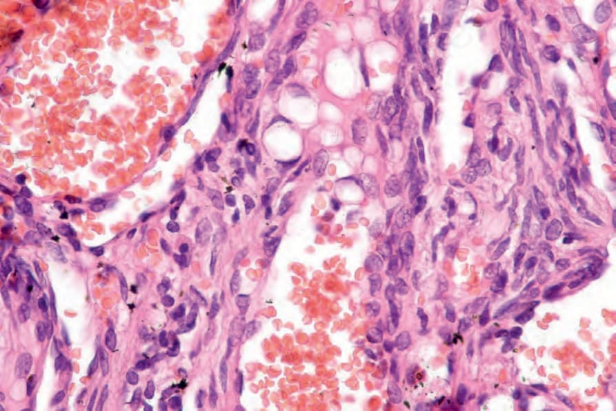
圖 35-514:梭形細胞血管瘤 (spindle cell hemangioma):胞質內腔 (intracytoplasmic lumina) 是重要的診斷特徵。
Fig. 35.514 Spindle cell hemangioma: intracytoplasmic lumina are an important diagnostic feature.
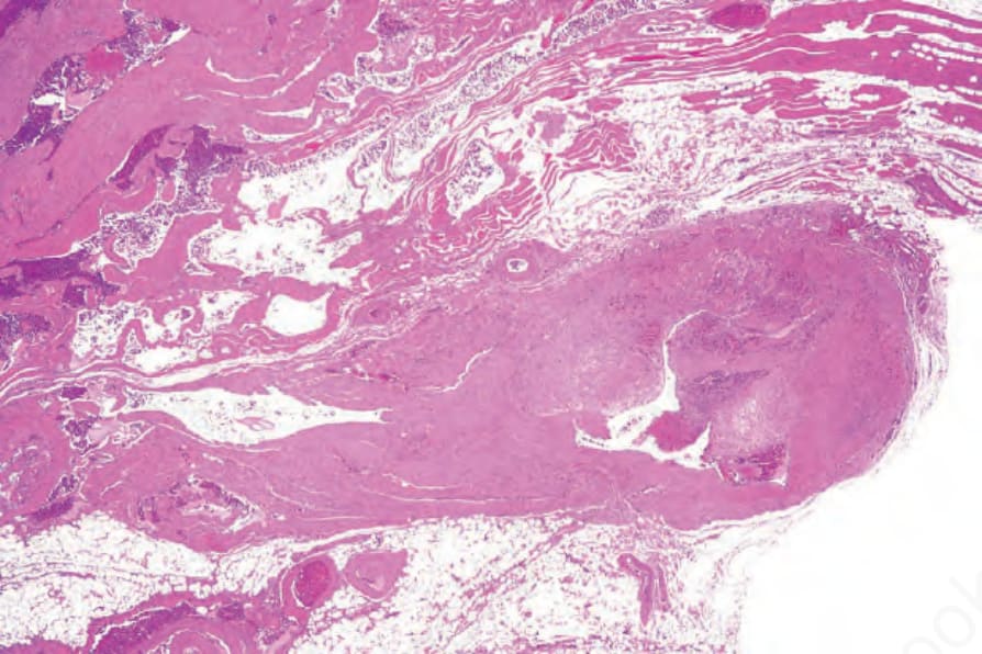
圖 35-515:血管瘤病 (angiomatosis):此例由大小不一的充血海綿狀血管 (congested cavernous vessels) 所構成。
Fig. 35.515 Angiomatosis: this example consists of variably sized congested cavernous vessels.