Spindle cell hemangioma
Spindle cell hemangioma
Clinical features Spindle cell hemangioendothelioma was first described as a variant of low-grade angiosarcoma in 1986.1,2 This proposal was based on the fact that one of the patients in the series developed a metastasis. However, it is almost certain that this metastasis originated from a radiation-induced sarcoma and not from the original lesion. More recent evidence strongly supports the notion that this condition is probably a vascular malformation or a benign process superimposed upon a malformation (spindle cell hemangioma).3–6
It most commonly affects the dermis or subcutis of the distal extremities and presents as single or (in 50% of cases) multiple red–blue nodules, which are quite often painful (Fig. 35.509). Rare lesions develop in the head and neck including the oral cavity.7,8 When multiple, lesions develop slowly over years and the clinical course is indolent. Most patients are in their first three decades and there is an equal sex incidence. Some cases are associated with early-onset varicose veins, congenital lymphedema, Klippel-Trenaunay syndrome or Maffucci syndrome.9.
Pathogenesis and histologic features An IDH R132C mutation have been found in both sporadic and Maffucci syndrome associated lesions, confirming that this tumor represents a neoplasm.10,11
Lesions are poorly circumscribed and consist of thin-walled, congested cavernous vascular spaces intermixed with varying proportions of bland spindled to epithelioid cells with vesicular nuclei (Figs 35.510–35.513). Intracytoplasmic lumina are often present and are a helpful diagnostic feature (Fig. 35.514). The vascular spaces are lined by a single layer of bland endothelial cells, which can rarely show degenerative nuclear pleomorphism. Thrombosis and papillary projections resembling those seen in Masson tumor are common features. Bundles of smooth muscle are quite often present around the blood vessels and in the spindled cell areas. In the periphery of many lesions, there are thick-walled, irregular blood vessels resembling a localized arteriovenous shunt. Rare cases can be associated with epithelioid hemangioendothelioma.
Immunohistochemically, vascular markers label mainly the endothelium of the blood vessels and the more epithelioid cells in the stroma. Admixed with the latter are actin-positive pericytes.3 Reticulin staining in the solid areas reveals a vasoformative architecture.
Differential diagnosis In nodular Kaposi sarcoma there are usually no cavernous vascular spaces or vacuolated epithelioid cells, and hyaline globules are often present in
1844 Connective tissue tumors
the spindled cells. The latter cells in Kaposi sarcoma are consistently CD34 positive.
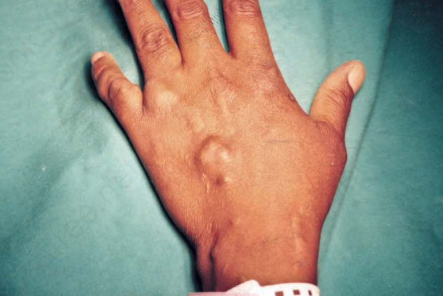
Fig. 35.509 Spindle cell hemangioma: multiple nodules are present at a characteristic site.
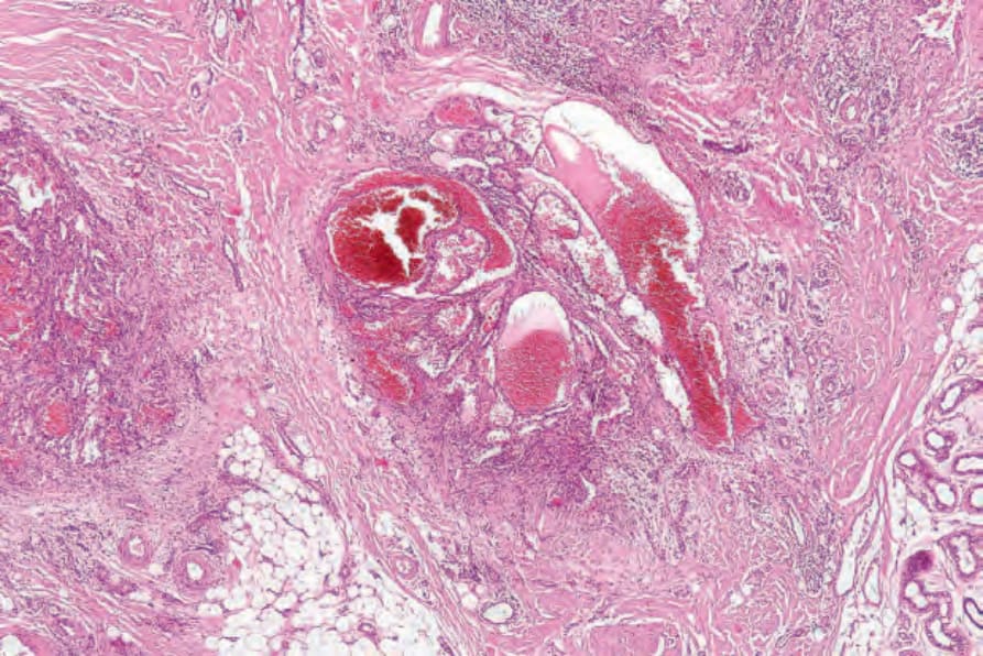
Fig. 35.510 Spindle cell hemangioma: low-power view showing conspicuous dilated vascular channels
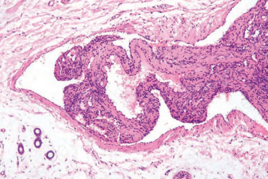
Fig. 35.511 Spindle cell hemangioma: there is an intravascular component.
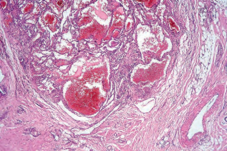
Fig. 35.512 Spindle cell hemangioma: the tumor is composed of an admixture of spindle cells and, often, cavernous vascular channels.
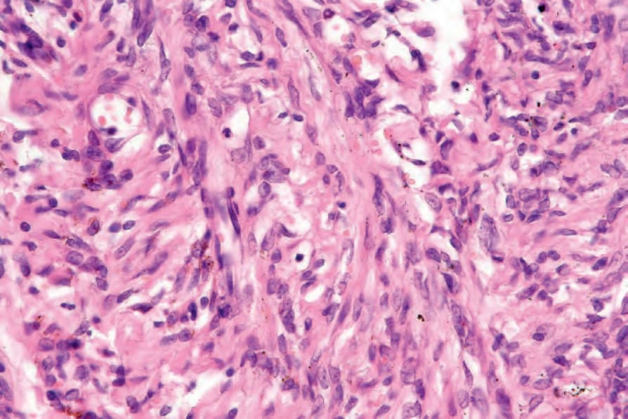
Fig. 35.513 Spindle cell hemangioma: the spindle cells are bland and have fairly regular oval or elongated nuclei.
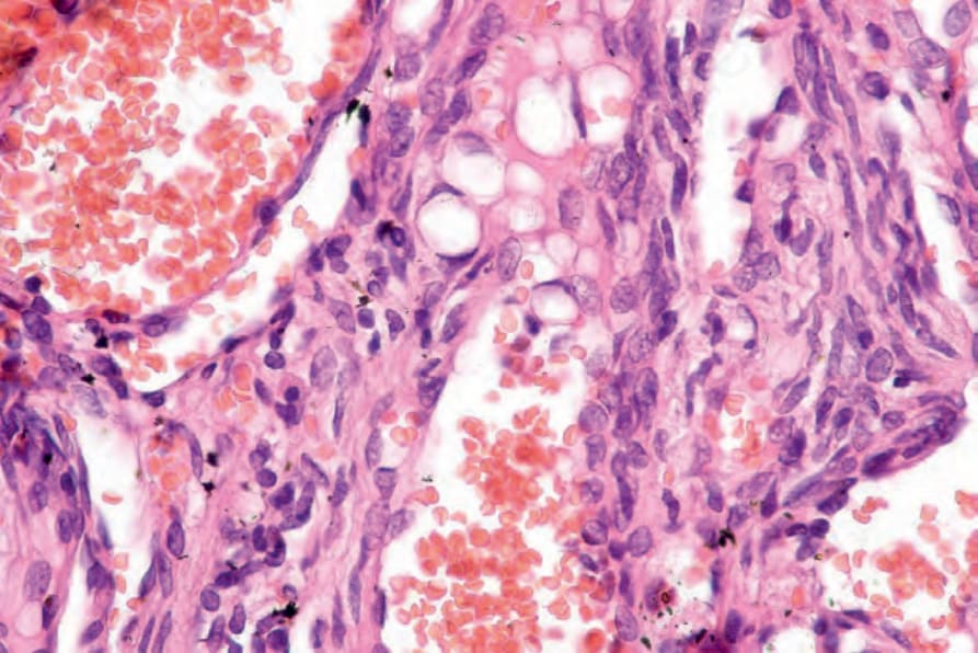
Fig. 35.514 Spindle cell hemangioma: intracytoplasmic lumina are an important diagnostic feature.
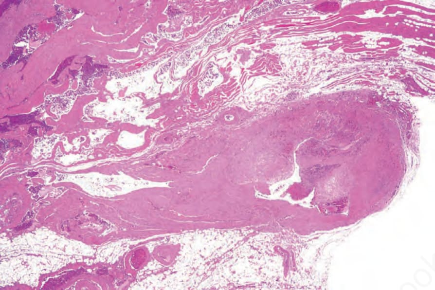
Fig. 35.515 Angiomatosis: this example consists of variably sized congested cavernous vessels.