疾病定義與分類
- 上皮樣血管瘤 (epithelioid hemangioma)(伴嗜酸性球之血管淋巴樣增生,angiolymphoid hyperplasia with eosinophilia)是一群良性血管腫瘤的首選名稱,其特徵為內皮細胞 (endothelial cells) 具有豐富的嗜酸性 (eosinophilic) 細胞質。
- 在一名 HIV 感染患者中,曾記載於 HHV-8 原發感染後出現短暫性 angiolymphoid hyperplasia 與 Kaposi sarcoma。然而,在散發性 epithelioid hemangioma 病灶中並未找到 HHV-8。
- 曾有報告記載具有與 epithelioid hemangioma 相似特徵的病灶與動靜脈畸形 (arteriovenous malformations) 相關。
致病機轉與組織學特徵 (Pathogenesis and histologic features)
- FOS 基因重排 (FOS gene rearrangements) 已在骨骼與軟組織的 epithelioid hemangioma 中被發現,但僅極例外地出現於軟組織與皮膚。FOSB 融合 (FOSB fusion) 曾被描述於一部分骨內 (intraosseous) epithelioid hemangioma,這些病例具有某些令人擔憂的組織病理特徵,例如較高的細胞密度 (cellularity)、細胞學異型性 (cytologic atypia) 與壞死 (necrosis)。與此融合相關的皮膚病例通常富含細胞 (cellular),且發生於陰莖 (penis)。
組織病理特徵 (Histopathology)
- 腫瘤主要位於真皮內 (intradermal),雖然偶爾會遇到皮下 (subcutaneous) 變異型。它們表現為界限不清、呈分葉狀 (lobulated) 的腫塊,由眾多血管腔隙 (vascular spaces) 構成 (Fig. 35.498)。
- 這些血管腔隙的管腔直徑大小不一,內襯著大而圓的內皮細胞,具豐富、相當嗜酸性的細胞質與卵圓形泡狀核 (oval vesicular nuclei) (Fig. 35.499)。部分細胞顯示細胞質內空泡,代表原始管腔 (primitive lumina) (Fig. 35.500)。也可能出現實性的細胞索 (solid cords of cells)。
- 雖然內皮細胞醒目,但它們不顯示多形性 (pleomorphism) 或核分裂活性 (mitotic activity)。相當比例的病例為部分或全部位於血管內 (intravascular),最常起源於靜脈 (vein) 內。一小部分皮膚病例較為實性,內皮細胞增生更為旺盛,可能被誤認為惡性,且好發於陰莖 (penis)。
- 圍繞這些小血管的是程度不一、頗為醒目的發炎細胞浸潤,主要由淋巴球 (lymphocytes)、眾多嗜酸性球 (eosinophils) 與組織球 (histiocytes) 組成 (Fig. 35.501)。淋巴管道 (lymphatic channels) 增多,已由 podoplanin 的免疫組化染色加以凸顯。某些病例中可能出現淋巴濾泡 (lymphoid follicles) (Fig. 35.502)。長期存在的病灶顯示間質硬化 (stromal sclerosis)。
- 不尋常的發現包括多核巨細胞 (multinucleated giant cells)、肉芽腫反應 (granulomatous reaction) 與毛囊黏液變性 (follicular mucinosis)。
免疫組化與特殊染色 (Immunohistochemistry & Special Stains)
- 在免疫組化方面,腫瘤細胞對內皮標記 (endothelial markers) 呈程度不一的陽性,但與其他部位(如骨骼)的 epithelioid hemangioma 不同,皮膚病灶為 cytokeratin 陰性,僅陰莖病灶有局部陽性。HHV-8 為陰性。雖然 FOSB 融合幾乎從未在皮膚病灶中被發現,但此標記的免疫組化在略多於 50% 的病例中呈陽性。
- 罕有報告 T 細胞克隆 (T-cell clone)。此發現的意義尚不確定。
靜脈內非典型血管增生 (Intravenous atypical vascular proliferation)
- 靜脈內非典型血管增生代表 epithelioid hemangioma 的一種靜脈內變異型。它們通常表現為頭部、頸部或上肢的單發結節,好發於年輕至中年成人。與一般的 epithelioid hemangioma 不同,它們通常具有醒目的紡錘細胞(周細胞)成分 (spindle cell (pericytic) component)(與上皮樣內皮管道緊密混雜),這增強了這些病灶的假惡性 (pseudomalignant) 外觀。然而,它們沒有復發傾向。
鑑別診斷 (Differential Diagnosis)
- Kimura disease(木村病):epithelioid hemangioma 常與之混淆。Kimura disease 較常發生於東方人 (Orientals) 的第一與第二個十年。它也表現於軀幹或四肢,且病灶常有壓痛 (tender) (Figs 35.503 and 35.504)。大多數病例顯示組織學上獨特的淋巴結病變 (lymphadenopathy)、循環中嗜酸性球增多 (circulating eosinophilia) 與 IgE 濃度升高;部分患者合併腎臟疾病與青少年型顳動脈炎 (juvenile temporal arteritis)。僅有一名 epithelioid hemangioma 患者合併腎病症候群 (nephrotic syndrome)。
- 在組織學上,Kimura disease 具有醒目的發炎細胞浸潤,伴眾多淋巴濾泡 (lymphoid follicles)、嗜酸性球微膿瘍 (eosinophil microabscesses)、嗜酸性球對生發中心 (germinal centers) 的浸潤、高內皮微靜脈 (high endothelial venules)(並非由上皮樣內皮細胞內襯)的增生,以及大面積的間質硬化 (stromal sclerosis) (Figs 35.505–35.508)。已有罕見病例報告 epithelioid hemangioma 與 Kimura disease 同時出現於同一患者。
- 皮膚淋巴瘤侵犯 (cutaneous involvement by lymphoma):缺乏此種獨特的血管增生。
- 持續性昆蟲叮咬反應 (persistent insect-bite reaction):顯示較多由正常扁平內皮 (flattened endothelium) 內襯的小微血管。
- 注射部位「肉芽腫」(injection-site ‘granuloma’)(鋁「肉芽腫」,aluminum ‘granuloma’):上皮樣內皮細胞並非特徵,可見組織球含有代表鋁 (aluminum) 的藍色顆粒狀細胞質物質。
- 桿菌性血管瘤病 (bacillary angiomatosis):上皮樣細胞呈淡染,且有豐富的嗜中性球 (neutrophils) 伴核塵 (nuclear dust) 與嗜鹼性的細菌團塊 (basophilic clumps of bacteria)。
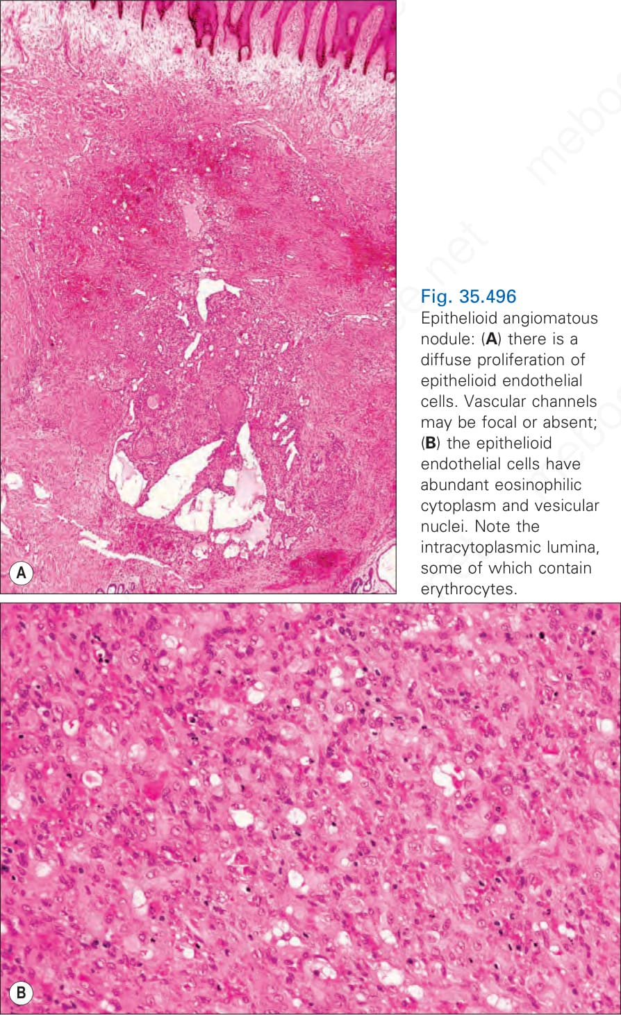
圖 35.496:上皮樣血管性結節 (epithelioid angiomatous nodule):(A) 可見上皮樣內皮細胞 (epithelioid endothelial cells) 瀰漫性增生。血管管道可能為局灶性或缺如;(B) 上皮樣內皮細胞具豐富的嗜酸性細胞質 (eosinophilic cytoplasm) 與泡狀核 (vesicular nuclei)。注意細胞質內管腔 (intracytoplasmic lumina),其中部分含有紅血球 (erythrocytes)。
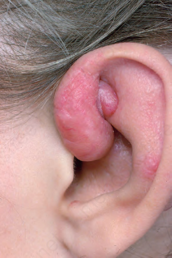
圖 35.497:上皮樣血管瘤 (epithelioid hemangioma):耳部常受侵犯。可見多個融合性病灶。From the collection of the late N.P. Smith, MD, the Institute of Dermatology, London, UK.
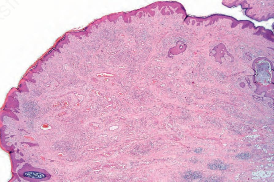
圖 35.498:上皮樣血管瘤 (epithelioid hemangioma):血管性結節的掃描視野,周邊可見淋巴樣聚集 (lymphoid aggregates)。
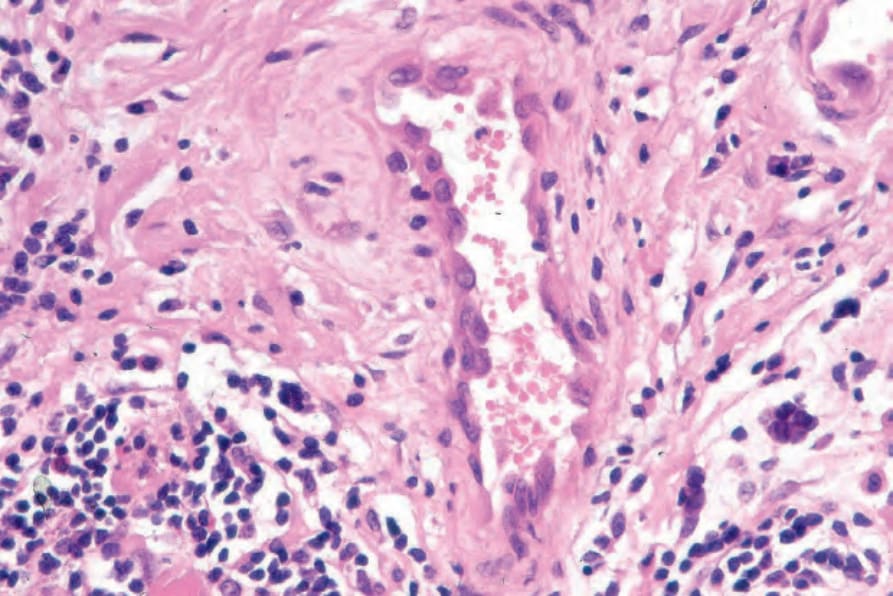
圖 35.499:上皮樣血管瘤 (epithelioid hemangioma):血管由大型內皮細胞內襯,呈明顯的組織球樣 (histiocytoid) 外觀。
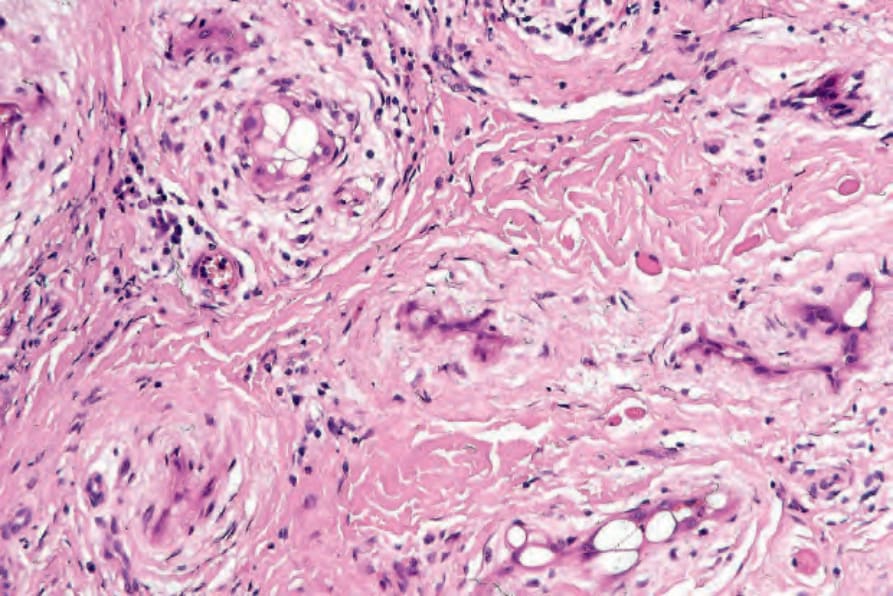
圖 35.500:上皮樣血管瘤 (epithelioid hemangioma):內皮細胞的細胞質內管腔 (intracytoplasmic lumina) 是其特徵性表現。
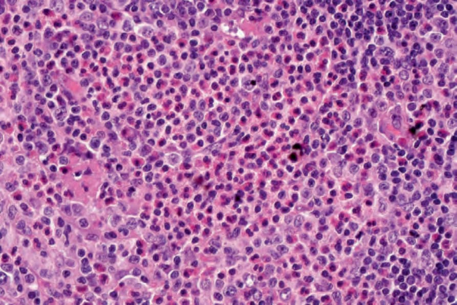
圖 35.501:上皮樣血管瘤 (epithelioid hemangioma):嗜酸性球 (eosinophils) 顯著可見。
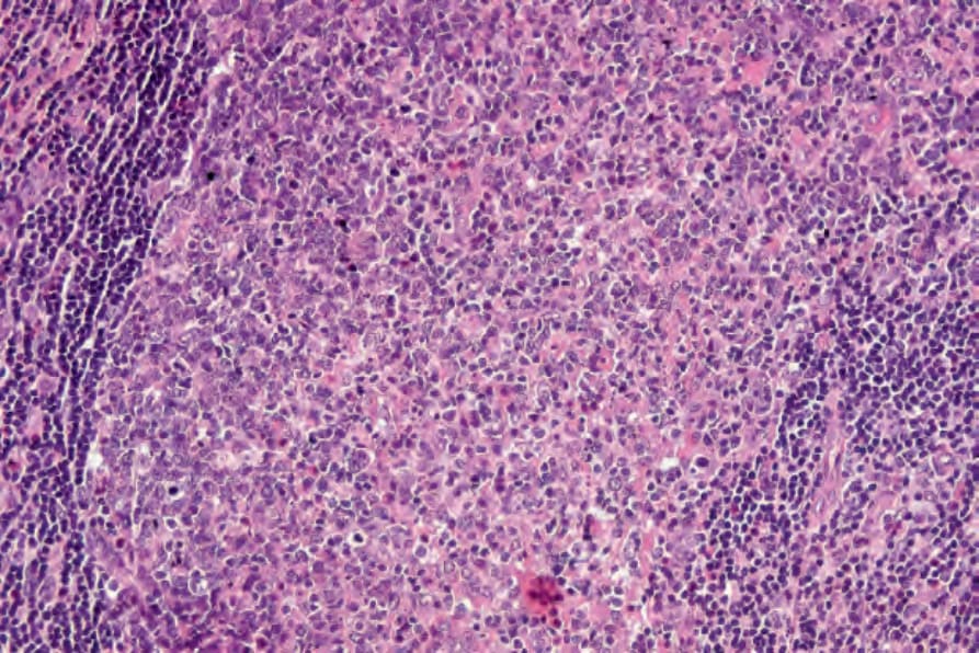
圖 35.502:上皮樣血管瘤 (epithelioid hemangioma):有時可見淋巴濾泡 (lymphoid follicles)。
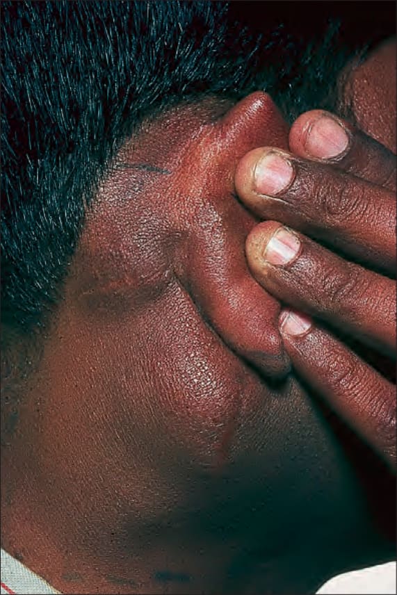
圖 35.503:Kimura disease(木村病):此患者表現為頸部顯著腫脹。By courtesy of the Institute of Dermatology, London, UK.
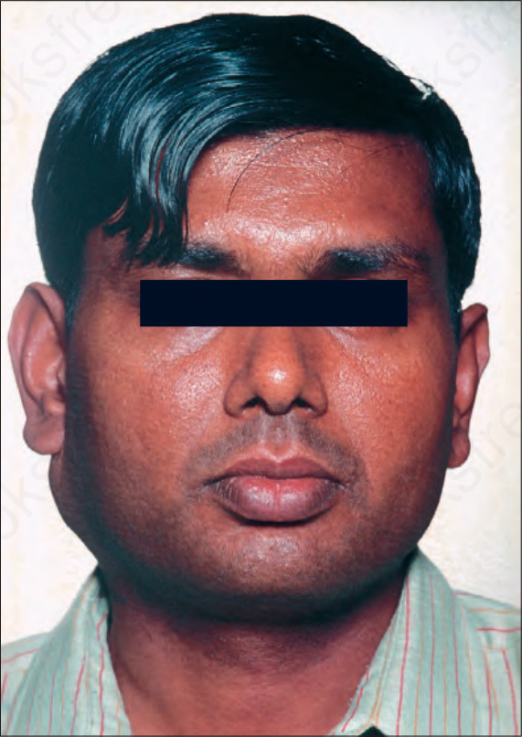
圖 35.504:Kimura disease(木村病):可見軟組織與淋巴結 (nodal) 侵犯。By courtesy of the Institute of Dermatology, London, UK.
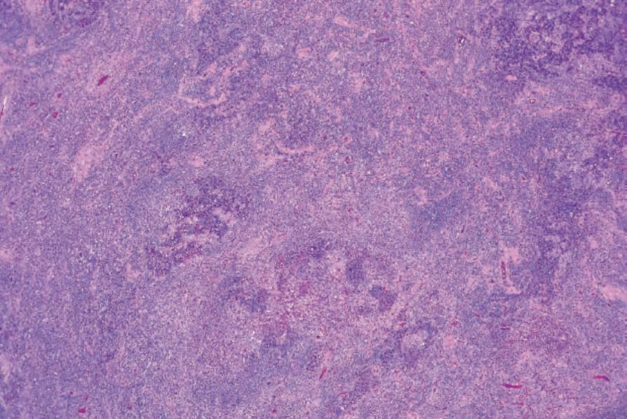
圖 35.505:Kimura disease(木村病):低倍視野顯示密集的細胞浸潤。
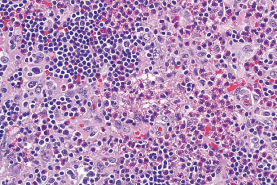
圖 35.506:Kimura disease(木村病):浸潤由淋巴球 (lymphocytes) 與眾多嗜酸性球 (eosinophils) 組成。
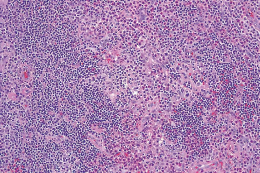
圖 35.507:Kimura disease(木村病):背景可見高微血管微靜脈 (high capillary venules) 增生。
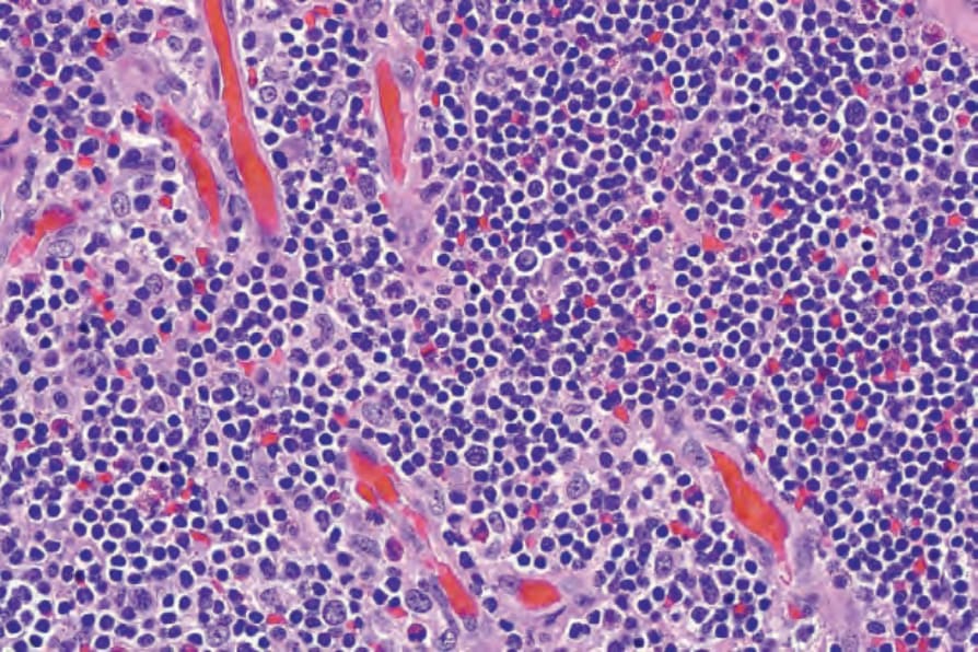
圖 35.508:Kimura disease(木村病):內皮細胞醒目,但不含細胞質內管腔 (intracytoplasmic lumina)。
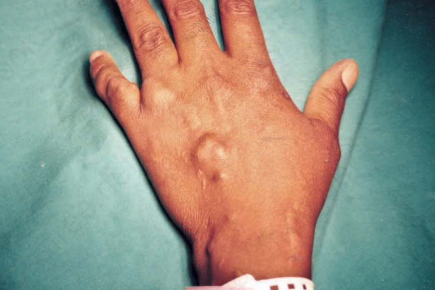
圖 35.509:紡錘細胞血管瘤 (spindle cell hemangioma):於特徵性部位可見多個結節。