Epithelioid hemangioma
Epithelioid hemangioma
Epithelioid hemangioma (angiolymphoid hyperplasia with eosinophilia) is the preferred term for a group of benign vascular tumors characterized by the presence of endothelial cells with abundant eosinophilic, sometimes
Transient angiolymphoid hyperplasia and Kaposi sarcoma have been documented after primary infection with HHV-8 in a patient with HIV infection.41 However, HHV-8 is not found in lesions of sporadic epithelioid hemangioma.42
Lesions with features similar to those seen in epithelioid hemangioma have been documented in association with arteriovenous malformations.43,44
1840 Connective tissue tumors
A
B
Pathogenesis and histologic features
FOS gene rearrangements have been found in epithelioid hemangioma of bone and soft tissue but only exceptionally in soft tissues and skin.45–47 FOSB fusion has been described in a subset of intraosseous epithelioid hemangioma with some worrisome histopathological features such as higher cellularity, cytologic atypia, and necrosis.45,46 Cases in the skin associated with the fusion are usually cellular and occur on the penis.45,46
Tumors are predominantly intradermal although occasionally subcutaneous variants are encountered. They present as an ill-defined, lobulated mass composed of numerous vascular spaces (Fig. 35.498). The latter, of varying luminal diameter, are lined by large rounded endothelial cells with copious, rather eosinophilic cytoplasm and oval vesicular nuclei (Fig. 35.499). Some show cytoplasmic vacuoles, representing primitive lumina (Fig. 35.500). Solid cords of cells may also be present. Although the endothelial cells are prominent, they do not show pleomorphism or mitotic activity. A significant proportion of cases are partially or totally intravascular, most often arising within a vein. A small percentage of skin cases are more solid with a more exuberant proliferation of endothelial cells that may be confused with malignancy and has predilection for the penis.48
Surrounding these small vessels is a variably prominent inflammatory cell infiltrate composed largely of lymphocytes, numerous eosinophils and histiocytes (Fig. 35.501). Increased lymphatic channels have been highlighted by immunohistochemistry for podoplanin.49 Lymphoid follicles may be present in some cases (Fig. 35.502). Long-standing lesions show stromal sclerosis.
Unusual findings include multinucleated giant cells, a granulomatous reaction and follicular mucinosis.50–52
Immunohistochemically, the tumor cells are variably positive for endothelial markers but, in contrast to epithelioid hemangiomas in other locations such as bone, cutaneous lesions are cytokeratin negative except for focal positivity in penile lesions.23,48 HHV-8 is negative.53 Although the FOSB fusion is hardly ever found in cutaneous lesions, immunohistochemistry for this marker is positive in slightly more than 50% of cases.24,54
A T-cell clone has been rarely reported.18,55 The significance of this finding is uncertain.
1841 Capillary hemangioma and its variants
The intravenous atypical vascular proliferation56 represent an intravenous variant of epithelioid hemangioma. They usually present as a solitary nodule on the head, neck or upper limbs with a predilection for young to middle-aged adults. Unlike, conventional epithelioid hemangioma they usually have a prominent spindle cell (pericytic) component (closely admixed with the epithelioid endothelial channels), which enhances the pseudomalignant appearance of these lesions. Although, they have no tendency to recur.56
Differential diagnosis Kimura disease, with which epithelioid hemangioma is frequently confused, tends to occur more commonly in Orientals in their first and second decades. It also presents on the trunk or limbs and the lesions are frequently tender (Figs 35.503 and 35.504).10,11,57 The majority of cases show a histologically distinctive lymphadenopathy, a circulating eosinophilia and raised IgE levels; some patients have associated renal disease and juvenile temporal
1842 Connective tissue tumors
arteritis.58 Only one patient with epithelioid hemangioma has had associated nephrotic syndrome.59 Histologically, Kimura disease has a prominent inflammatory cell infiltrate with numerous lymphoid follicles, eosinophil microabscesses, infiltration of germinal centers by eosinophils, proliferation of high endothelial venules (not lined by epithelioid endothelial cells) and large areas of stromal sclerosis (Figs 35.505–35.508). Rare cases of epithelioid hemangioma and Kimura disease presenting in the same patient have been reported.60
Cutaneous involvement by lymphoma lacks the distinctive vascular proliferation, while a persistent insect-bite reaction shows a greater number of small capillaries lined by normal flattened endothelium. In injection-site ‘granuloma’ ” (aluminum ‘granuloma’ ”), epithelioid endothelial cells are not a feature and histiocytes with bluish granular cytoplasmic material representing aluminum are found.61,62 In bacillary angiomatosis, the epithelioid cells are pale and there are abundant neutrophils with nuclear dust and basophilic clumps of bacteria.
1843 Capillary hemangioma and its variants
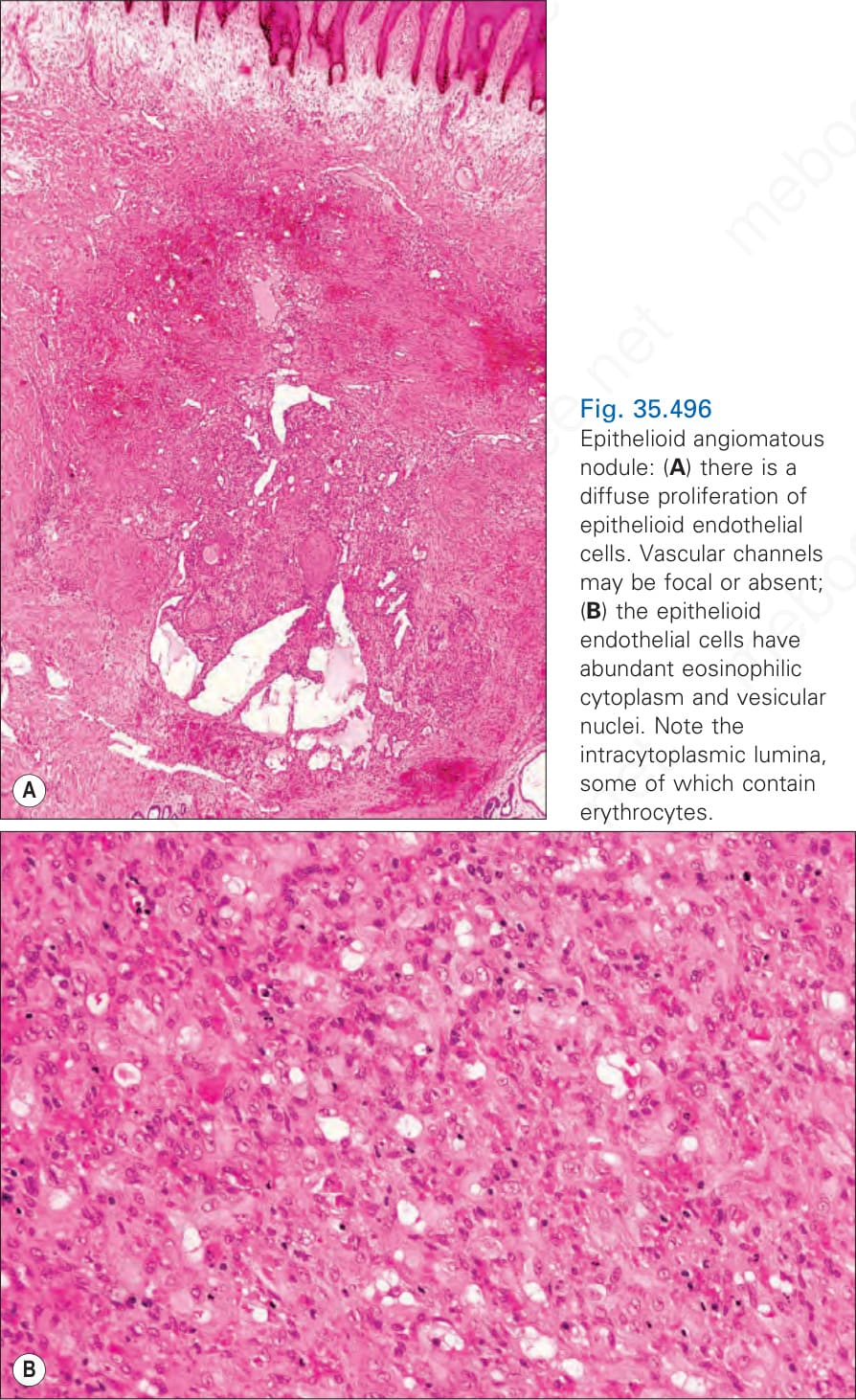
Fig. 35.496 Epithelioid angiomatous nodule: (A) there is a diffuse proliferation of epithelioid endothelial cells. Vascular channels may be focal or absent; (B) the epithelioid endothelial cells have abundant eosinophilic cytoplasm and vesicular nuclei. Note the intracytoplasmic lumina, some of which contain erythrocytes.
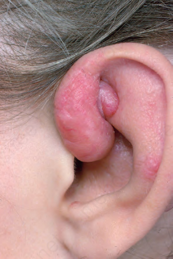
Fig. 35.497 Epithelioid hemangioma: the ear is commonly involved. There are multiple confluent lesions. From the collection of the late N.P. Smith, MD, the Institute of Dermatology, London, UK.
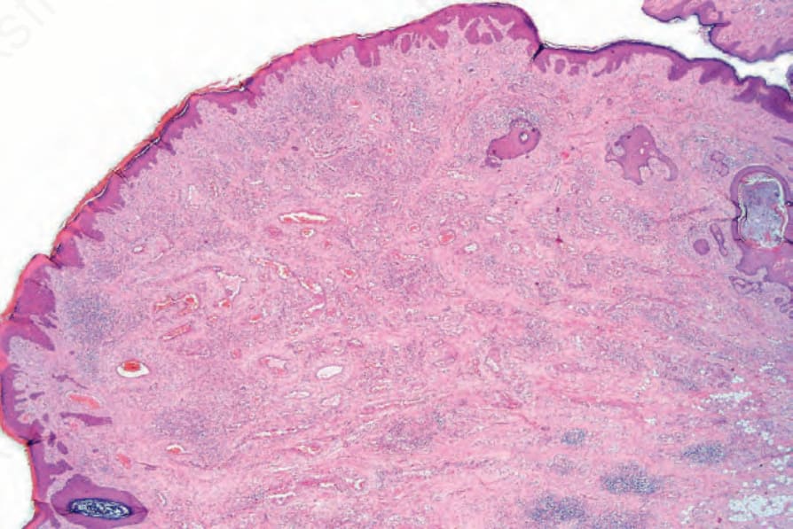
Fig. 35.498 Epithelioid hemangioma: scanning view of a vascular nodule with lymphoid aggregates at the periphery.
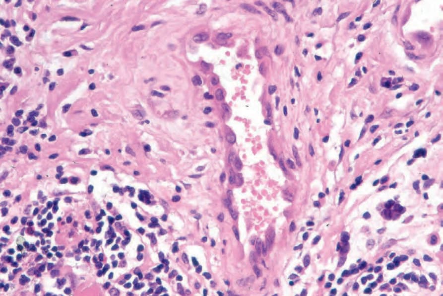
Fig. 35.499 Epithelioid hemangioma: the vessels are lined by large endothelial cells with markedly histiocytoid appearances.
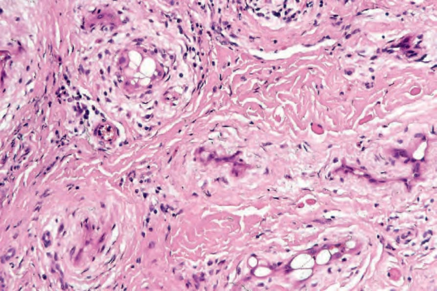
Fig. 35.500 Epithelioid hemangioma: endothelial cell intracytoplasmic lumina are a characteristic feature.
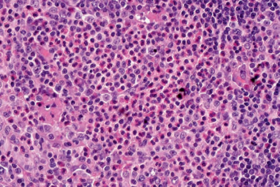
Fig. 35.501 Epithelioid hemangioma: eosinophils are conspicuous.
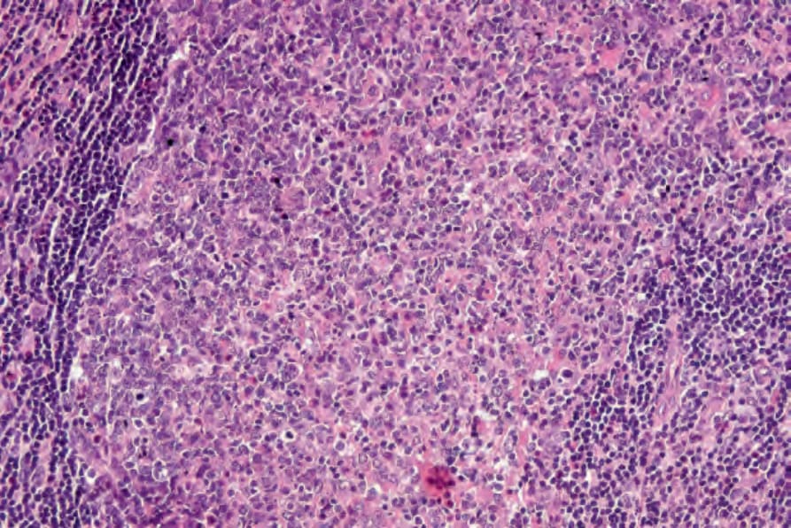
Fig. 35.502 Epithelioid hemangioma: lymphoid follicles are sometimes present
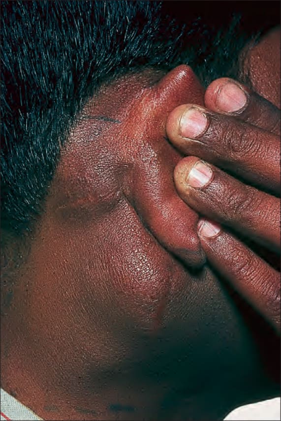
Fig. 35.503 Kimura disease: this patient presented with striking swelling of the neck. By courtesy of the Institute of Dermatology, London, UK.
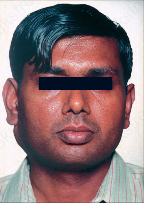
Fig. 35.504 Kimura disease: there is soft tissue and nodal involvement. By courtesy of the Institute of Dermatology, London, UK.
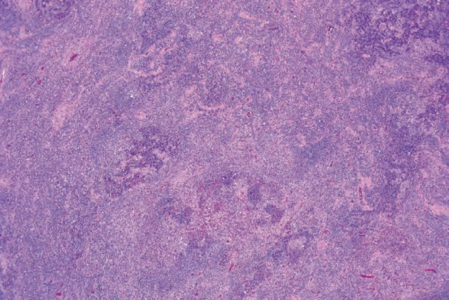
Fig. 35.505 Kimura disease: low-power view showing an intense cellular infiltrate.
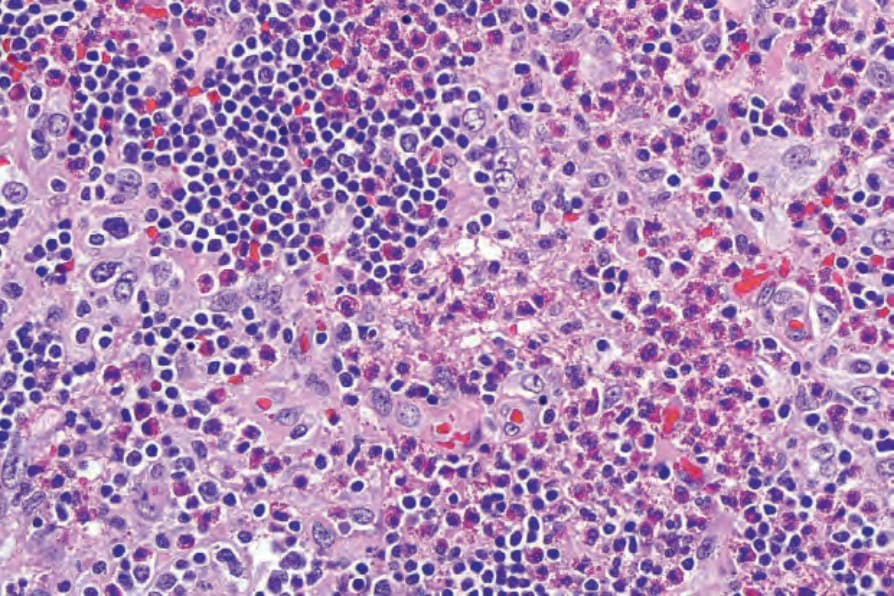
Fig. 35.506 Kimura disease: the infiltrate consists of lymphocytes and numerous eosinophils.
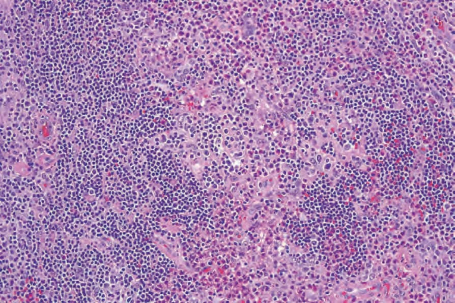
Fig. 35.507 Kimura disease: there is a background proliferation of high capillary venules.
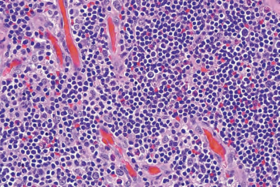
Fig. 35.508 Kimura disease: the endothelial cells are prominent but do not contain intracytoplasmic lumina.
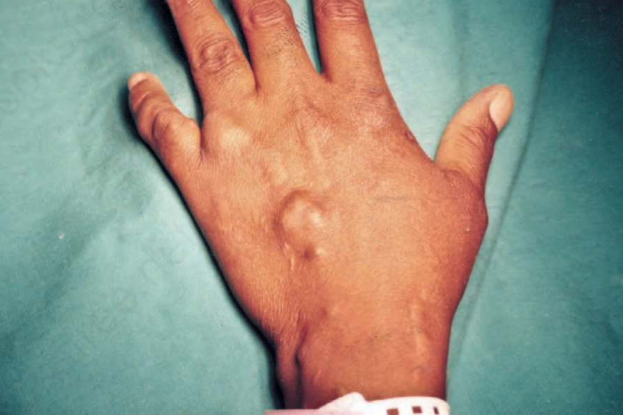
Fig. 35.509 Spindle cell hemangioma: multiple nodules are present at a characteristic site.