疾病定義與分類
- 微靜脈性血管瘤 (microvenular hemangioma)。
致病機轉/分子 (Pathogenesis / Molecular)
- 致病機轉與組織學特徵:僅依據免疫組化對 WT1 呈陰性,曾有人提出本病灶代表一種表淺淋巴管畸形 (superficial lymphatic malformation)。12,13
臨床特徵 (Clinical Features)
- Microvenular hemangioma 為一種無症狀病灶,常見於年輕成人的四肢,表現為紅—藍色 (red–bluish) 的丘疹、結節或斑塊。1–3
- 少數病人曾記錄到多發、有時數量眾多的病灶。3–6
- 一篇報告的個案中描述了多發、雙側的斑 (macules)、片狀斑 (patches) 與斑塊。7
- 兒童的表現罕見。8,9
- 曾於 POEMS syndrome 的脈絡中記錄到一例 human herpesvirus-8 陽性的個案。10
- 復發極為罕見。
組織病理特徵 (Histopathology)
-
組織學上,由不規則、分支、薄壁的小靜脈 (venules) 組成,內襯形態溫和、含有飽滿核 (plump nuclei) 的內皮細胞 (Fig. 35.489)。
-
腫瘤廣泛延伸至整個真皮,在某種程度上呈玻璃樣變 (hyalinized) 的膠原束之間穿插剝離 (dissecting) (Fig. 35.490)。
-
血管腔隙浸潤立毛肌 (arrector pili muscles) 是常見的發現。每一腔隙周圍環繞著一層
-
低倍視野下最顯著的特徵是楔形 (wedge-shaped) 的血管增生,其基底朝向表皮。血管腔隙不規則、薄壁、擴張,內襯帶有溫和突出核 (bland protruding nuclei) 與稀少胞質的內皮細胞(鞋釘狀細胞 / hobnail cells)(Figs 35.492 and 35.493)。
-
局灶性乳頭狀突起 (focal papillary projections) 是一項特徵性表現。隨著病灶向深部真皮延伸,血管腔隙變得較不明顯,外觀上在膠原束之間穿插剝離,並由較扁平的內皮細胞內襯。
-
紅血球外滲 (extravasation of red blood cells) 與含鐵血黃素沉積 (hemosiderin deposition) 可以很明顯,但這取決於病灶的分期。
-
炎症通常不是特徵之一,但有時可見散在的淋巴球與漿細胞 (plasma cells)。
-
本腫瘤很可能代表一組以鞋釘狀內皮細胞 (hobnail endothelial cells) 為特徵的病灶譜系中的良性端,這組病灶包括乳頭狀淋巴管內血管內皮瘤 (papillary intralymphatic angioendothelioma, PILA, Dabska tumor) 與網狀血管內皮瘤 (retiform
1838 Connective tissue tumors
A
B
hemangioendothelioma)。14
- 外傷 (trauma) 可能誘發類似於 hobnail hemangioma 所見的繼發性變化。15
免疫組化與特殊染色 (Immunohistochemistry & Special Stains)
- Hobnail hemangioma 中的內皮細胞對血管標記呈瀰漫性染色,包括 CD31 與 ERG。
- CD34 通常為陰性或僅極局灶性陽性。
- 一層 alpha-SMA 陽性的周細胞 (pericytes) 環繞著部分血管腔隙。
- 儘管有與月經週期相關的變化,內皮細胞對動情素 (estrogen) 與黃體素 (progesterone) 受體呈陰性。9
- 對血管內皮生長因子受體 3 (vascular endothelial growth factor receptor 3, VEGFR-3) 與 D2-40 呈陽性染色,這導致有人提出 hobnail hemangioma 顯示淋巴管分化 (lymphatic differentiation)。4,12,13,16
- 然而,VEGFR-3 對淋巴管內皮並非完全特異。
- 對 HHV-8 的染色一致呈陰性。17
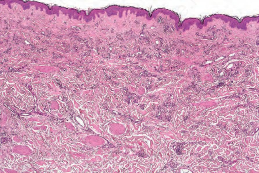
圖 35-489:Microvenular hemangioma:血管不規則浸潤真皮的方式有時會被誤認為 Kaposi sarcoma。
Fig. 35.489 Microvenular hemangioma: the manner in which the vessels irregularly infiltrate the dermis is sometimes mistaken for Kaposi sarcoma.
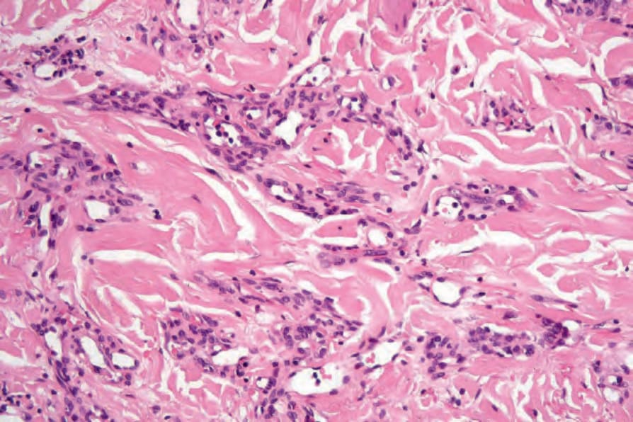
圖 35-490:Microvenular hemangioma:分支的血管由一層飽滿的內皮單層 (plump endothelial monolayer) 與外層較為梭形的周細胞 (pericytes) 內襯。
Fig. 35.490 Microvenular hemangioma: the ramifying vessels are lined by a plump endothelial monolayer and an outer layer of more spindled pericytes.
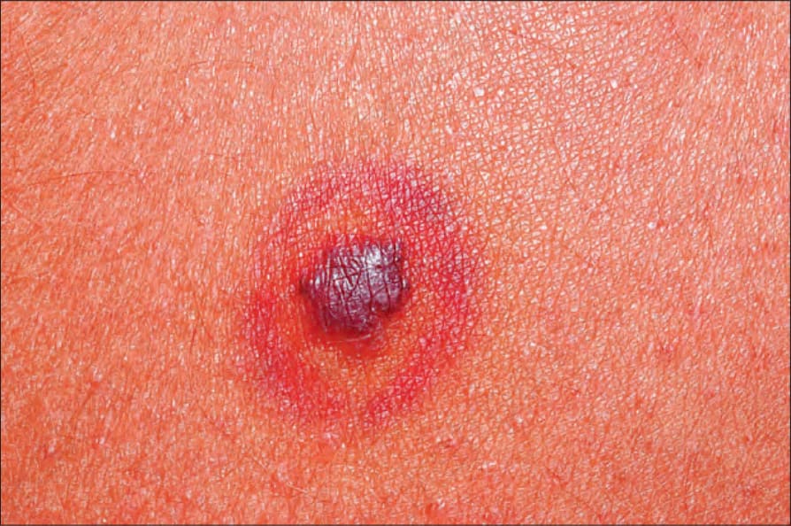
圖 35-491:Hobnail hemangioma:此例顯示特徵性的標靶樣外觀 (targetoid appearance)。
Fig. 35.491 Hobnail hemangioma: this example shows the characteristic targetoid appearance.
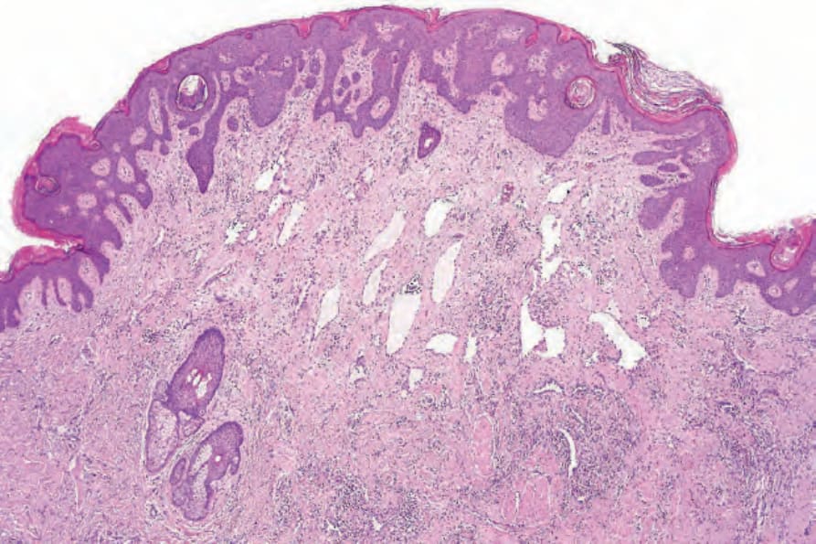
圖 35-492:Hobnail hemangioma:薄壁血管腔隙存在於表淺真皮。生長型態呈楔形 (wedge shaped)。
Fig. 35.492 Hobnail hemangioma: thin-walled vascular channels are present in the superficial dermis. The growth pattern is wedge shaped.
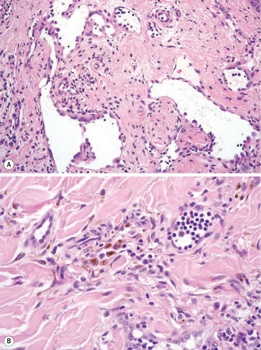
圖 35-493:Hobnail hemangioma:(A) 內皮細胞顯著並突入管腔。注意乳頭狀突起 (papillary processes);(B) 有大量含鐵血黃素色素 (hemosiderin pigment)。
Fig. 35.493 Hobnail hemangioma: (A) the endothelial cells are prominent and protrude into the lumen. Note the papillary processes; (B) there is abundant hemosiderin pigment.