疾病定義與分類
- 小葉狀微血管瘤 (lobular capillary hemangioma / pyogenic granuloma) 是一種非常常見的良性血管病灶,多年來一直被視為反應性或感染性的過程。此一假設是基於這些病灶幾乎一律存在的廣泛次發性變化。然而,其潛在過程其實是一種微血管的小葉狀增生 (lobular proliferation of capillaries),這更可能屬於腫瘤性 (neoplastic) 性質,因此被重新命名為 lobular capillary hemangioma。
臨床特徵 (Clinical Features)
- 本病灶可發生於任何年齡、任一性別,好發於頭頸部(尤其是黏膜)與四肢(特別是上臂與手部)(Fig. 35.470)。口腔病灶在女性較為常見。lobular capillary hemangioma 亦可發生於胃腸道及其他器官。
- 典型上,病灶演變迅速,於數個月內即達到其最大尺寸(通常直徑小於 2 cm)。其表現為一帶蒂 (pedunculated) 的紅色或藍色結節,易於潰瘍或出血 (Fig. 35.471)。完全的自發性消退並不會發生,少數病人會以多發性病灶表現,可呈瀰漫性或局限性分布。
- 曾有描述伴隨藥物過敏反應 (drug hypersensitivity reaction)、地雷傷害 (landmine injury)、燒傷,以及與後天性動靜脈畸形 (acquired arteriovenous malformation) 相關之噴發性 (eruptive) 病灶。先天性病灶極為罕見,曾有一例以瀰漫性病灶表現。病灶可發生於葡萄酒色斑 (port-wine stain) 之內,更罕見地與單側皮節性淺表毛細血管擴張 (unilateral dermatomal superficial telangiectasia) 相關聯。
- 多發性類似 pyogenic granuloma 的病灶已被記錄與以下藥物相關:BRAF inhibitors、capecitabine、外用 tretinoin、isotretinoin、gefitinib、afatinib、5-fluorouracil、levothyroxine、EGFR tyrosine kinase inhibitors,以及 anti-TNF-alpha therapy。曾有一例被認為與 erythropoietin 相關。甲下 (subungual) 病灶不僅可與藥物及創傷相關發生,亦可於周邊神經損傷 (peripheral nerve injury) 之後發生。lobular capillary hemangioma 亦曾被記錄為用以治療葡萄酒色斑之脈衝染料雷射 (pulse dye laser) 的併發症、發生於眼眶 hydroxyapatite 植入物之後、骨髓移植 (bone marrow transplant) 之後,以及發生於一名同時患有 NF1 與 von Hippel-Lindau syndrome 的病人。
- 切除後的局部復發相對頻繁,少數病例會復發並伴隨多發性衛星病灶 (satellite lesions),在臨床上可能令人擔憂 (Fig. 35.472)。後者此一現象傾向發生於較年輕的個體,其原發病灶非常常見於軀幹。
- Granuloma gravidarum 為一變異型,表現於孕婦的牙齦 (gingivae),並於分娩後消退。懷孕亦可能於其他部位誘發病灶。
- 皮下或深層真皮型 lobular capillary hemangioma (subcutaneous or deep dermal lobular capillary hemangioma) 好發於上肢。由於其從不潰瘍,因此不伴隨次發性發炎變化。
- 靜脈內型 lobular capillary hemangioma (intravenous lobular capillary hemangioma) 不常見,但傾向發生於年輕成人的頸部與上肢。
組織病理特徵 (Histopathology)
- Lobular capillary hemangioma 通常由一外生性 (exophytic)、分葉狀的真皮腫塊組成,其由眾多小微血管構成,這些微血管常從較大、較居中的血管放射狀延伸而出,置身於一疏鬆水腫的膠原基質 (loose edematous collagenous matrix) 之中 (Figs 35.473 and 35.474)。
- 內皮細胞 (endothelial cells) 的細胞核可由溫和 (bland) 到飽滿 (plump) 不等,並可局灶性地呈類上皮樣 (epithelioid),尤其在黏膜腫瘤中。有絲分裂 (mitoses) 常見且可能為數眾多 (Fig. 35.475)。可見因退化所致之局灶性細胞學異型性 (focal cytologic atypia) (Figs 35.476 and 35.477)。有時可見化生性骨化 (metaplastic ossification),並極罕見地記錄到髓外造血 (extramedullary hematopoiesis)。
- 大量急性與慢性發炎細胞的淺表浸潤是常見的發現,但此僅見於潰瘍性病灶。在此類情況下,鄰接的表皮常呈棘層肥厚 (acanthotic) 並傾向形成一界線分明的領圈 (collarette)。當發炎明顯時,整體特徵與肉芽組織 (granulation tissue) 極為相似,唯一例外是病灶基部深層真皮中存在微血管小葉 (capillary lobules)。
- 那些發展出衛星病灶的病例常顯示向皮下脂肪延伸。
- Intravenous lobular capillary hemangioma 在組織學上與較常規的病灶相似,唯其缺乏顯著的發炎成分 (Figs 35.478 and 35.479)。
致病機轉/分子 (Pathogenesis / Molecular)
- 在某些 lobular capillary hemangioma 病例中曾發現 human papillomavirus type 2,暗示可能存在病因學上的關聯。在偶發性與伴隨葡萄酒色斑的 lobular capillary hemangiomas 中曾發現 RAS 與 BRAF 突變,特別是 BRAF V600E,暗示 RAS / ERK pathway 在其致病機轉中可能扮演的角色。
鑑別診斷 (Differential Diagnosis)
- 最重要的鑑別診斷是 bacillary angiomatosis,這是一種由 rickettsial 微生物 Rochalimaea henselae 所引起的感染性血管增生。後者主要發生於 AIDS 病人,較罕見地發生於其他免疫抑制 (immunosuppressed) 宿主,或極例外地發生於正常個體。雖然兩種病灶在結構上非常相似,但 bacillary angiomatosis 由淡嗜伊紅性內皮細胞 (pale eosinophilic endothelial cells) 組成,且整個病灶中可見多形核白血球 (polymorphs),並在嗜鹼性顆粒狀聚集物 (basophilic granular aggregates) 附近更為明顯。後者以 Giemsa 或 Warthin-Starry 染色時,可顯示成簇的短桿菌 (short bacilli)。此桿菌亦可藉由免疫組織化學 (immunohistochemistry) 來證實。
- Lobular capillary hemangioma 有時在臨床上需要與其他類型的微血管瘤 (capillary hemangioma) 區分。具有非常飽滿的內皮細胞與高有絲分裂率的黏膜病灶,可藉由其小葉狀結構 (lobular architecture) 而輕易地與血管肉瘤 (angiosarcoma) 區分。
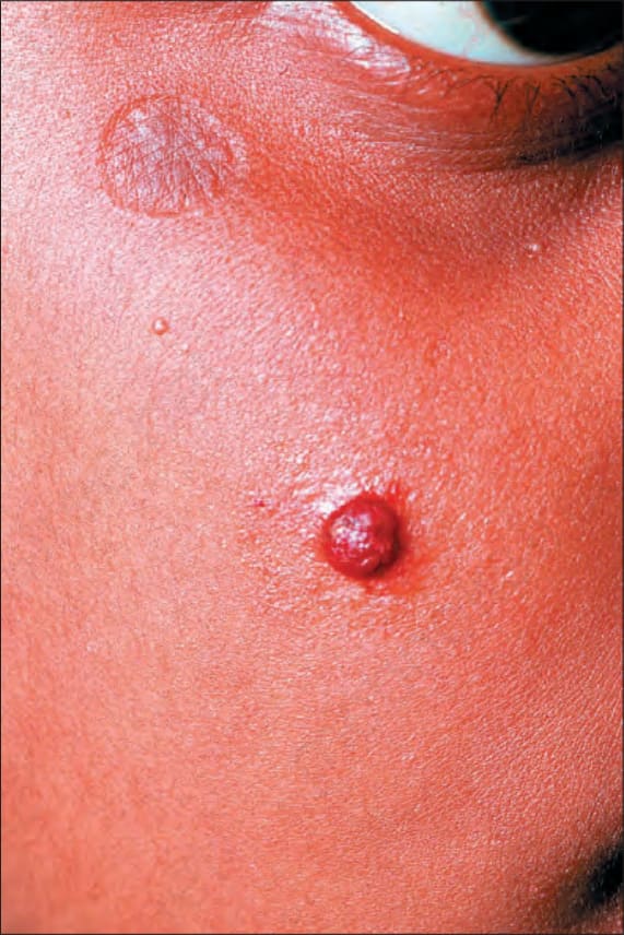
圖 35.470:Lobular capillary hemangioma:年輕女性病人臉部一典型隆起的紅色結節。By courtesy of M.M. Black, MD, St Thomas’ Hospital, London, UK.
Fig. 35.470 Lobular capillary hemangioma: a typical raised red nodule on the face of a young female patient. By courtesy of M.M. Black, MD, St Thomas’ Hospital, London, UK.
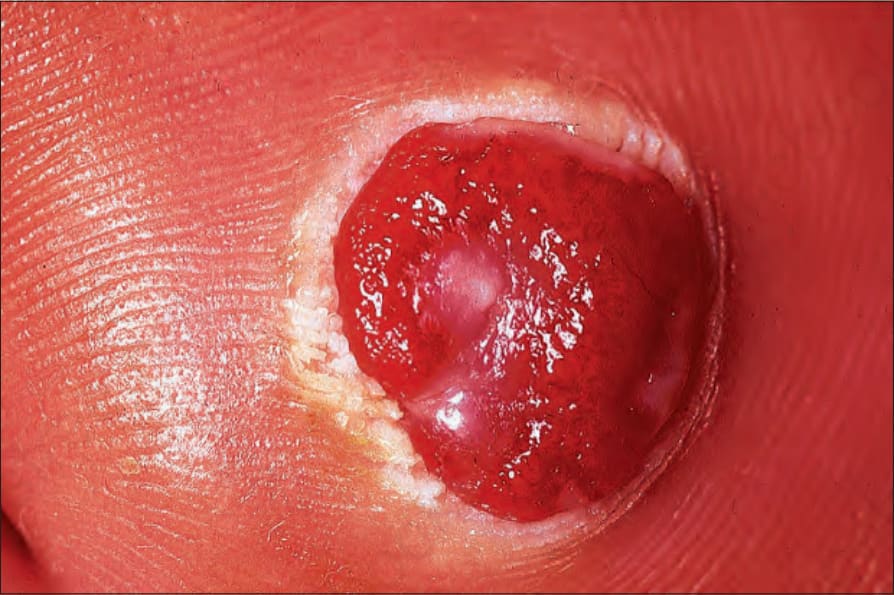
圖 35.471:Lobular capillary hemangioma:這些病灶具有特徵性的潰瘍。By courtesy of the Institute of Dermatology, London, UK.
Fig. 35.471 Lobular capillary hemangioma: these lesions are characteristically ulcerated. By courtesy of the Institute of Dermatology, London, UK.
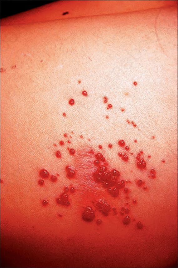
圖 35.472:Lobular capillary hemangioma (satellitosis):軀幹上多發性衛星病灶的特徵性外觀。By courtesy of E. Wilson Jones, MD, Institute of Dermatology, London, UK.
Fig. 35.472 Lobular capillary hemangioma (satellitosis): characteristic appearance of multiple satellite lesions on the trunk. By courtesy of E. Wilson Jones, MD, Institute of Dermatology, London, UK.
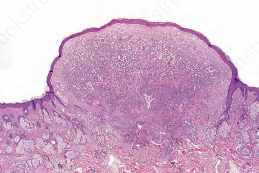
圖 35.473:Lobular capillary hemangioma:此掃描切面顯示病灶的息肉狀結構 (polypoid structure) 與發育良好的領圈 (collarette)。
Fig. 35.473 Lobular capillary hemangioma: this scanning section shows the polypoid structure of the lesion and the well-formed collarette.
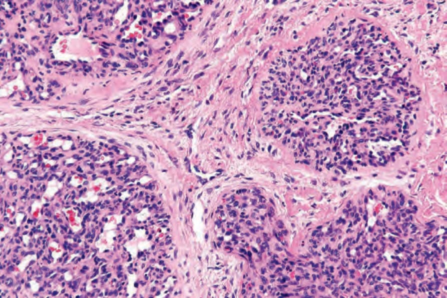
圖 35.474:Lobular capillary hemangioma:注意發育良好的小葉狀結構 (lobular architecture)。
Fig. 35.474 Lobular capillary hemangioma: note the well-developed lobular architecture.
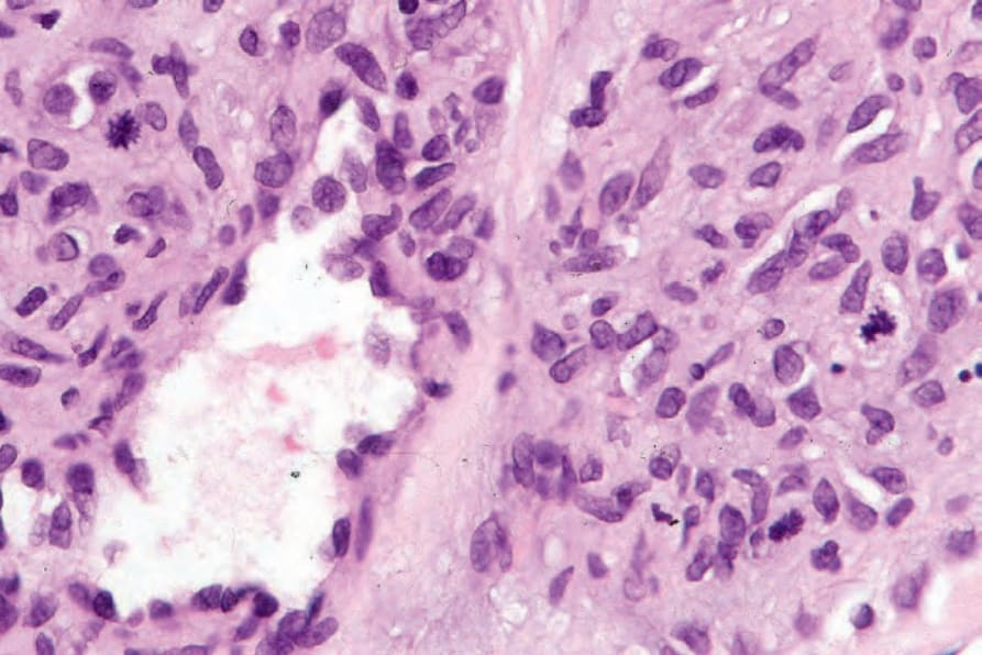
圖 35.475:Lobular capillary hemangioma:常存在顯著的有絲分裂活性 (mitotic activity),特別是在演變中的病灶。
Fig. 35.475 Lobular capillary hemangioma: conspicuous mitotic activity is often present, particularly in evolving lesions.
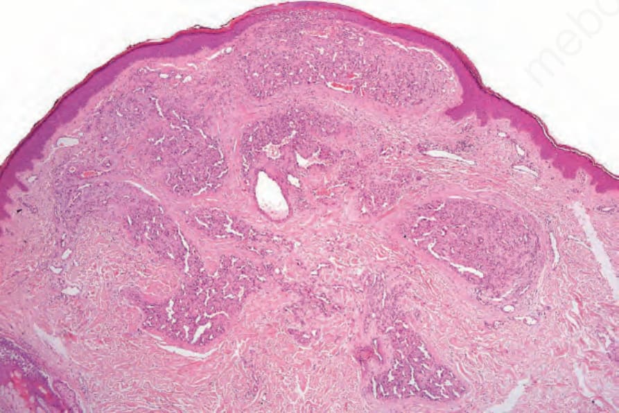
圖 35.476:伴異型性的 lobular capillary hemangioma:掃描視野顯示多個小葉 (lobules) 與伴隨的纖維性基質 (fibrous stroma)。
Fig. 35.476 Lobular capillary hemangioma with atypia: scanning view showing multiple lobules with an associated fibrous stroma.
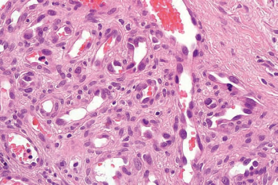
圖 35.477:伴異型性的 lobular capillary hemangioma:可見細胞核多形性 (nuclear pleomorphism),並存在一個異型性有絲分裂 (atypical mitosis)。
Fig. 35.477 Lobular capillary hemangioma with atypia: there is nuclear pleomorphism and an atypical mitosis is present.
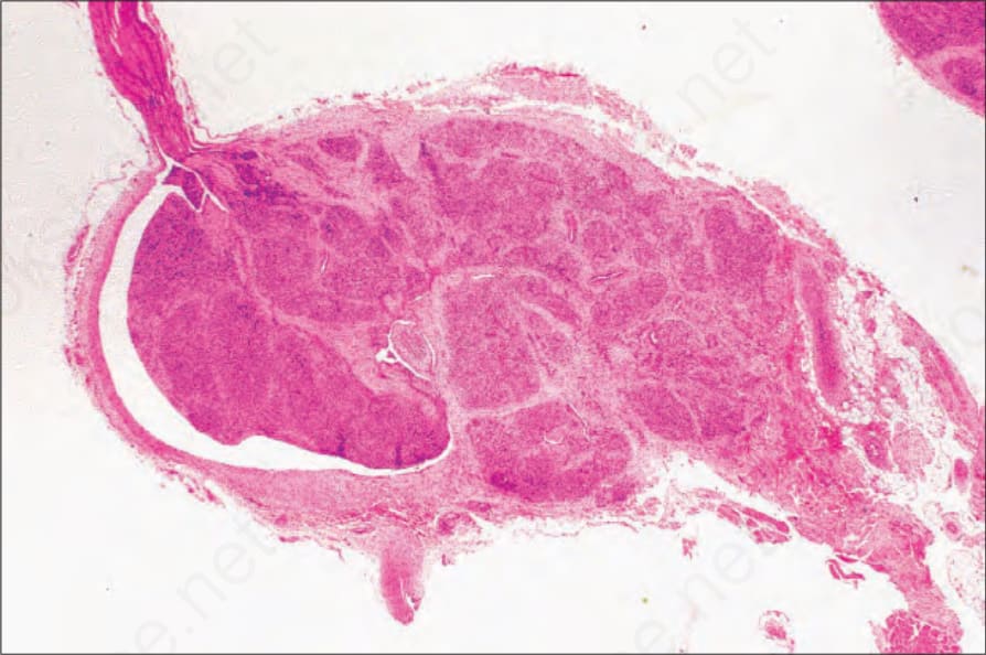
圖 35.478:Intravascular lobular capillary hemangioma:這是一種罕見的病灶。注意其薄的血管壁與顯著的小葉性 (lobularity)。
Fig. 35.478 Intravascular lobular capillary hemangioma: this is a rare lesion. Note the thin vessel wall and prominent lobularity.
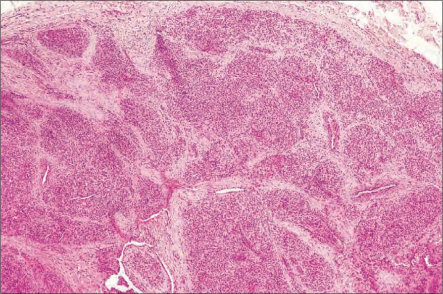
圖 35.479:Intravascular lobular capillary hemangioma:高倍視野顯示血管小葉 (vascular lobules)。
Fig. 35.479 Intravascular lobular capillary hemangioma: higher-power view showing the vascular lobules.
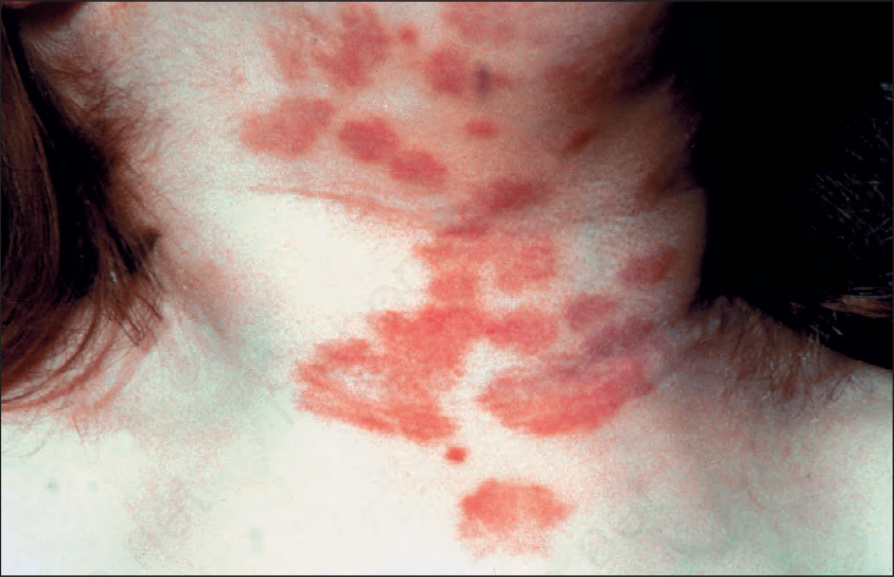
圖 35.480:Tufted angioma:病灶常表現於頸部與上軀幹。注意廣泛的斑 (macules) 與斑塊樣病灶 (plaque-like lesions) 的存在。By courtesy of the Institute of Dermatology, London, UK.
Fig. 35.480 Tufted angioma: lesions commonly present on the neck and upper trunk. Note the presence of extensive macules and plaque-like lesions. By courtesy of the Institute of Dermatology, London, UK.