非消退性先天性血管瘤 (Non-involuting Congenital Hemangioma, NICH)
非消退性先天性血管瘤 (non-involuting congenital hemangioma, NICH)
臨床特徵 (Clinical Features)
- Non-involuting congenital hemangioma 在出生時即已完全發育成熟,但不會消退;相反地,它會隨時間傾向於進展。男性與女性受影響的程度相當。其解剖分布廣泛,但好發於頭部與四肢。3
組織病理特徵 (Histopathology)
- 嬰兒型血管瘤 (infantile hemangiomas) 具有相當一致的顯微鏡下表現,特徵為真皮內或皮下的多葉狀 (multilobular) 增生,由眾多襯覆飽滿內皮細胞 (plump endothelial cells) 的小血管腔隙所組成,這些內皮細胞可能具有有絲分裂活性 (Figs 35.467 and 35.468)。在早期階段,血管腔隙傾向於不明顯,腫瘤的血管本質可能無法立即顯現。然而,reticulin stain(網狀纖維染色)可凸顯眾多管腔化不良 (poorly canalized) 血管通道的存在。隨著成熟,血管增大並擴張,內皮顯得較為扁平且成熟。在病灶的深部邊緣,常可見到一條大型的供血小動脈 (feeding arteriole)。一個偶發但完全屬於良性的特徵是神經周圍侵犯 (perineural invasion) 的存在。5,6 較陳舊的病灶逐漸變得更為纖維化,顯示血管成分逐步消失,而對大致已消退的病例做出組織學診斷可能會有困難。
1831 Capillary hemangioma and its variants
- 透過免疫組織化學 (immunohistochemistry) 與電子顯微鏡 (electron microscopy) 已證實,腫瘤的細胞族群是異質性的,不僅由內皮細胞組成,還包含纖維母細胞 (fibroblasts) 與周細胞 (pericytes)。7,8 這支持其為一種錯構瘤過程 (hamartomatous process),而非真正的腫瘤。它具有一種獨特的免疫表型,與表達 GLUT-1、LeY 及 WT-1 的胎盤微血管 (placental microvessels) 所共有。9 GLUT-1 為紅血球型葡萄糖轉運蛋白 (erythrocyte-type glucose transporter protein),在這些血管瘤演化的所有階段皆有表達。9,10 由於 GLUT-1 與 WT-1 並不表達於兒童發生的其他血管腫瘤,此標記的存在對於鑑別診斷是一項有價值的輔助,尤其是在血管畸形 (vascular malformations) 的情境中。11,12 增生性病灶中的內皮細胞共同表達 LYVE-1 與 CD34,且 Prox-1 為陰性,而 LYVE-1 在消退中的病灶為陰性,這顯示增生性嬰兒型血管瘤中的內皮細胞被停滯於血管分化的早期發育階段。13 此外,capillary hemangiomas 已被證實為單株性 (clonal)。14,15
鑑別診斷 (Differential Diagnosis)
- 與先天性血管瘤 (congenital hemangiomas) 的鑑別診斷於後者項下討論。
伴隨輕微或停滯生長的嬰兒型血管瘤(流產型血管瘤)(Infantile hemangiomas with minimal or arrested growth, abortive hemangiomas)
臨床特徵 (Clinical Features)
- 這些近期被描述的病灶,特徵為帶有周邊丘疹的微血管擴張性斑片 (telangiectatic patches with peripheral papules),並好發於下半身。1–3 它們被定義為具有增生成分等於或小於病灶總表面 25% 的血管病灶。1 略少於 50% 的患者在其他部位有典型的嬰兒型血管瘤。1 病灶傾向於持續存在。
組織病理特徵 (Histopathology)
- 取自微血管擴張性斑片的切片顯示,淺層真皮中散布著擴張的血管通道。在深部真皮中,可見微血管小葉 (lobules of capillaries)。取自丘疹的切片顯示 capillary hemangioma 的特徵。內皮細胞 GLUT-1 為陽性。此特徵,以及與典型嬰兒型血管瘤共存的現象,證實它們彼此密切相關。
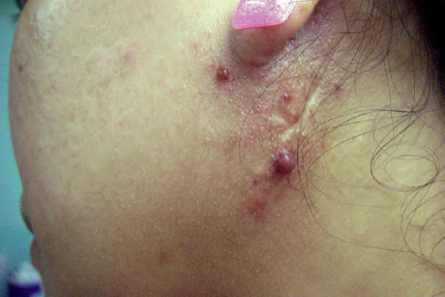
圖 35-465:嬰兒型血管瘤 (infantile hemangioma):在此孩童耳部與頸部周圍可見多個隆起的紅斑性結節。By courtesy of J. Dayrit, MD, Manila, The Philippines.
Fig. 35.465 Infantile hemangioma: multiple raised erythematous nodules are present around this child’s ear and neck. By courtesy of J. Dayrit, MD, Manila, The Philippines.

圖 35-466:嬰兒型血管瘤 (infantile hemangioma):此女嬰前額上可見兩個隆起的結節。By courtesy of M.M. Black, MD., The Institute of Dermatology, London, UK.
Fig. 35.466 Infantile hemangioma: two raised nodules are present on the forehead of this female infant. By courtesy of M.M. Black, MD., The Institute of Dermatology, London, UK.
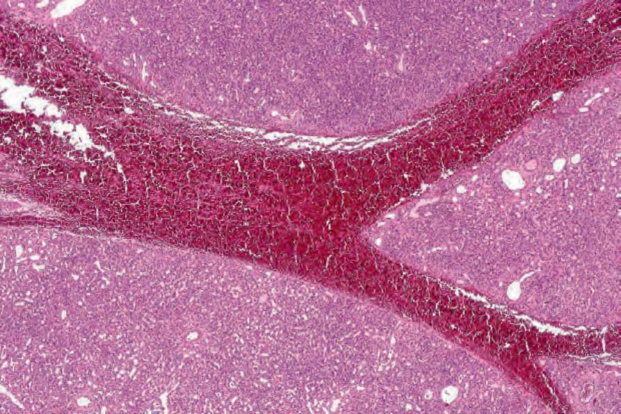
圖 35-467:嬰兒型血管瘤 (infantile hemangioma):此為一演化中的病灶,由管腔化不良血管 (poorly canalized blood vessels) 的分葉狀聚集所組成。
Fig. 35.467 Infantile hemangioma: this is an evolving lesion composed of lobulated aggregates of poorly canalized blood vessels.
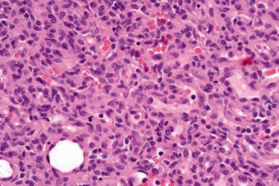
圖 35-468:嬰兒型血管瘤 (infantile hemangioma):血管由飽滿內皮細胞 (plump endothelial cells) 所襯覆。注意多處有絲分裂象 (multiple mitoses)。
Fig. 35.468 Infantile hemangioma: the blood vessels are lined by plump endothelial cells. Note the multiple mitoses.