皮膚膠原性血管病變 (Cutaneous Collagenous Vasculopathy)
皮膚膠原性血管病變 (cutaneous collagenous vasculopathy)
臨床特徵 (Clinical Features)
- 這是一種罕見、但可能診斷不足 (underdiagnosed) 的疾病,特徵為全身性微血管擴張 (generalized telangiectasia),類似全身性原發性微血管擴張 (generalized essential telangiectasia)。
- 好發於第五至第九個十年齡層 (between the 5th and 9th decades of life) 的病人,無性別偏好,特徵為進行性微血管擴張 (progressive telangiectasia),常始於下肢並以進行性方式擴展。1–6
- 偶見病灶呈瘀斑樣 (echimotic)。
致病機轉與組織學特徵 (Pathogenesis and Histologic Features)
- 病因不明 (unknown)。
- 組織學顯示淺層真皮血管腔 (superficial dermal vascular channels) 輕度擴張,血管壁增厚,並伴有由基底膜重複增生 (reduplication of the basement membrane) 所形成的玻璃樣嗜伊紅物質 (hyaline eosinophilic material) (Figs 35.460 and 35.461)。
- 此物質 PAS 染色陽性,並對 collagen type IV 染色呈陽性。1–6
- 在單一病例中,於血管腔內發現纖維蛋白血栓 (fibrin thrombi)。7
口腔血管角化瘤 (oral angiokeratomas) 可作為孤立現象出現,或與其他類型的血管角化瘤 (angiokeratomas) 相關,包括 Fabry disease。33,34
1829 先天性血管瘤 (Congenital hemangiomas)
組織學特徵 (Histologic Features)
- 所有變異型的組織學特徵相似,由乳頭層真皮 (papillary dermis) 內眾多擴張且充血的微血管 (dilated and congested capillaries) 組成,其上覆蓋棘層肥厚 (acanthosis) 與過度角化 (hyperkeratosis) (Fig. 35.462)。
- 在 Anderson-Fabry disease 中,曾於內皮細胞 (endothelial cells)、周細胞 (pericytes) 及纖維母細胞 (fibroblasts) 內描述到胞質內脂質空泡 (intracytoplasmic lipid vacuoles)。35
鑑別診斷 (Differential Diagnosis)
- 相同的特徵可見於疣狀血管瘤 (verrucous hemangioma) 的淺層部分,然而疣狀血管瘤總是具有深層真皮及皮下成分 (deep dermal and subcutaneous component)。
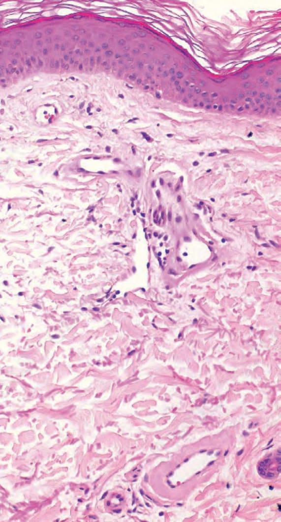
圖 35-460:皮膚膠原性血管病變 (cutaneous collagenous vasculopathy):擴張的淺層真皮血管腔,伴有增厚的血管壁。
Fig. 35.460 Cutaneous collagenous vasculopathy: dilated superficial dermal vascular channels with thickened walls.
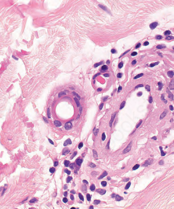
圖 35-461:皮膚膠原性血管病變 (cutaneous collagenous vasculopathy):血管腔周圍的無定形明亮嗜伊紅物質 (amorphous bright eosinophilic material)。
Fig. 35.461 Cutaneous collagenous vasculopathy: amorphous bright eosinophilic material around vascular channels.
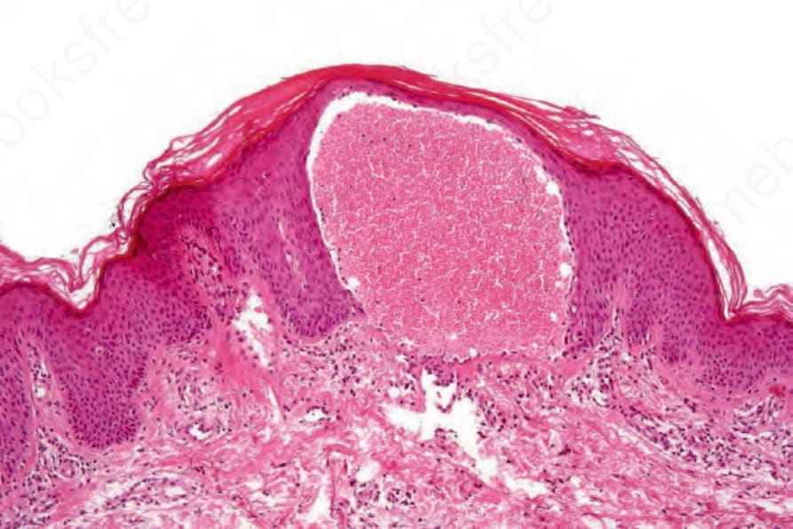
圖 35-462:血管角化瘤 (angiokeratoma):病灶由小而擴張的血管組成,這些血管常看似位於表皮 (epidermis) 之內。
Fig. 35.462 Angiokeratoma: the lesion consists of small dilated vessels that often appear to be within the epidermis.
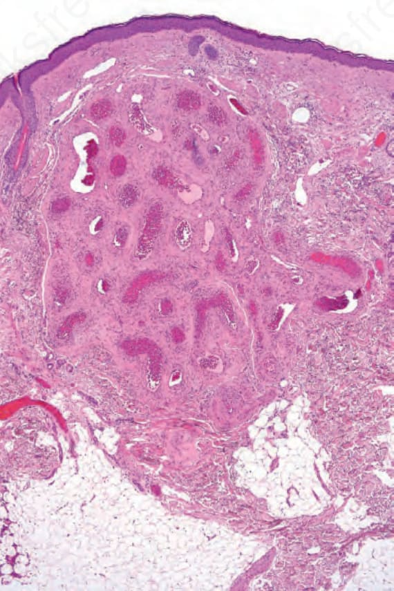
圖 35-463:動靜脈血管瘤 (arteriovenous hemangioma):真皮內可見一團厚壁血管 (thick-walled blood vessels),包含動脈與靜脈兩者。
Fig. 35.463 Arteriovenous hemangioma: within the dermis is a collection of thick-walled blood vessels comprising both arteries and veins.