Cutaneous collagenous vasculopathy
Cutaneous collagenous vasculopathy
Clinical features This is a rare but probably underdiagnosed condition is characterized by generalized telangiectasia mimicking generalized essential telangiectasia. It presents in patients between the 5th and 9th decades of life with no sex predilection and characterized by progressive telangiectasia that often starts on the lower limbs and spreads in a progressive manner.1–6 Occasionally, lesions are echimotic.
Pathogenesis and histologic features The etiology is unknown. Histology shows mild dilatation of superficial dermal vascular channels with thickening of the walls associated with hyaline eosinophilic material resulting from the reduplication of the basement membrane (Figs 35.460 and 35.461). The material is PAS positive and stains for collagen type IV.1–6 In a single case fibrin thrombi were identified within the lumina of vascular channels.7
Oral angiokeratomas are seen either as an isolated phenomenon or in association with other types of angiokeratomas including Fabry disease.33,34
1829 Congenital hemangiomas
Histologic features The histologic features in all variants are similar and consist of numerous dilated and congested capillaries in the papillary dermis with overlaying acanthosis and hyperkeratosis (Fig. 35.462). In Anderson- Fabry disease, intracytoplasmic lipid vacuoles have been described in endothelial cells, pericytes and fibroblasts.35
Differential diagnosis Identical features can be seen in the superficial portion of a verrucous hemangioma, which always, however, has a deep dermal and subcutaneous component.
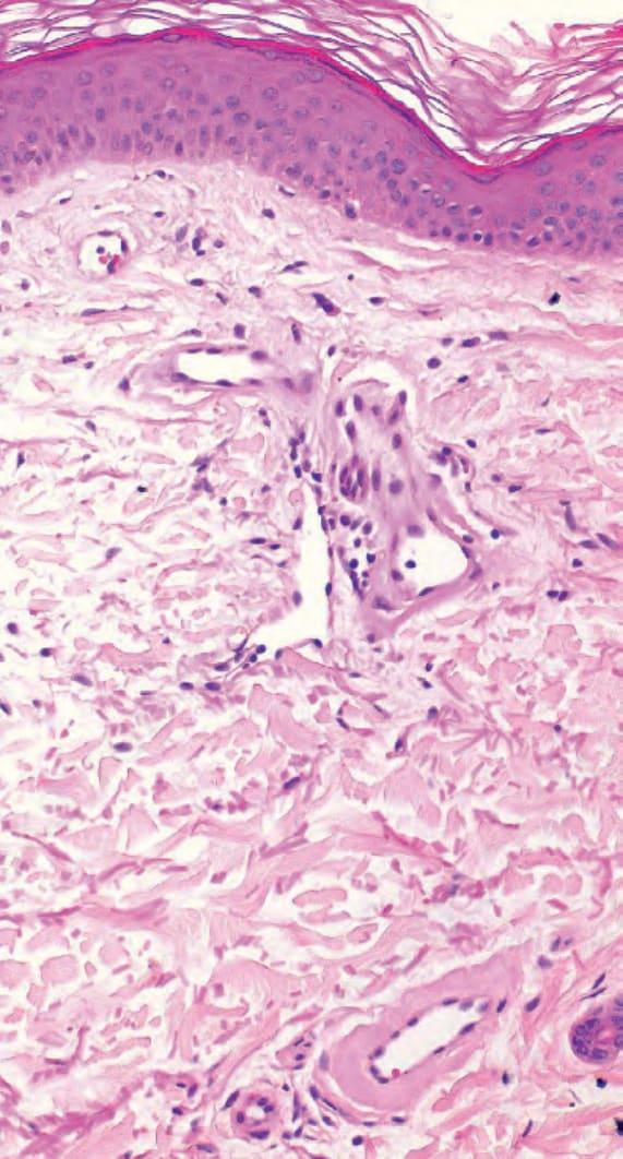
Fig. 35.460 Cutaneous collagenous vasculopathy: dilated superficial dermal vascular channels with thickened walls.
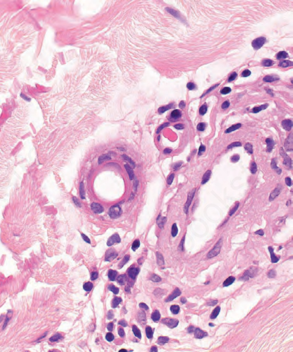
Fig. 35.461 Cutaneous collagenous vasculopathy: amorphous bright eosinophilic material around vascular channels.
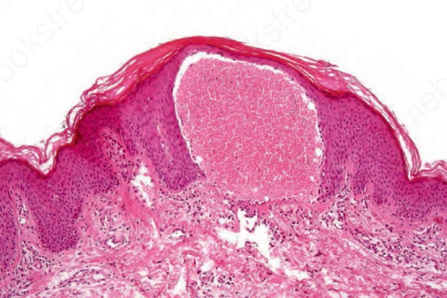
Fig. 35.462 Angiokeratoma: the lesion consists of small dilated vessels that often appear to be within the epidermis.
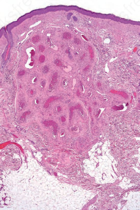
Fig. 35.463 Arteriovenous hemangioma: within the dermis is a collection of thick-walled blood vessels comprising both arteries and veins.