疾病定義與分類
橫紋肌肉瘤 (rhabdomyosarcoma) 極少出現於皮膚科醫師的診間,原因其一為此腫瘤本身罕見,其二為其鮮少發生於真皮,儘管偶爾確會出現皮膚轉移。傳統上描述有四種主要亞型:embryonal(包含 botryoid 變異型)、alveolar、pleomorphic 與 spindle cell/sclerosing。近期更記載了一種極為罕見的 epithelioid 變異型。前兩者為最常見者。
臨床特徵 (Clinical Features)
橫紋肌肉瘤以原發腫瘤或轉移形式出現於皮膚者極為罕見,僅占所有 rhabdomyosarcoma 的 0.7%(Fig. 35.432)。在已記載的皮膚病例中,男性略占多數,好發於頭頸部,發病時年齡呈雙峰分布(兒科病例平均年齡為 10 歲,成人病例平均年齡為 74 歲)。alveolar 型其次為 embryonal 型,是兒科族群中最常見的組織學亞型,而 pleomorphic 變異型則在成人中遠為常見。曾有與 epidermal nevus syndrome 相關的病例報告。關於皮膚病灶預後的資訊極為有限,但曾報告其具有侵襲性病程的潛能。雖然關於 epithelioid rhabdomyosarcoma 的可用資料有限,18 例中有 4 例發生於表淺部位,兩性分布相等,平均年齡為 69.5 歲。
致病機轉與組織學特徵 (Pathogenesis and Histologic Features)
在分子遺傳學層次,alveolar 型的特徵為 t(2;13)(q35;q14),較少見者為 t(1;13)(p36;q14),分別將 PAX3 或 PAX7 與 FOXO1A 融合。更近期的研究顯示,NCOA1 (2p23) 與 AFX/FOXO4 (Xq13.1) 在罕見情況下可取代 FOXO1A 並與 PAX3 配對。PAX3-FOXO1 腫瘤相較於 PAX7-FOXO1 腫瘤似乎具有較差的預後。在一部分病灶中,已辨識出與 low grade liposarcoma 相似的重排(MDM2 與 CDK4)。
Embryonal 病灶通常在其他細胞遺傳學異常之中具有 11p 缺失。
在 spindle cell rhabdomyosarcoma 中,已辨識出 8q13 重排所導致的 SRF-NCOA2 與 TEAD1-NCOA2 融合基因。一種反覆出現的新形態 (neomorphic) MYOD1 突變與較差的預後相關。Pleomorphic rhabdomyosarcoma 顯示複雜的核型。
Embryonal 型
Embryonal 型主要由小型圓形或紡錘形的未分化細胞組成,常鬆散地排列於 myxoid stroma 之中。明顯的 rhabdomyoblasts 突顯程度不一,呈嗜伊紅性細胞質並具有「帶狀 (strap)」或「蝌蚪狀 (tadpole)」形態(Figs 35.433–35.435)。此型 rhabdomyosarcoma 的變異型包括 botryoid、spindle cell 與 anaplastic。
Alveolar 型
Alveolar 變異型的典型表現為腫瘤細胞排列成由纖維間隔分隔的離散巢狀結構,並在細胞聚集體中央呈現細胞解離的 alveolar 排列型態(Figs 35.436 與 35.437)。然而,某些病例幾乎完全呈實心生長型態(Fig. 35.438)。腫瘤細胞傾向於相對較大且呈圓形,或易於辨識為 rhabdomyoblasts,並可能排列成纖細的乳頭狀、實心團塊狀,或游離於各細胞巢內。其細胞核較 embryonal 型更大且更具深染性。多核(花環狀,wreath-like)巨細胞為常見特徵(Fig. 35.439)。在一例原發性 alveolar rhabdomyosarcoma 中曾記載到 epidermotropism。Alveolar 型的組織學表現可見於缺乏特徵性融合基因的病例中。此類病例在遺傳學與臨床上究竟較類似 embryonal 還是 fusion-positive alveolar 病例,目前文獻中仍有爭議。
Pleomorphic 型
Pleomorphic 型為一異質性腫瘤,其特徵為怪異的紡錘狀細胞與易於辨識的多角形 rhabdomyoblasts 混雜,後者常數量眾多(Fig. 35.440)。此變異型大致僅見於成人,於皮膚中幾乎不為人知。
Spindle cell 型
雖然 embryonal rhabdomyosarcoma 包含一種 spindle cell 變異型,但成人中已描述了一種獨特的 spindle cell rhabdomyosarcoma 變異型。在這些病灶中,可見非典型紡錘狀細胞與 rhabdomyoblasts 混雜。
Epithelioid 型
此罕見類型由具有 epithelioid 特徵(豐富的細胞質與大型空泡狀細胞核)的均一細胞組成,呈瀰漫性片狀生長。核仁顯著,腫瘤細胞可能類似 melanoma 細胞。可見多核細胞。不存在明顯的 rhabdomyoblasts。典型可見顯著的壞死。
免疫組化 (Immunohistochemistry)
免疫組化在 rhabdomyosarcoma 的診斷中極為有用,因為腫瘤細胞傾向於至少局部呈 desmin、muscle-specific actin (HHF-35) 陽性,以及在診斷上更具特異性的 myogenin 與 MyoD1 陽性(Fig. 35.441)。
在所有 rhabdomyosarcoma 變異型中,腫瘤細胞對 desmin、myogenin (myf-4) 與 MyoD-1 的陽性程度不一。Rhabdomyosarcoma 可能顯示對 cytokeratins 的局部陽性。此外,偶爾可見對 S100 的局部陽性。Myogenin、Ap2beta、NOS-1 與 HMGA1 為可用的免疫組化標記,似乎與融合狀態 (fusion status) 相關。
鑑別診斷 (Differential Diagnosis)
Rhabdomyosarcomas 應與其他 small round cell neoplasms 區分:
- neuroblastoma 含有 neurofibrils 並顯示 rosette 形成;
- primitive neuroectodermal tumor 常具有成簇 (packeted) 的外觀,顯示細胞質內 PAS 陽性程度不一,缺乏類似 rhabdomyoblasts 的細胞,且呈瀰漫性 CD99 陽性;
- malignant lymphomas 最常(但非總是)呈 PAS 陰性,並對 LCA 染色陽性。Pleomorphic 型應藉由免疫組化與 rhabdomyoblasts 的辨識,與其他 pleomorphic 腫瘤區分。
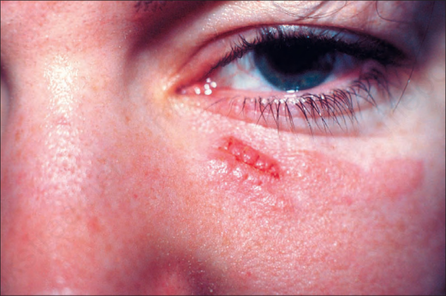
圖 35-432:皮膚 rhabdomyosarcoma:出現於皮膚屬例外情形,且見於兒童。此為一 embryonal 變異型。承蒙 Institute of Dermatology, London, UK 提供。
Fig. 35.432 Cutaneous rhabdomyosarcoma: presentation in the skin is exceptional and occurs in children. This was an embryonal variant. By courtesy of the Institute of Dermatology, London, UK.
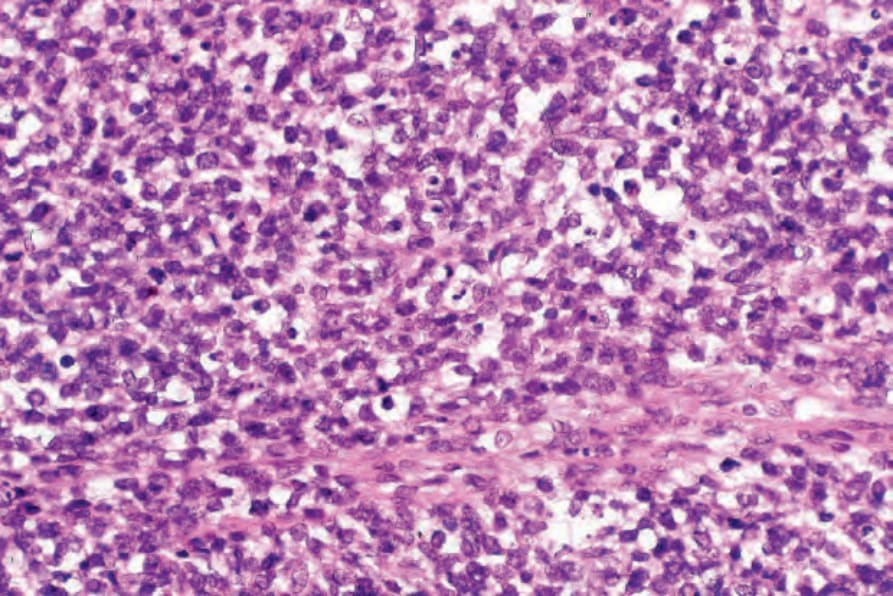
圖 35-433:Embryonal rhabdomyosarcoma:一例未分化 rhabdomyosarcoma。腫瘤細胞為小型且含嗜鹼性細胞核。診斷有賴免疫細胞化學 (immunocytochemistry),或在標本他處辨識出較典型的 rhabdomyoblasts。
Fig. 35.433 Embryonal rhabdomyosarcoma: an example of undifferentiated rhabdomyosarcoma. The tumor cells are small and contain basophilic nuclei. Diagnosis depends upon immunocytochemistry or identifying more typical rhabdomyoblasts elsewhere in the specimen.
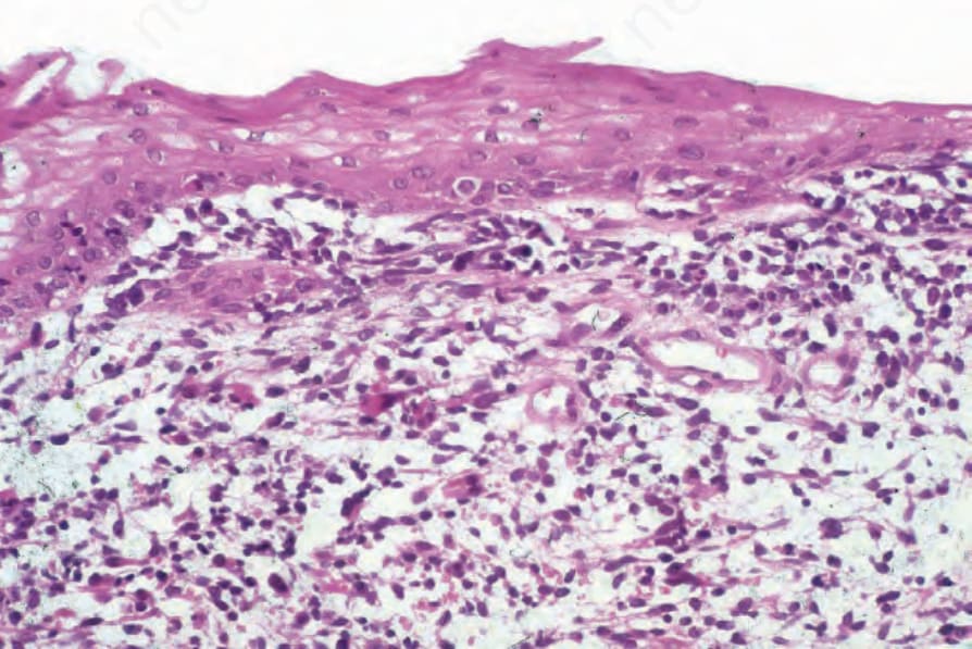
圖 35-434:Embryonal rhabdomyosarcoma:上皮下方為小型嗜鹼性細胞的浸潤,並可見偶發的原始 rhabdomyoblasts,後者具有更為明顯的嗜伊紅性細胞質。
Fig. 35.434 Embryonal rhabdomyosarcoma: beneath the epithelium is an infiltrate of small basophilic cells, and there are also occasional primitive rhabdomyoblasts with more obvious eosinophilic cytoplasm.
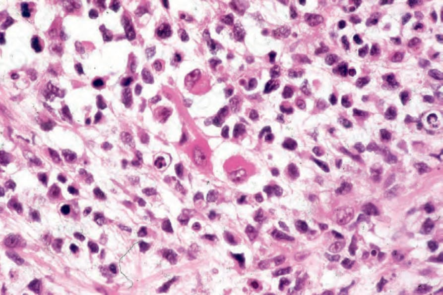
圖 35-435:Embryonal rhabdomyosarcoma:視野中央為一典型的「蝌蚪狀 (tadpole)」rhabdomyoblast,帶有逐漸變細的嗜伊紅性細胞質突起。
Fig. 35.435 Embryonal rhabdomyosarcoma: in the center of the field is a typical ‘tadpole’ rhabdomyoblast with a tapering eosinophilic, cytoplasmic process.
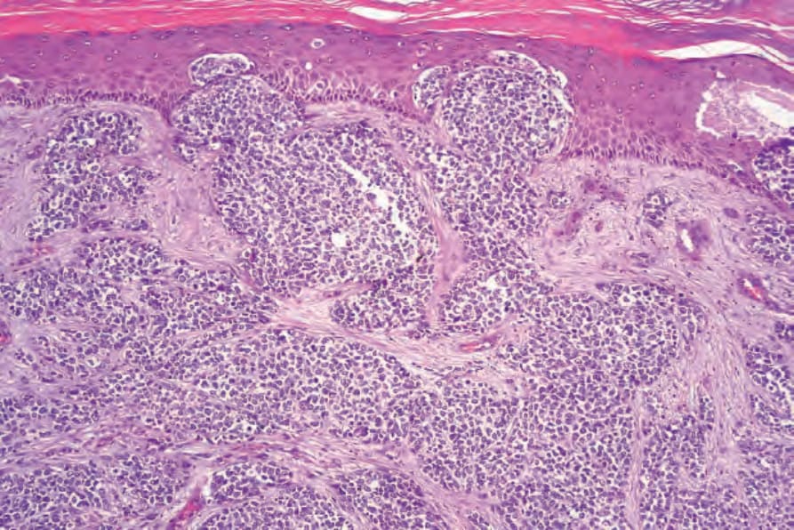
圖 35-436:Alveolar rhabdomyosarcoma:此為一極罕見的例子,顯示 epidermotropism。其特徵類似 neuroendocrine (Merkel cell) carcinoma。
Fig. 35.436 Alveolar rhabdomyosarcoma: this is a very rare example showing epidermotropism. The features mimic neuroendocrine (Merkel cell) carcinoma.
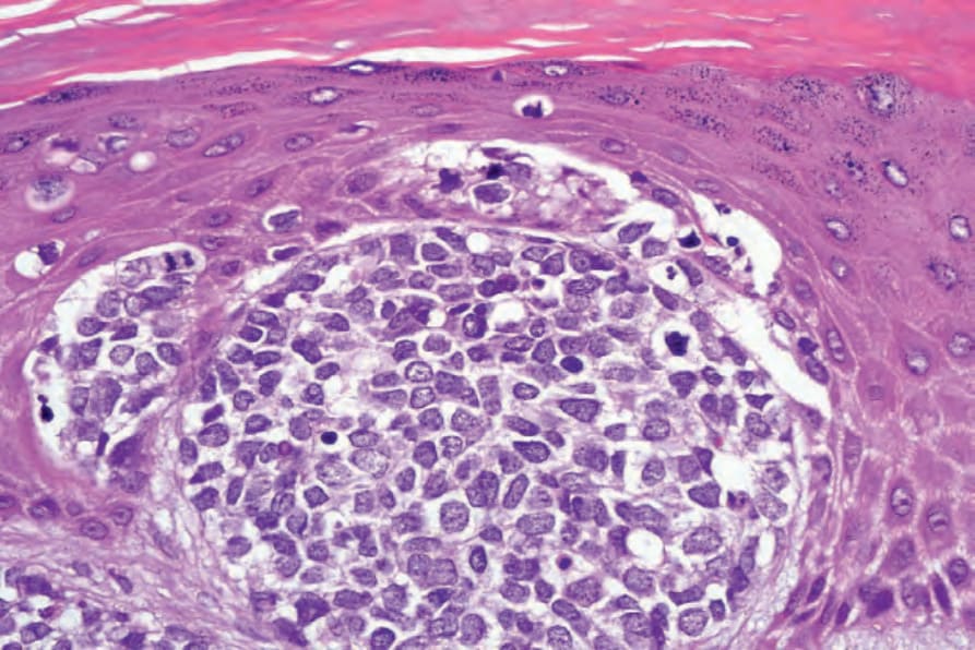
圖 35-437:Alveolar rhabdomyosarcoma:高倍視野。
Fig. 35.437 Alveolar rhabdomyosarcoma: high-power view.
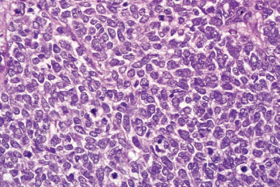
圖 35-438:Alveolar rhabdomyosarcoma:腫瘤細胞具有 pleomorphic 細胞核,並存在多個有絲分裂。此例中無 skeletal muscle differentiation 的證據。診斷有賴免疫組化。
Fig. 35.438 Alveolar rhabdomyosarcoma: the tumor cells have pleomorphic nuclei and multiple mitoses are present. There is no evidence of skeletal muscle differentiation in this example. Diagnosis depends upon immunohistochemistry.
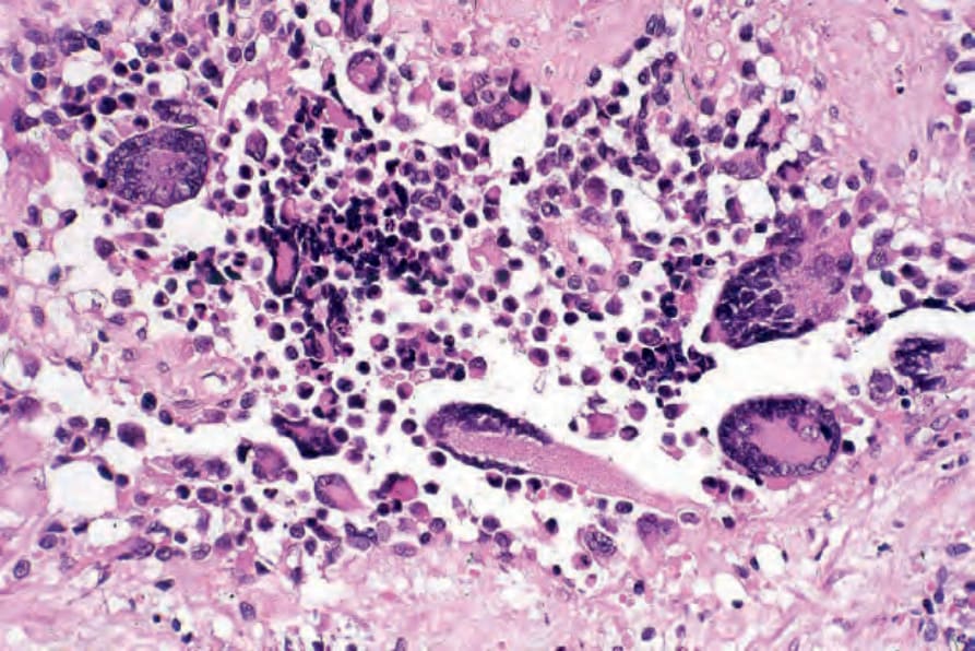
圖 35-439:Alveolar rhabdomyosarcoma:花環狀 (wreath-like) 巨細胞為一特徵性表現。
Fig. 35.439 Alveolar rhabdomyosarcoma: wreath-like giant cells are a characteristic feature.
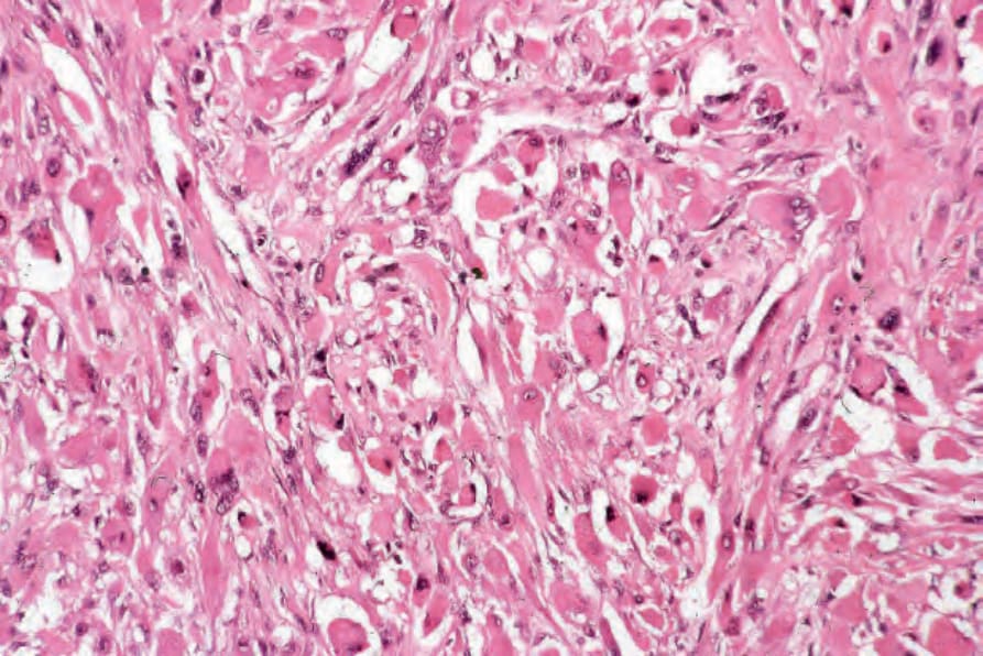
圖 35-440:Pleomorphic rhabdomyosarcoma:rhabdomyoblasts 極度 pleomorphic。
Fig. 35.440 Pleomorphic rhabdomyosarcoma: the rhabdomyoblasts are extremely pleomorphic.
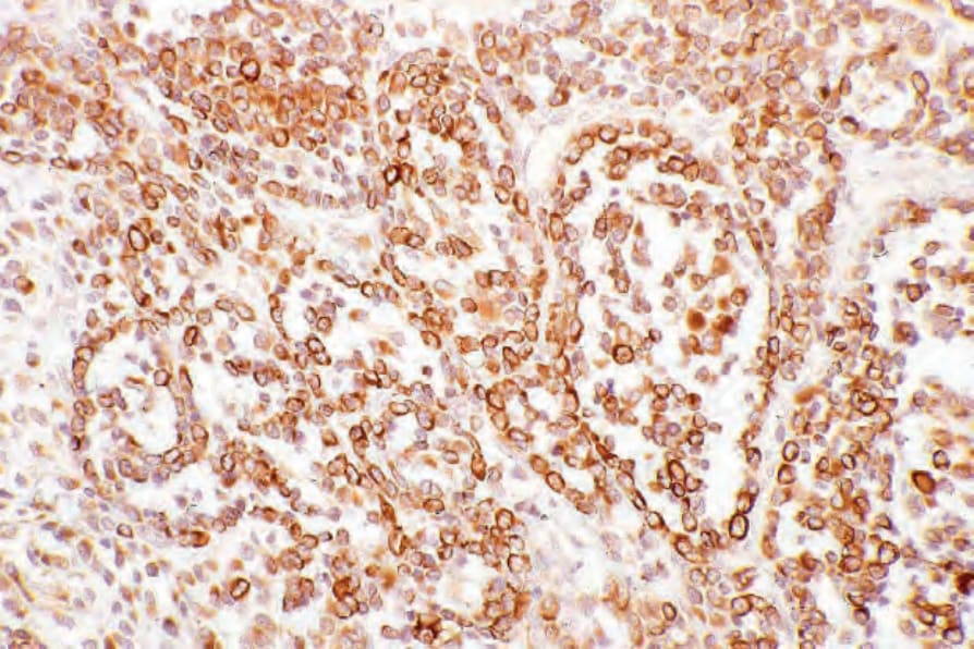
圖 35-441:Alveolar rhabdomyosarcoma:在此例中,腫瘤細胞強烈表現 desmin。
Fig. 35.441 Alveolar rhabdomyosarcoma: in this example, the tumor cells strongly express desmin.