臍息肉與臍肉芽腫 (Umbilical Polyp and Granuloma)
致病機轉與組織學特徵 (Pathogenesis and Histologic Features)
-
黏液性汗管化生 (mucinous syringometaplasia) 最初被描述時,被認為代表一種良性腫瘤——muciparous epidermal tumor(產黏液性表皮腫瘤)。然而目前一般認為它代表一種化生現象 (metaplastic phenomenon),主要影響淺層 eccrine ducts,極罕見影響 apocrine duct。其病因不明,但曾有人提出慢性創傷、壓力與發炎為可能原因。
-
先天性臍息肉 (congenital umbilical polyp) 代表 vitelline (omphalomesenteric) duct(卵黃〔臍腸繫膜〕管)最遠端節段的殘存,此管在早期胎兒時連接小腸與卵黃囊 (yolk sac)。它通常在妊娠約第七週時消失。此管殘存最常見的表現為 Meckel diverticulum(梅克爾憩室),而最嚴重的後果為腸—臍瘻管 (intestinal–umbilical fistula)。皮膚表現包括息肉 (polyps)、竇道 (sinuses) 與囊腫 (cysts)。較少見的情況下,臍息肉可源自 urachal remnants(臍尿管殘餘,即 urachal sinus 或 cyst)。
臨床特徵 (Clinical Features)
-
病灶通常在出生時即被注意到,但竇道與囊腫的表現可能延遲數日或數年。例外情況下,病灶可能直到成年晚期才出現。本病有明顯的男性好發傾向 (6:1)。患者常以臍部一個鮮紅色、直徑 1 至 4 cm、類似化膿性肉芽腫 (pyogenic granuloma-like) 的息肉表現;分泌物有時使其觸感相當黏稠或呈黏液性 (mucinous),且周圍皮膚可能因酸或酵素而受損。
-
組織學上,它通常以一個表皮內陷 (epidermal invagination) 為特徵,該內陷在其基底處與 eccrine ducts 相連續,後者由非角化性鱗狀上皮 (nonkeratinizing squamous) 及含黏液上皮 (mucin-containing epithelium) 所襯(圖 34.42 與 34.43)。後者有時可見於表皮內,且常可見 goblet cells(杯狀細胞)。無顯著多形性 (pleomorphism),且核分裂象 (mitoses) 稀少或缺如。鄰近表皮呈現過度角化 (hyperkeratotic)、局部角化不全 (parakeratotic),並有明顯棘層肥厚 (acanthotic)。其下方真皮 (dermis) 常含有大量慢性發炎細胞浸潤,伴有顯著的漿細胞 (plasma cells);常有纖維化 (fibrosis)。在某些病例中,這些管道與下方真皮的 eccrine sweat ducts 相連續。在一個病例中,變化延伸至 eccrine secretory coil(分泌性盤管)。在其他病例中,則無法顯示此種連續性。
-
含黏液細胞對 diastase–PAS、mucicarmine、colloidal iron(無論有無 hyaluronidase 處理)以及 pH 1 與 2.5 的 Alcian blue 染色皆呈陽性。
-
臍肉芽腫 (umbilical granuloma) 代表一種肉芽組織息肉 (granulation tissue polyp),有時在臍帶分離後不久於臍部發生。它可能與感染有關,表現為直徑 1.0 cm 或更大的紅色息肉狀病灶。
組織學特徵 (Histologic Features)
-
此息肉伴隨從複層鱗狀上皮 (stratified squamous) 到胃型、小腸型或大腸型腺體上皮 (glandular epithelium) 的突然轉變(圖 34.44 與 34.45)。有時可見腸壁的平滑肌成分。亦曾辨識出胰臟 (pancreas)。Urachal lesions(臍尿管病灶)則由移行細胞上皮 (transitional cell epithelium) 組成。
-
此肉芽腫由發炎的血管性肉芽組織 (inflamed vascular granulation tissue) 組成。
1697 Pseudocyst of the auricle
A
B
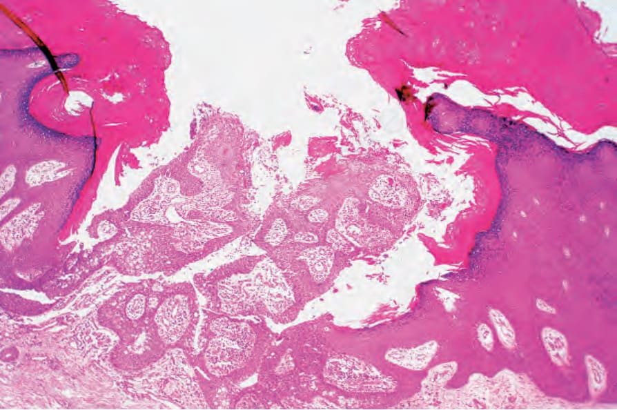
圖 34-42:黏液性汗管化生 (mucinous syringometaplasia):此病灶取自手掌。缺損中央內可見兩個由上皮襯覆的乳頭 (epithelial-lined papillae)。承蒙 J. Grant, MD, Worthing Hospital, Worthing, UK 提供。
Fig. 34.42 Mucinous syringometaplasia: this lesion comes from the palm of the hand. Within the center of the defect are two epithelial-lined papillae. By courtesy of J. Grant, MD, Worthing Hospital, Worthing, UK.
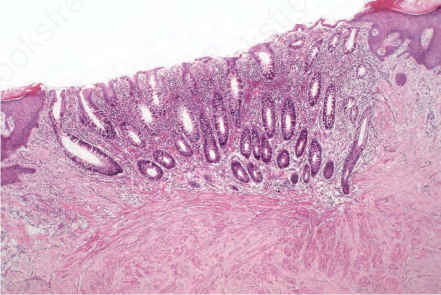
圖 34-44:先天性臍息肉 (congenital umbilical polyp):在此例中,息肉表面由大腸黏膜 (large intestinal mucosa) 所覆蓋。注意管狀腺體 (tubular glands)。上皮深部的平滑肌束 (fascicles of smooth muscle) 代表黏膜肌層 (muscularis mucosae)。
Fig. 34.44 Congenital umbilical polyp: in this example, the surface of the polyp is covered by large intestinal mucosa. Note the tubular glands. The fascicles of smooth muscle deep to the epithelium represent muscularis mucosae.
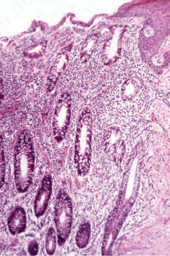
圖 34-45:先天性臍息肉 (congenital umbilical polyp):注意柱狀上皮與鱗狀上皮 (columnar and squamous epithelium) 之間的連續性。
Fig. 34.45 Congenital umbilical polyp: note the continuity between the columnar and squamous epithelium.
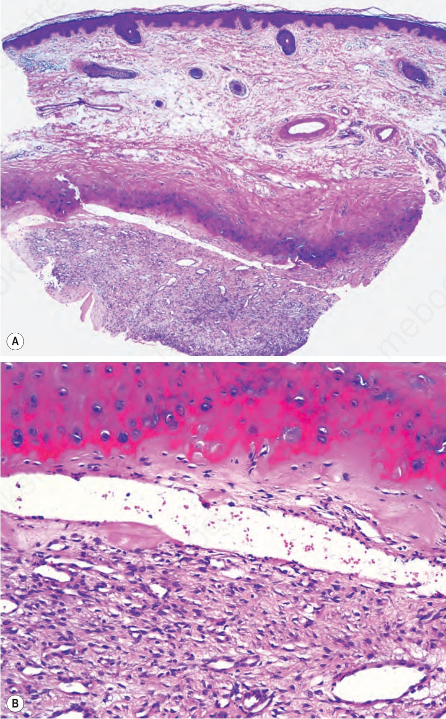
圖 34-46:耳廓假性囊腫 (pseudocyst of the auricle):(A) 軟骨內的囊狀腔隙 (cystic space within the cartilage);(B) 腔內被肉芽組織 (granulation tissue) 所占據。
Fig. 34.46 Pseudocyst of the auricle: (A) cystic space within the cartilage; (B) the cavity is occupied by granulation tissue.