正中縫囊腫 (Median Raphe Cyst)
臨床特徵 (Clinical Features)
- 正中縫囊腫 (median raphe cyst)(又稱 genitoperineal raphe cyst、parameatal cyst)通常直徑可達 1 cm,內含清澈液體,最常在生命的前三十年(前三個十年)被注意到,表現為一無症狀結節,有時呈半透明狀,位於陰莖的腹側面。
- 罕見的病灶可表現為條索狀 (cordlike) 或管道狀 (canal-like) 的硬結。
- 龜頭 (glans) 是最常受影響的部位。
- 病灶亦可見於陰囊 (scrotum) 的腹側面以及會陰 (perineum)。
- 肛周 (perianal) 病灶極為罕見。
- 此囊腫不與尿道 (urethra) 相通。
- 復發少見。
- 罕見情況下,囊腫會合併 split median raphe(正中縫裂開)發生。
致病機轉與組織學特徵 (Pathogenesis and Histologic Features)
-
一般認為此囊腫是由 genitourethral folds(生殖泌尿皺襞)與 urethral plate(尿道板)的異常融合所致,導致尿道上皮 (urethral epithelium) 錯置形成的巢於腹側中線。
-
另一種解釋是,某些病例可能源自錯置的 periurethral glands(尿道周圍腺體)(mucoid cyst,黏液囊腫)或迷走的 urethral buds(尿道芽)。
-
labial–scrotal folds(陰唇—陰囊皺襞)的類似錯誤融合則造成陰囊與會陰的變異型。
-
組織學上,囊腫的內襯多變。大多數報告中,其由 pseudostratified columnar epithelium(偽複層柱狀上皮)組成,厚度為 1–4 層細胞(Fig. 34.35)。
-
較少見的情況下,可見 diastase-resistant(抗澱粉酶)、periodic acid-Schiff (PAS)-positive(PAS 陽性)的黏液性上皮,以及 stratified squamous epithelia(複層鱗狀上皮)(Fig. 34.36)。
-
腺體形成 (glandular formation) 非常罕見。
-
化生性纖毛變異型 (metaplastic ciliated variants) 及混雜的 goblet cells(杯狀細胞)非常偶爾可見。
-
例外情況下,可能會遇到與 intraepithelial dendritic melanocytes(上皮內樹突狀黑色素細胞)相關的色素性變異型。
-
有一篇文獻描述了沿著一名男嬰陰囊正中縫 (median raphe of the scrotum) 延伸至肛緣 (anal verge) 的線狀小型表皮樣囊腫 (linear small epidermoid cysts)。這些可能是在 raphe scroti(陰囊縫)發育過程中被卡住的鱗狀上皮殘餘 (squamous epithelial rests) 所造成。
-
上皮細胞為 CK7 與 CEA 陽性,CK20 陰性。
-
已有兩個案例記錄到以 chromogranin 與 synaptophysin 表現為特徵的局部神經內分泌分化 (focal neuroendocrine differentiation)。
鑑別診斷 (Differential Diagnosis)
- 正中線部位、以偽複層化 (pseudostratification) 為主,以及缺乏斷頭分泌 (decapitation secretion) 與肌上皮層 (myoepithelial layer),可將 median raphe cyst 與 apocrine cystadenoma(頂泌汗腺囊腺瘤)區分開來。
- 此外,median raphe cysts 不表現 human milk fat globulin 1 (HMFG-1)。
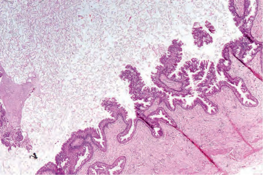
圖 34-35:正中縫囊腫 (Median raphe cyst):低倍視野,顯示由柱狀上皮 (columnar epithelium) 覆蓋的乳頭狀突起 (papillary processes)。
Fig. 34.35 Median raphe cyst: low-power view showing papillary processes covered by columnar epithelium.
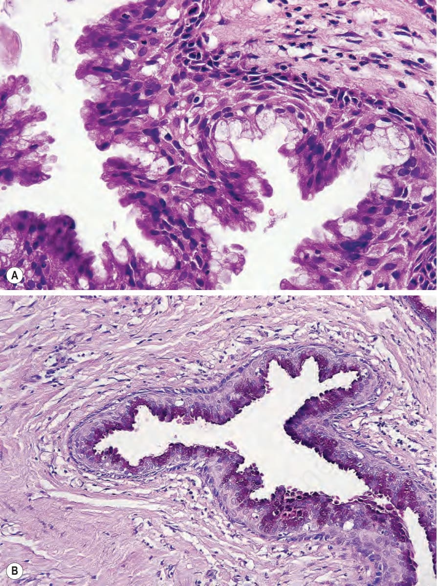
圖 34-36:正中縫囊腫 (Median raphe cyst):(A) 囊腫局部由含黏液上皮 (mucus-containing epithelium) 內襯;(B) 後者為 PAS 陽性 (PAS positive)(抗澱粉酶 diastase resistant)。
Fig. 34.36 Median raphe cyst: (A) the cyst is focally lined by mucus-containing epithelium; (B) the latter is PAS positive (diastase resistant).
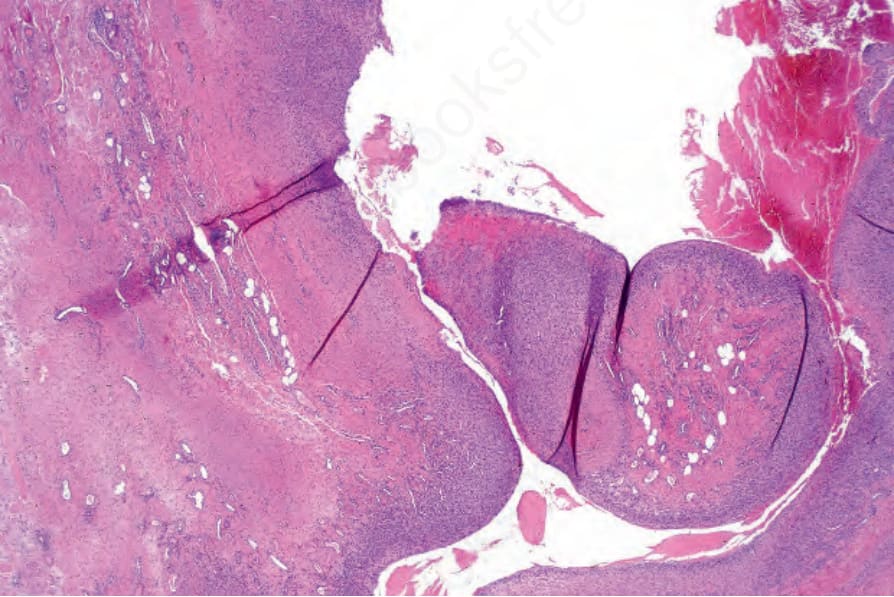
圖 34-37:化生性滑膜囊腫 (Metaplastic synovial cyst):此例在腹部手術後以瘻管 (fistulous tract) 形式表現。可見絨毛狀突起 (villous processes)。
Fig. 34.37 Metaplastic synovial cyst: this example presented as a fistulous tract following abdominal surgery. Villous processes are evident.