臨床特徵 (Clinical Features)
- 毳毛囊腫 (vellus hair cysts) 最初被報告於兩性的兒童與年輕成人。性別分布相等,且無種族偏好。
- 病人表現為眾多無症狀、散在、柔軟、膚色或紅棕色的丘疹,直徑 1–5 mm,特別好發於胸骨旁 (parasternal) 區域,但分布範圍可相當廣泛。亦曾記載有泛發性 (generalized) 分布。
- 曾描述一例外案例其外觀類似太田痣 (nevus of Ota)。病灶罕見呈單側性。
- 偶見病灶呈臍狀凹陷 (umbilicated),擠壓時可擠出白色乾酪樣 (caseous) 物質。
- 進一步的病例已將本病擴展至包含一種遺傳性(autosomal dominant,體染色體顯性)變異型,此型出生時可能呈現或不呈現,且較常發生於四肢的伸側 (extensor aspects)。曾報告發生於雙胞胎的病例。
- 曾描述一種顏面型 (facial form)、一名以眼周 (periorbital) 分布表現的病人,以及另一名具單一眼眶 (orbital) 病灶的病人。
- 自發性退化 (spontaneous involution) 並不少見。
A
- 毳毛囊腫偶爾與腎衰竭 (renal failure) 及若干遺傳性皮膚病 (genodermatoses) 相關聯,包括 pachyonychia congenita、anhidrotic ectodermal dysplasia、hidrotic ectodermal dysplasia,以及罕見的 Lowe syndrome(眼腦腎症候群 (oculocerebrorenal syndrome),其特徵為 Fanconi 型腎衰竭、智能不足與眼部異常)。
- 偶爾會遇到孤立性 (solitary) 病灶,且可能體積較大。
致病機轉與組織學特徵 (Pathogenesis and Histologic Features)
- 噴發性毳毛囊腫 (eruptive vellus hair cysts) 最可能是因毳毛 (vellus hairs) 漏斗部 (infundibulum) 阻塞,導致囊狀擴張並滯留角質碎屑與毳毛而形成。阻塞的原發原因不明。
- 亦有人提出它們代表毛囊錯構瘤 (follicular hamartomas)。
- 研究顯示噴發性毳毛囊腫與多發性皮脂囊腫 (steatocystomas) 兩者皆表現 keratin 17,而後者亦表現 keratin 10。此種 keratin 表現的重疊,或可協助解釋此兩種病灶的潛在相似性,乃至可能重疊的特徵;然而此一觀點並非普世共識,兩者確切的關係仍有待闡明。
B
- 特徵性組織學為真皮中層 (mid-dermal) 的囊腫,內含層狀角質 (laminated keratin) 與許多毳毛 (Figs 34.25 與 34.26)。
- 上皮襯裡 (epithelial lining) 由數層鱗狀上皮 (squamous epithelium) 構成,常具顆粒細胞層 (granular cell layer)。
- 有時囊腫與表皮、萎縮的毛囊 (atrophic follicle),或立毛肌 (pilomotor muscle) 相連續。在先天型 (congenital variant) 中,毳毛囊腫較可能開口至表面。
- 偶爾囊腫破裂,伴隨異物巨細胞反應 (foreign body giant cell reaction),並可能伴有膽固醇裂隙 (cholesterol clefts) 的形成。
鑑別診斷 (Differential Diagnosis)
- 噴發性毳毛囊腫與 steatocystoma multiplex(多發性皮脂囊腫)在臨床上有非常顯著的重疊,僅能藉組織學分析加以區分。
- Steatocystoma 的特徵為表皮樣 (epidermoid) 襯裡且無顆粒細胞層。囊壁最內側覆有波浪狀的嗜伊紅性角質層 (undulating eosinophilic cuticle)。皮脂腺 (sebaceous glands) 存在於囊壁內或其緊鄰處。
- 然而,有時病人同時具有兩種類型的囊腫,且偶爾會出現重疊的組織學特徵,有時構成混合性囊腫 (hybrid cyst)。
1689 Follicular cysts
- 如上所述,差異性 keratin 表現已被證明可區分這兩種囊腫。因此 vellus hair cyst 表現 K17 但不表現 K10,而 steatocystoma 則同時表現 K17 與 K10。
- 有趣的是,KRT17 基因的突變可同時造成 pachyonychia congenita type 2,以及一種與 steatocystoma multiplex 非常相似或相同的病況。這些發現對 steatocystoma simplex 與 vellus hair cysts 的相關性(若有的話)仍有待確定。
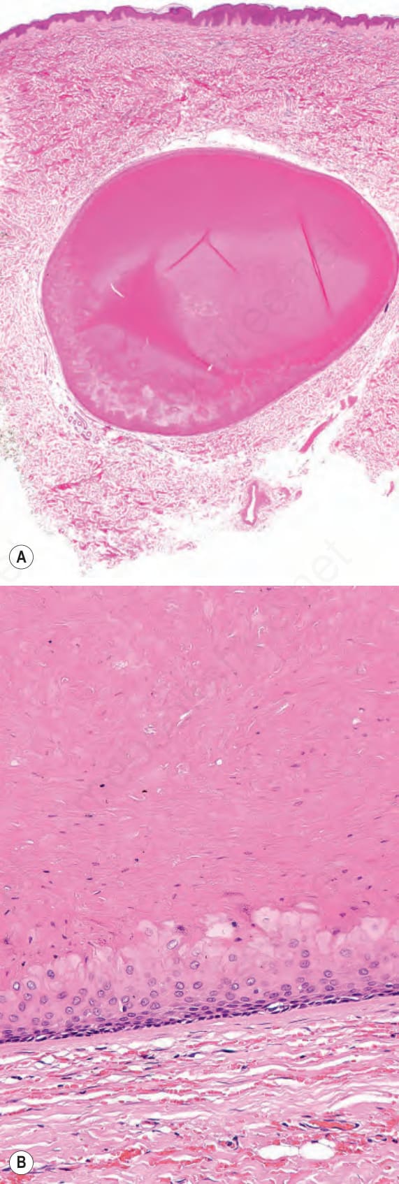
圖 34-21(A、B):毛根鞘囊腫 (trichilemmal cyst):此等視野顯示均質的嗜伊紅性內容物。注意明顯的基底細胞層 (basal cell layer)。
Fig. 34.21 (A, B) Trichilemmal cyst: these views show the homogeneous eosinophilic contents. Note the distinct basal cell layer.
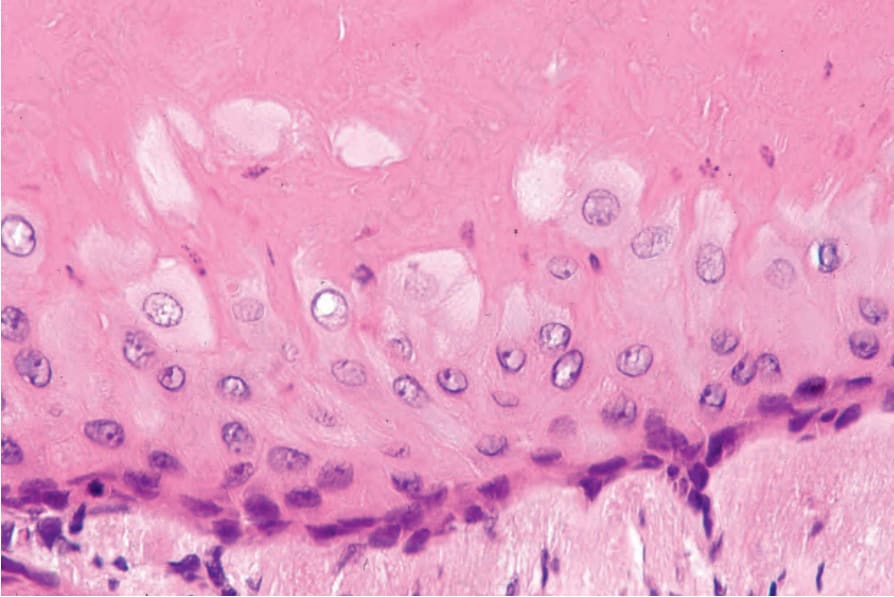
圖 34-22:毛根鞘囊腫 (trichilemmal cyst):囊壁由鱗狀上皮 (squamous epithelium) 構成,且無顆粒細胞層 (granular cell layer)。最表淺的細胞較大、呈垂直走向,且具有豐富的細胞質。角化 (keratinization) 呈突然性。
Fig. 34.22 Trichilemmal cyst: the cyst wall is composed of squamous epithelium and a granular cell layer is not present. The most superficial cells are larger, vertically orientated, and have abundant cytoplasm. Keratinization is abrupt.
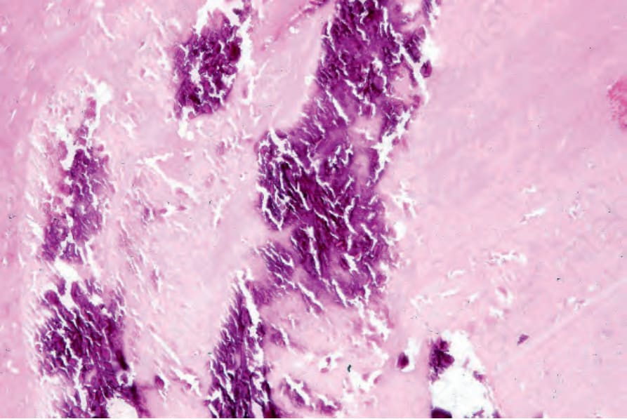
圖 34-23:毛根鞘囊腫 (trichilemmal cyst):嗜鹼性顆粒狀鈣化 (basophilic granular calcification) 為常見的組織學發現。
Fig. 34.23 Trichilemmal cyst: basophilic granular calcification is a frequent histologic finding.
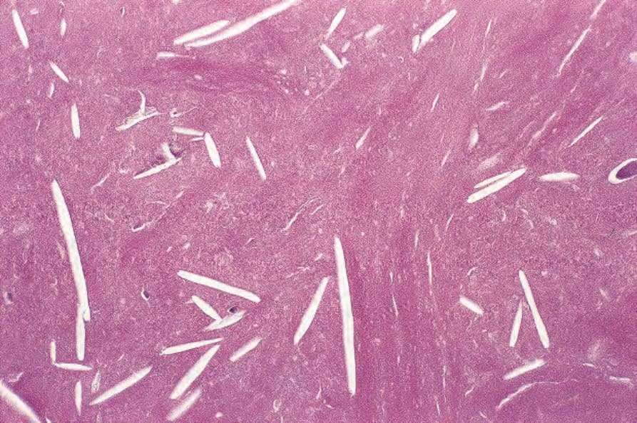
圖 34-24:毛根鞘囊腫 (trichilemmal cyst):空腔(膽固醇裂隙 (cholesterol clefts))為此病灶的常見特徵。
Fig. 34.24 Trichilemmal cyst: the empty spaces (cholesterol clefts) are a common feature of this lesion.
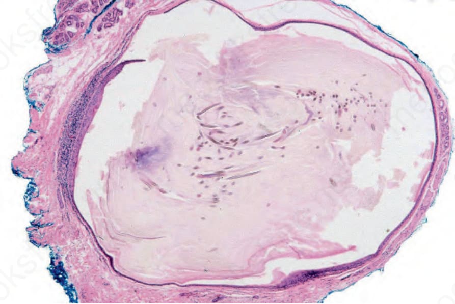
圖 34-25:毳毛囊腫 (vellus hair cyst):此薄壁囊腫位於真皮中層 (mid-dermis)。
Fig. 34.25 Vellus hair cyst: this thin-walled cyst is present in the mid-dermis.
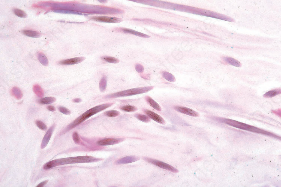
圖 34-26:毳毛囊腫 (vellus hair cyst):高倍視野下,囊腔內含眾多毳毛 (vellus hairs)。
Fig. 34.26 Vellus hair cyst: on high power, the lumen contains numerous vellus hairs.