臨床特徵 (Clinical Features)
- 表皮樣囊腫 (epidermoid cyst,亦稱 epidermal cyst、infundibular cyst) 特別好發於臉部、頸部與上軀幹,一般認為是毛皮脂腺單位 (pilosebaceous units) 受損所致。
- 外陰大陰唇 (vulval labia majora) 與陰囊 (scrotum) 亦為好發部位。罕見病灶可發生於無毛區域,如足底 (soles)(見下文)。
- 以年輕與中年成人最常受影響,男女比例相當。
- Epidermoid cyst 表現為平滑、圓頂狀的腫脹,直徑從數毫米到數公分不等 (Fig. 34.1)。通常可見一個開口 (punctum) (Fig. 34.2)。
致病機轉與組織病理特徵 (Pathogenesis and Histologic Features)
-
病灶之發生係毛囊上部阻塞,或繼發於外傷(植入性囊腫,inclusion cysts)的結果。
-
Epidermoid cyst 為單房 (unilocular)、球形 (spherical),由類表皮上皮 (epidermis-like epithelium) 襯覆,並含有顆粒細胞層 (granular cell layer) (Figs 34.4–34.6)。例外情況下可見多房 (multilocular) 病灶。
-
囊內容物為層狀角質 (laminated keratin),一般認為代表毛囊漏斗部 (follicular infundibular) 衍生(即非植入型,nonimplantation variant)。在較舊的病灶中,襯覆上皮常略為變薄。可見囊壁的苔癬樣發炎 (lichenoid inflammation),與 lichen planus 無法區別。
-
急性發炎可導致囊壁隨後破裂,並產生強烈的異物巨細胞反應 (foreign body giant cell reaction) (Fig. 34.7)。有時此反應如此顯著,以致完全破壞囊腫,僅留下真皮內局灶性的角質碎片聚集 (Fig. 34.8)。
-
細菌是否在 epidermoid cyst 發炎的形成中扮演重要角色尚不明確。一項來自日本的研究發現,相較於無發炎者,發炎病灶中厭氧菌 (anaerobes) 的發生率增加。然而,這究竟是定殖 (colonization) 抑或真正感染 (true infection) 的結果,仍有待確立。
-
偶爾,囊腫襯覆上皮可顯示類表皮 (epidermoid) 與局灶性毛根鞘 (trichilemmal) 角化。在罕見的混合性囊腫 (hybrid cyst) 中,囊腫上半部為類表皮角化、下半部為毛根鞘角化。例外情況下,可遇到含有多個終毛毛幹碎片 (terminal hair shaft fragments) 的色素變異型(即色素性毛囊囊腫,pigmented follicular cyst)。亦曾報告一例含有眾多角質球 (keratin spherules) 者。
-
多發性病灶之存在可能提示 Gardner syndrome 的可能性,該症候群除皮膚囊腫外,還包括結腸息肉症 (polyposis coli)、頜骨骨瘤 (jaw osteomas) 與腸纖維瘤病 (intestinal fibromatoses)。較少見的情形,患者可表現脂肪瘤 (lipomas)、毛母質瘤 (pilomatrixomas,包括具毛母質襯覆的 epidermoid cysts) 與平滑肌瘤 (leiomyomas)。多發性病灶亦見於 Gorlin-Goltz syndrome(見第 24 章),並可能為該疾病的首發表現。結膜下 (subconjunctival) epidermoid cysts 似乎僅發生於患有此症候群的患者。
-
多發且常為大型的 epidermoid cysts 有時可見於移植受贈者接受 ciclosporin 治療的併發症。
-
在 Gardner syndrome 患者中,囊腫襯覆上皮偶爾顯示局灶性基底樣細胞增生 (basaloid cell proliferation) 伴影細胞變化 (ghost cell change),如同毛母質瘤 (pilomatrixoma) 所見 (Fig. 34.9)。此情境外的病灶亦可發生,包括一例發生於皮脂腺痣 (nevus sebaceous) 背景者。因此,雖然高度提示,這些特徵不能視為病理特異性 (pathognomonic)。
-
Gorlin-Goltz syndrome 中的囊腫通常為混合性 (hybrid),兼具 epidermoid cyst 與 steatocytoma 的特徵。
-
曾報告一例結節性硬化症 (tuberous sclerosis) 患者出現多發性 epidermoid cysts 合併血管纖維瘤 (angiofibroma) 病灶,與阻塞/外傷有關。
-
Epidermoid cysts 與黑色素細胞痣 (melanocytic nevi) 並存並不罕見。此點具特別重要性,因為由此導致囊腫體積增大,可能在臨床上引起對黑色素瘤 (melanoma) 的懷疑。多數痣為平凡的真皮內痣,但亦曾記載與複合痣 (compound nevi)、先天性痣 (congenital nevi)、發育不良痣 (dysplastic nevi)、藍痣 (blue nevi) 及 Reed 紡錘細胞痣 (spindle cell nevi of Reed) 相關的囊腫。
-
惡性腫瘤罕見地可於 epidermoid cyst 的囊壁內發生,包括基底細胞癌 (basal cell carcinoma)、鱗狀細胞癌 (squamous cell carcinoma) 及原位鱗狀細胞癌 (squamous cell carcinoma in situ) (Fig. 34.10)。亦有罕見病例報告描述 epidermoid cyst 與 Paget disease 及皮膚神經內分泌癌 (cutaneous neuroendocrine carcinoma,即 Merkel cell carcinoma) 相關。曾有一例報告黑色素瘤發生於病灶中、一例原位黑色素瘤與一鄰近皮膚黑色素瘤相關並定殖於 epidermoid cyst,以及另有單一報告描述原位黑色素瘤發生於非皮膚的小腦橋腦角 (cerebellopontine angle) epidermoid cyst 中。亦曾記載毛母質瘤 (pilomatrixoma)(在無 Gardner syndrome 的情況下)與 epidermoid cyst 並存。由於發生惡性變化的 epidermoid cysts 在臨床上無法可靠地與其極為常見的良性對應者區分,故建議對所有此類囊腫進行組織學檢查。
-
曾有描述顯示一系列皮膚病變特徵的 epidermoid cysts。這些包括 pemphigus、psoriasis、lichen planus 與 Darier disease。亦曾描述表皮鬆解性過度角化 (epidermolytic hyperkeratosis) 的變化,以及受傳染性軟疣 (molluscum contagiosum) 侵犯。人類乳突病毒 (human papillomavirus, HPV) 之關聯敘述於下文(見疣狀囊腫 verrucous cyst 與足底表皮樣囊腫 epidermoid cyst of the sole)。
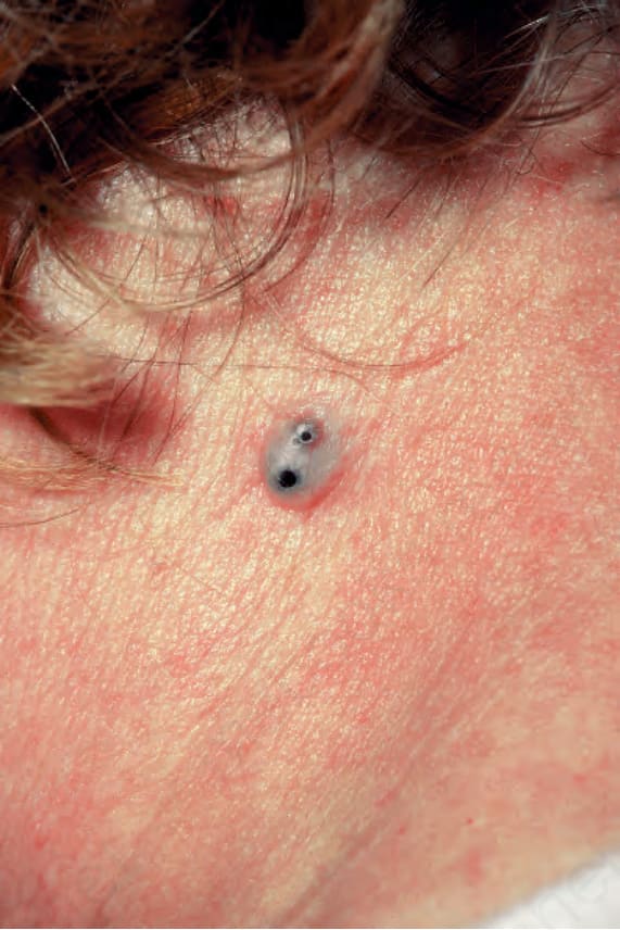
圖 34-1:表皮樣囊腫 (epidermoid cyst):典型的圓頂狀腫脹,具兩個開口 (puncta)。承蒙 R.A. Marsden, MD(英國倫敦 St George’s Hospital)惠贈。
Fig. 34.1 Epidermoid cyst: a typical dome-shaped swelling with two puncta. By courtesy of R.A. Marsden, MD, St George’s Hospital, London, UK.
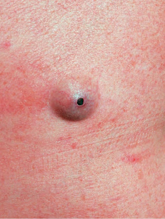
圖 34-2:表皮樣囊腫 (epidermoid cyst):開口 (punctum) 的特寫視野。承蒙倫敦 Institute of Dermatology 惠贈。
Fig. 34.2 Epidermoid cyst: close-up view of a punctum. By courtesy of the Institute of Dermatology, London, UK.
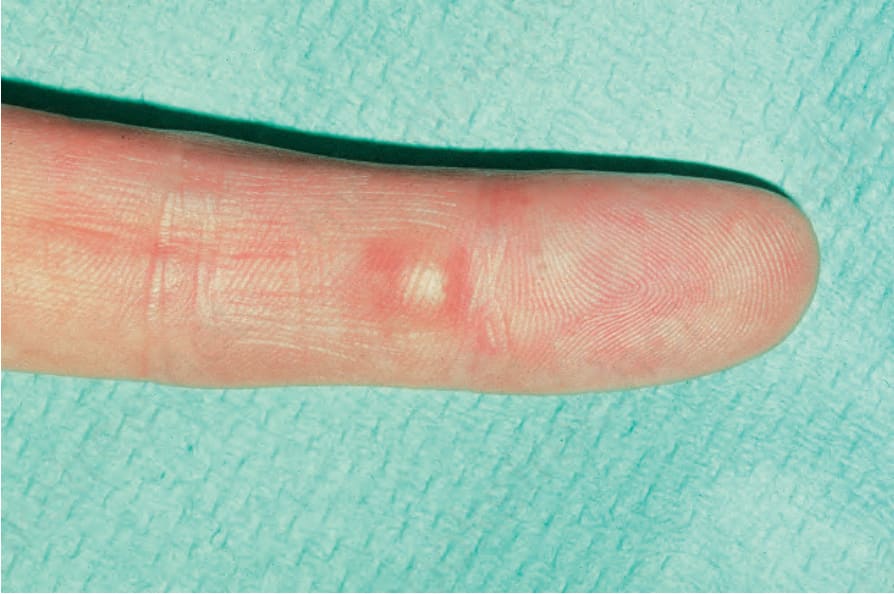
圖 34-3:表皮樣囊腫 (epidermoid cyst):此植入型 (implantation variant) 位於一特徵性部位。承蒙 R.A. Marsden, MD(英國倫敦 St George’s Hospital)惠贈。
Fig. 34.3 Epidermoid cyst: this implantation variant is at a characteristic site. By courtesy of R.A. Marsden, MD, St George’s Hospital, London, UK.
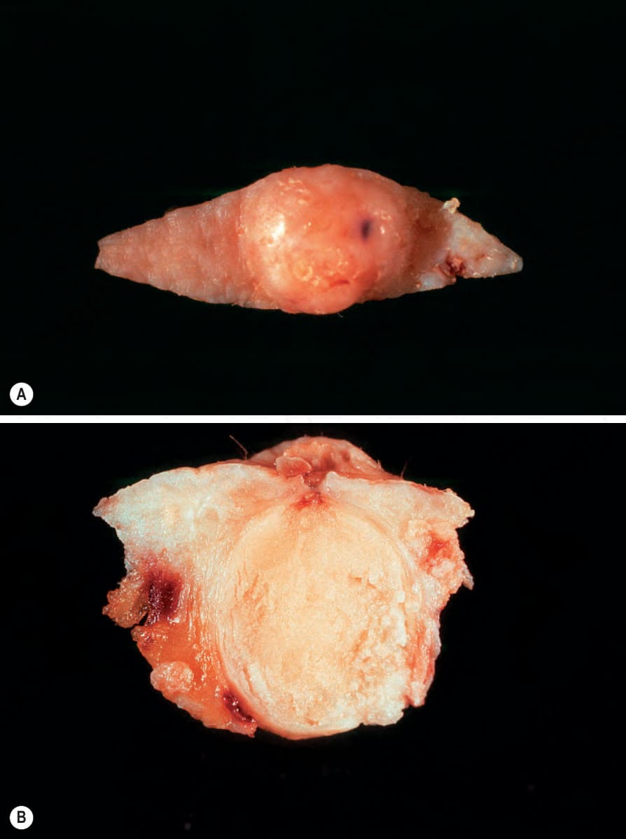
圖 34-4:(A, B) 表皮樣囊腫 (epidermoid cyst):在此切除標本中,開口 (punctum) 清晰可見。
Fig. 34.4 (A, B) Epidermoid cyst: in this excision specimen, the punctum is clearly visible.
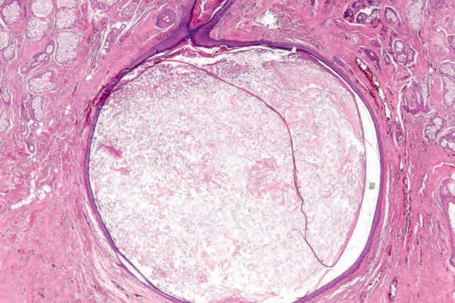
圖 34-5:表皮樣囊腫 (epidermoid cyst):真皮內可見一單發病灶。
Fig. 34.5 Epidermoid cyst: a solitary lesion is present in the dermis.
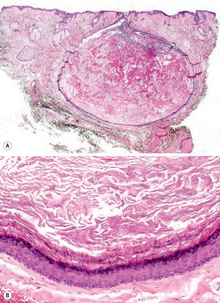
圖 34-6:表皮樣囊腫 (epidermoid cyst):(A) 此例可見開口 (punctum);(B) 囊壁由鱗狀上皮 (squamous epithelium) 組成並包含顆粒細胞層 (granular cell layer)。注意層狀角質 (laminated keratin)。
Fig. 34.6 Epidermoid cyst: (A) in this example, the punctum is present; (B) the cyst wall is composed of squamous epithelium and includes a granular cell layer. Note the laminated keratin.
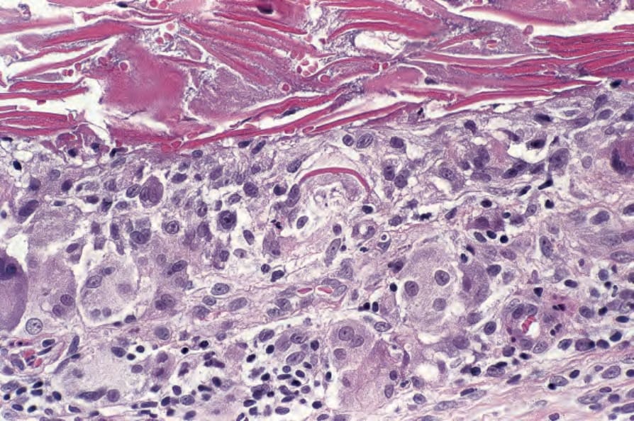
圖 34-7:表皮樣囊腫 (epidermoid cyst):破裂伴隨異物肉芽腫反應 (foreign body granulomatous response)。視野中央,一巨細胞 (giant cell) 含有角質碎片 (keratin fragment)。
Fig. 34.7 Epidermoid cyst: rupture is associated with a foreign body granulomatous response. In the center of the field, a giant cell contains a keratin fragment.
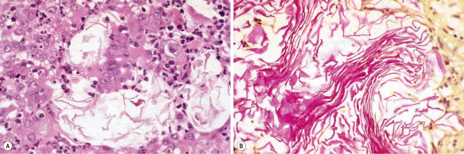
圖 34-8:表皮樣囊腫 (epidermoid cyst):(A) 在此近乎癒合的病灶中,視野中央所見的殘餘角質薄層 (residual keratin lamellae) 是破裂囊腫僅存的部分;(B) 這些可藉 Lendrum phloxine tartrazine 反應加以突顯。
Fig. 34.8 Epidermoid cyst: (A) in this almost healed lesion, residual keratin lamellae, as seen in the center of the field, are all that is left of the ruptured cyst; (B) these may be highlighted by the Lendrum phloxine tartrazine reaction.
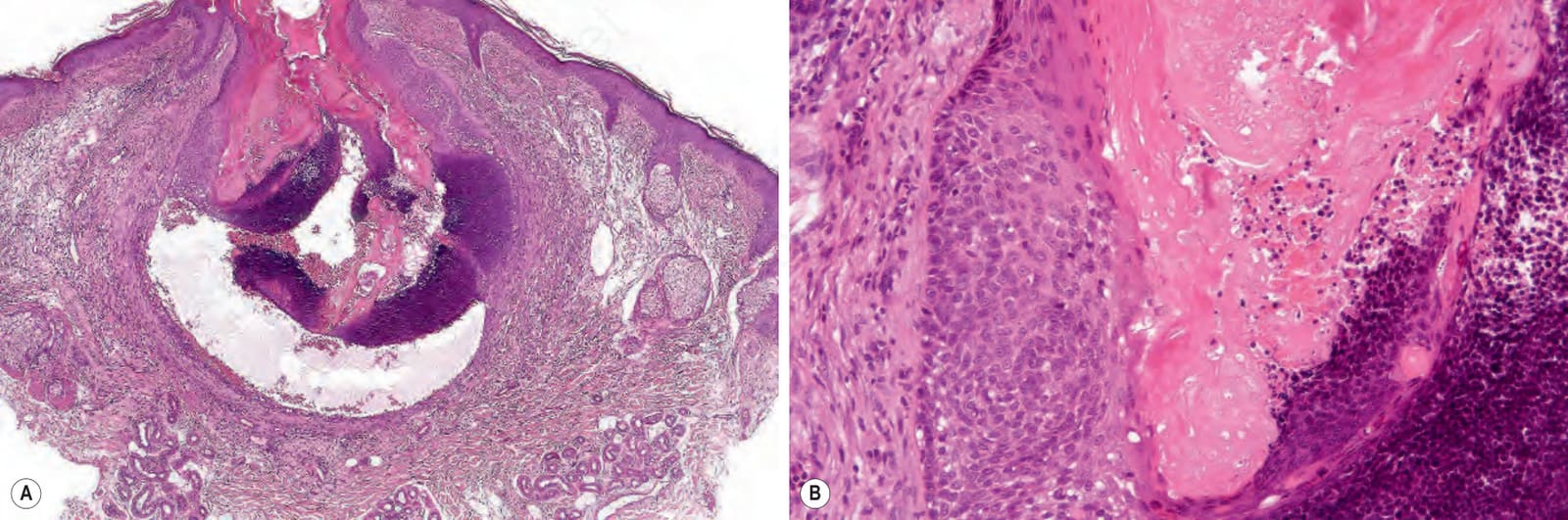
圖 34-9:(A, B) 表皮樣囊腫 (epidermoid cyst):囊壁下半部顯示毛母質分化 (matrical differentiation)。
Fig. 34.9 (A, B) Epidermoid cyst: the lower half of the cyst wall shows matrical differentiation.
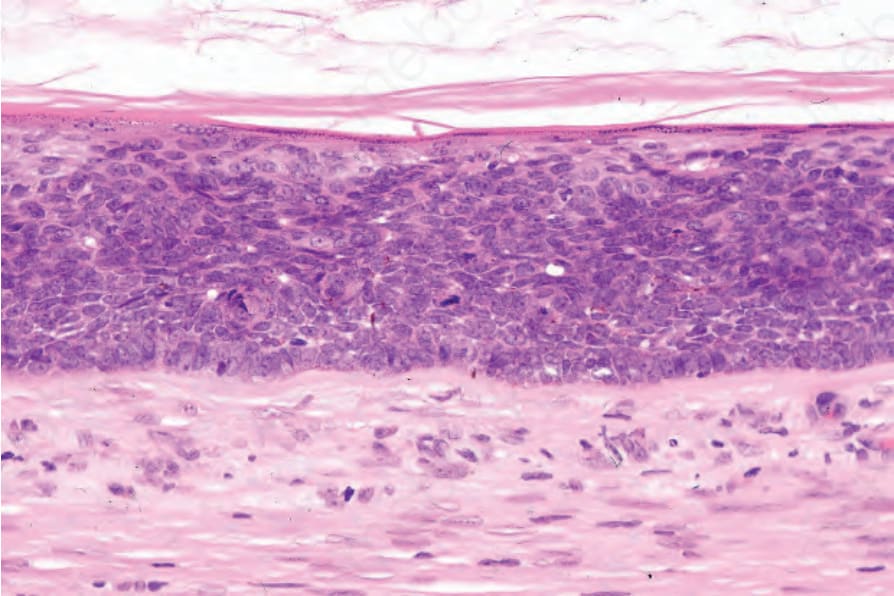
圖 34-10:表皮樣囊腫 (epidermoid cyst):此例中,上皮性囊壁顯示原位癌 (carcinoma in situ) 的特徵。
Fig. 34.10 Epidermoid cyst: in this example, the epithelial wall shows the features of carcinoma in situ.
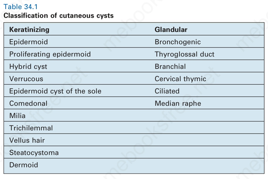
表 34-1:皮膚囊腫之分類 (Classification of cutaneous cysts)。
Table 34.1 Classification of cutaneous cysts