Syringoma
臨床特徵 (Clinical Features)
- 汗管瘤 (syringoma) 為常見腫瘤,最常表現為多發、對稱分布、通常無症狀的小丘疹 (1–3 mm),好發於下眼瞼與上頰部 (Fig. 33.119)。¹⋅²
- 病灶於青春期或成年早期出現,並有顯著的女性優勢 (female predominance)。
- 個別丘疹質地堅實、呈膚色或略帶黃色 (Fig. 33.120)。
- 然而 syringoma 可有多種不同的臨床表現。可單發或多發於頭皮、前額、頸部、腋窩、胸部、腹部、臀部、四肢或外生殖器(男性與女性皆可)、口周,或罕見地呈線狀痣樣 (linear nevoid) 單側、節段性 (segmental) 或斑塊樣 (plaquelike) 分布。¹⋅³⋅⁴⁻²⁶
- 另曾描述一種大小可達 1 cm 的巨型變異 (giant variant),且以粟丘疹 (milia) 形式表現屬罕見。²⁷⁻³¹
- 外陰部 (vulval) syringoma 的患者常同時合併眼瞼病灶,外陰搔癢 (vulval pruritus) 是常見的就診症狀。¹²
- 曾描述一種發疹性變異 (eruptive variant),特徵為年輕人前側體表陸續成批出現丘疹。³²⋅³³ 典型受累部位包括頸部、胸部、腋窩、肘前窩 (antecubital fossae)、上肢、下腹部與腹股溝。³²⋅³⁴ 偶有家族性病例的記載。³⁴⁻³⁹
- Eruptive syringoma 在東方人 (Orientals) 中較常見,並見於 18% 的成熟期 Down syndrome 患者。⁴⁰⁻⁴³
- 罕見情況下可與粟丘疹囊腫 (milium cysts) 及蟲蝕狀皮膚萎縮 (vermiculate atrophoderma) 相關 (Nicolau and Balus syndrome)。⁴⁴
致病機轉與組織學特徵 (Pathogenesis and Histologic Features)
- 組織化學 (histochemical) 與電子顯微鏡 (electron microscopic) 研究顯示,syringoma 代表起源自 acrosyringium(即表皮內的小汗腺汗管,intraepidermal eccrine sweat duct)的腺瘤 (adenoma)。⁴⁵⋅⁴⁶
- 免疫組織化學 (immunohistochemical) 研究進一步證實此腫瘤朝向 acrosyringium 或真皮內小汗腺汗管 (dermal eccrine duct) 分化。⁴⁷⁻⁵¹ 因此 syringoma 的上皮含有 succinic dehydrogenase、phosphorylase 與 leucine aminopeptidase。
- 腫瘤由相互連接的上皮索 (epithelial strands) 與汗管 (ducts) 組成,散布於上真皮的纖維性間質 (fibrous stroma) 中 (Fig. 33.121)。
- 汗管由兩層扁平的立方狀細胞 (flattened cuboidal cells) 襯覆。管腔可有一層角質膜 (cuticle) 襯覆,腔內常含嗜酸性顆粒狀碎屑 (eosinophilic granular debris)。
- 有時汗管會與一條上皮索相連,形成具特徵性的蝌蚪狀構型 (tadpole configuration) (Figs 33.122 and 33.123)。
- 偶可見單一富含肝醣 (glycogen-rich) 的汗管細胞,罕見情況下所有汗管細胞皆含肝醣,形成 syringoma 的透明細胞變異 (clear cell variant)。此變異似乎與糖尿病 (diabetes mellitus) 特別相關。⁵²⋅⁵³
- Syringoma 的粟丘疹樣變異 (milium-like variant) 在組織學上的特徵為大型、上皮襯覆的囊腫,內含角質性物質 (keratinaceous material),位於淺層真皮內。²⁸ 其餘特徵與 syringoma 相同,且充滿角質的囊腫對 CEA 呈免疫反應陽性。
鑑別診斷 (Differential Diagnosis)
- Syringoma 必須與 desmoplastic trichoepithelioma 區分,後者典型上具有眾多角質囊腫 (keratocysts)。
- 雖然與 eccrine epithelioma 在組織學上有明顯重疊,但臨床特徵卻相當不同。² Eccrine epithelioma 是一種範圍大得多的腫瘤,可侵犯皮下脂肪,並伴隨明顯的硬化性間質 (markedly desmoplastic stroma)。其 syringoma 樣的特徵通常範圍不廣,且常可見神經周圍間隙 (perineural space) 的浸潤。Syringoma 不具有有絲分裂 (mitoses) 的特徵。
- 在小型切片中,與 microcystic adnexal carcinoma 的區分可能無法達成,因此臨床病理對照 (clinicopathological correlation) 至為關鍵。
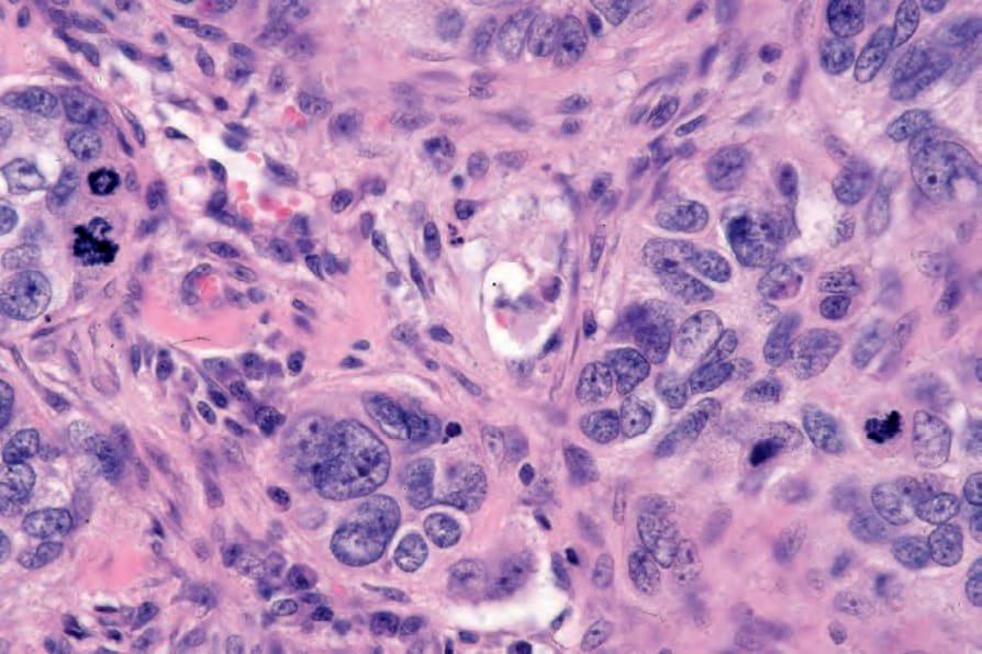
圖 33-107:Eccrine porocarcinoma:可見明顯的有絲分裂 (conspicuous mitoses)。
Fig. 33.107 Eccrine porocarcinoma: conspicuous mitoses are present.
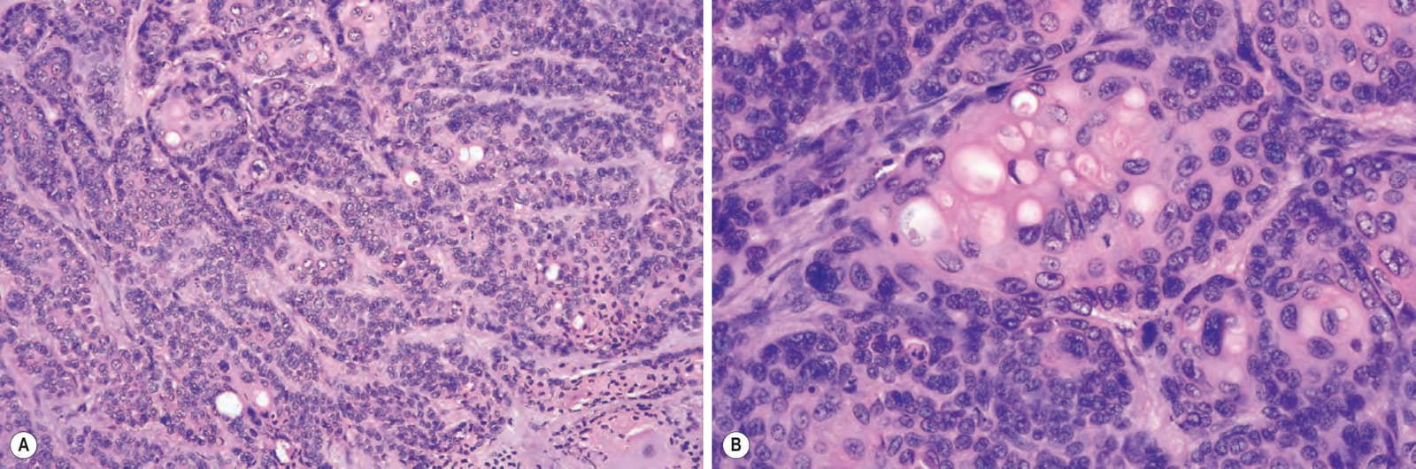
圖 33-108:(A, B) Eccrine porocarcinoma:汗管分化 (ductal differentiation) 為必要的診斷特徵。
Fig. 33.108 (A, B) Eccrine porocarcinoma: ductal differentiation is an essential diagnostic feature.
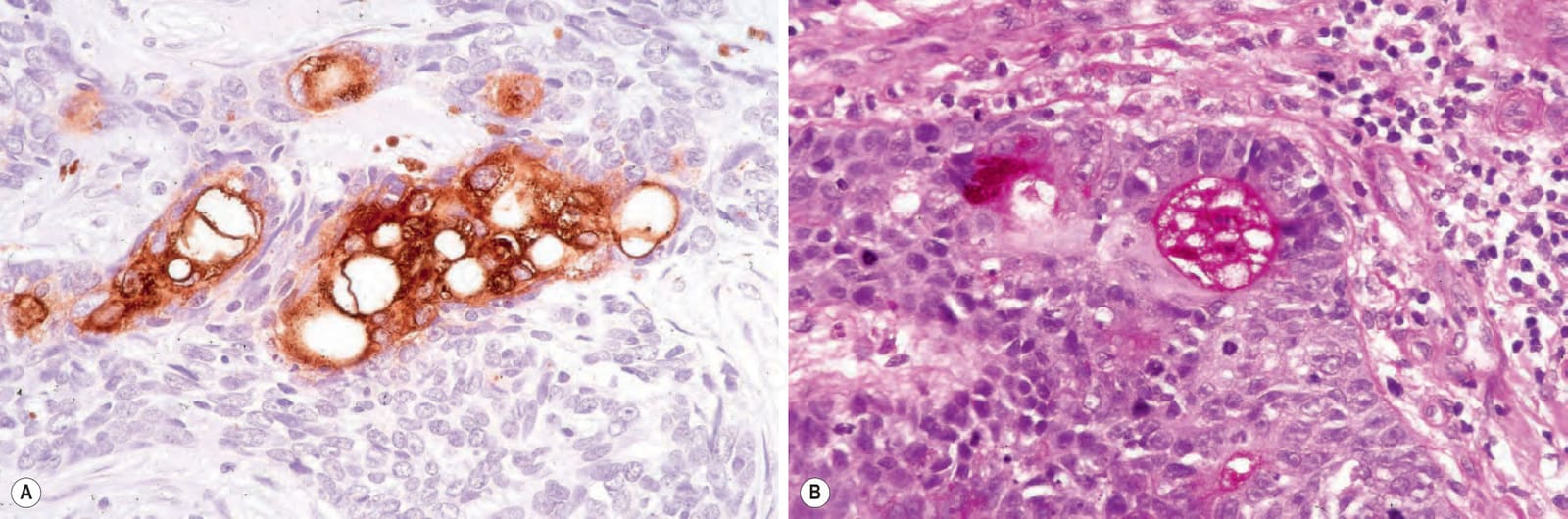
圖 33-109:Eccrine porocarcinoma:(A) 汗管分化與胞質內管腔 (intracytoplasmic lumina) 可用 EMA 或 CEA 免疫組化標示 (EMA);(B) 汗管的管腔緣對 diastase 具抗性、PAS 陽性 (diastase resistant, PAS positive)。
Fig. 33.109 Eccrine porocarcinoma: (A) ductal differentiation and intracytoplasmic lumina can be highlighted with EMA or CEA immunohistochemistry (EMA); (B) the luminal border of the duct is diastase resistant, PAS positive.
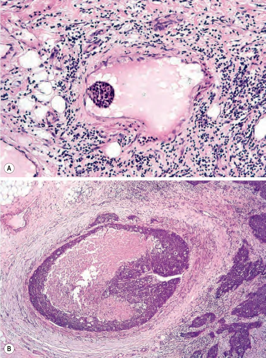
圖 33-110:(A, B) Eccrine porocarcinoma:淋巴血管侵犯 (lymphovascular invasion)。
Fig. 33.110 (A, B) Eccrine porocarcinoma: lymphovascular invasion.
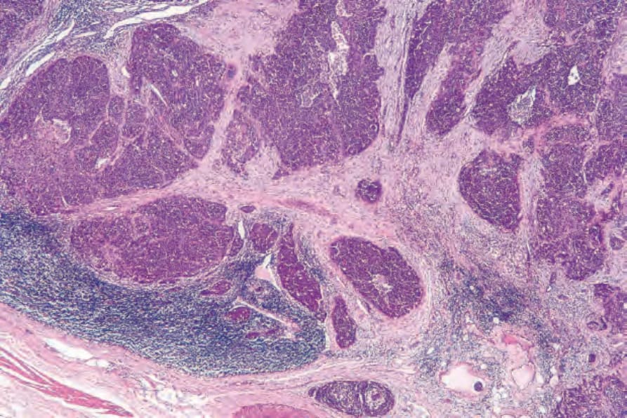
圖 33-111:Eccrine porocarcinoma:淋巴結轉移 (lymph node metastasis),與 Fig. 33.108B 所示為同一患者。
Fig. 33.111 Eccrine porocarcinoma: lymph node metastasis from the same patient as shown in Fig. 33.108B.
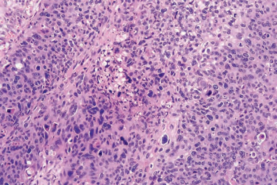
圖 33-112:Eccrine porocarcinoma:高惡性度腫瘤 (high-grade tumors) 常顯示壞死區域 (areas of necrosis)。
Fig. 33.112 Eccrine porocarcinoma: high-grade tumors commonly show areas of necrosis.
圖 33-113:Clear cell eccrine porocarcinoma:(A) 可見顯著的胞質空泡化 (striking cytoplasmic vacuolation);(B) 高倍視野。
Fig. 33.113 Clear cell eccrine porocarcinoma: (A) there is striking cytoplasmic vacuolation; (B) high-power view.
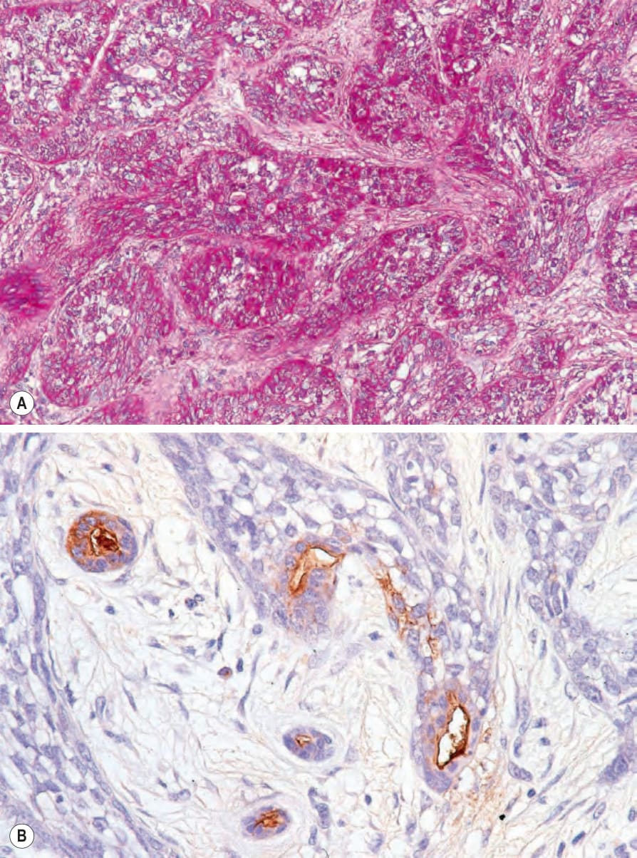
圖 33-114:Clear cell eccrine porocarcinoma:(A) 腫瘤細胞為 PAS 陽性 (PAS positive);(B) EMA。
Fig. 33.114 Clear cell eccrine porocarcinoma: (A) the tumor cells are PAS positive; (B) EMA.
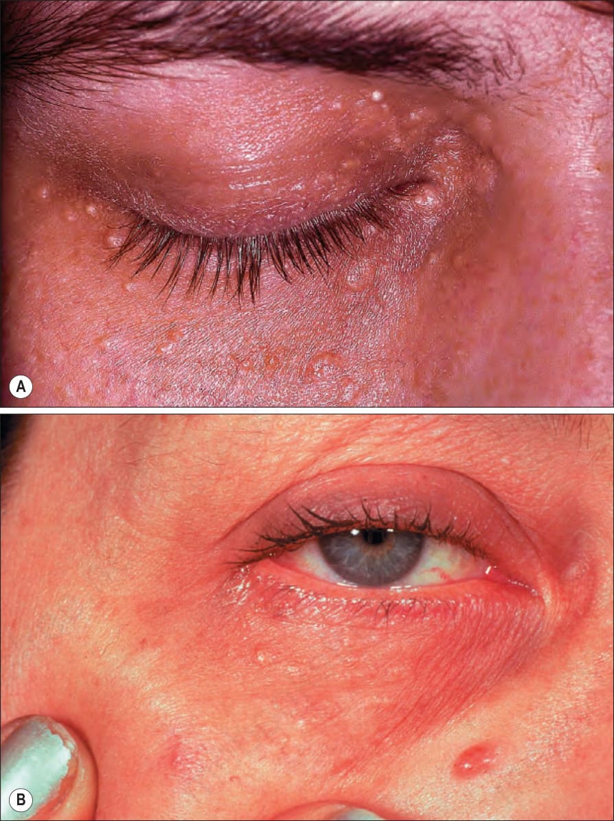
圖 33-119:(A, B) Syringoma:注意這些小丘疹典型的眼眶周圍分布 (periorbital distribution)。(A) 由 R.A. Marsden, MD, St George’s Hospital, London, UK 提供;(B) 由 Institute of Dermatology, London, UK 提供。
Fig. 33.119 (A, B) Syringoma: note the typical periorbital distribution of these small papules. (A) By courtesy of R.A. Marsden, MD, St George’s Hospital, London, UK; (B) by courtesy of the Institute of Dermatology, London, UK.
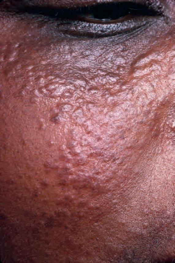
圖 33-120:Syringoma:頰部有廣泛受累。出自已故 N.P. Smith, MD, Institute of Dermatology, London, UK 之收藏。
Fig. 33.120 Syringoma: there is extensive involvement of the cheek. From the collection of the late N.P. Smith, MD, Institute of Dermatology, London, UK.
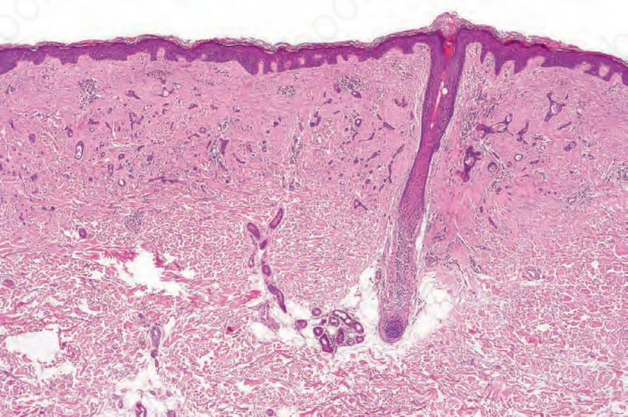
圖 33-121:Syringoma:真皮內可見具特徵性的上皮索 (epithelial strands) 與小囊腫。並有緻密、硬化的纖維性間質 (dense, sclerotic fibrous stroma)。
Fig. 33.121 Syringoma: characteristic epithelial strands and small cysts are present in the dermis. There is a dense, sclerotic fibrous stroma.
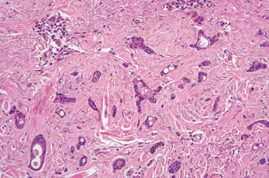
圖 33-122:Syringoma:注意上皮索 (epithelial strands) 與汗管分化 (ductal differentiation)。
Fig. 33.122 Syringoma: note the epithelial strands and ductal differentiation.