疾病定義與分類
- 生毛性腫瘤 (trichogenic tumors) 是一群重現生發性毛球 (germinative hair bulb) 及其相關間質的腫瘤。然而其命名極為混亂。
- 類比於牙源性腫瘤 (odontogenic neoplasms),Headington 依上皮成分與間質成分的相對比例,以及是否存在間質誘導性變化 (stromal inductive change),將這些腫瘤加以分類。純上皮性腫瘤被命名為 trichoblastoma;而當存在毛囊分化時,混合上皮與間質的腫瘤則被稱為 trichoblastic fibroma 或 trichogenic trichoblastoma。
- 然而,個別腫瘤本身即能展現不同程度的分化,原先提出的分類過於嚴格且受限。此外,後續關於生毛性腫瘤的報告使用了各式各樣的稱謂,使這些腫瘤的分類更加混亂。例如,類似 trichoblastic fibroma 的腫瘤曾被稱為 giant solitary trichoepithelioma、subcutaneous trichoepithelioma 或 immature trichoepithelioma。
- 較近期,亦有 trichogerminoma、rippled-pattern trichoblastoma、rippled-pattern trichomatricoma 與 melanotrichoblastoma 等實體被描述。這些腫瘤展現出某些獨特的組織學特徵,但也與傳統的生毛性腫瘤共有部分特徵。
- 雖然辨識一個腫瘤內的不同組織學型態很重要,但分類能被理解且具有意義同樣重要。由於前述各實體的臨床表現與行為相似,且任一腫瘤內均可見到多種組織學型態,本文將生毛性腫瘤統一歸於 trichoblastoma 此一單一標題之下。
- 皮膚淋巴腺瘤 (cutaneous lymphadenoma) 已被證實為 trichoblastoma 的一種變異型,且可能與 trichoblastoma with(原文於此中斷)為同義。
- Trichoblastoma 典型上表現為良性。然而,惡性變化曾被罕見地描述(見下文)。
組織病理特徵 (Histopathology)
-
Trichoblastoma 是一個界限清楚但無包膜的結節狀腫瘤,橫跨整個真皮,特徵性地延伸進入皮下組織 (Fig. 31.109)。罕見可見到純粹位於皮下的位置。它不具有表皮或毛囊的衍生來源,並以大小不一、極類似基底細胞癌 (basal cell carcinoma) 的上皮性巢團為特徵。
-
周邊柵欄狀排列 (peripheral palisading) 明顯,但腫瘤小葉周圍有間質凝聚 (stromal condensation),而裂隙人工假象 (cleft artifact) 並非顯著特徵。周圍間質的量以及「間質誘導 (stromal induction)」在不同腫瘤之間、甚至同一病灶之內均有變化。
-
被稱為 trichoblastic fibroma 的腫瘤特徵性地為雙相性 (biphasic),由基底樣細胞 (basaloid cells) 小葉與明顯的纖維黏液樣間質 (fibromyxoid stroma) 緊密相伴組成 (Fig. 31.110)。較大的小葉及其間質常排列成鑲嵌狀型態 (mosaic pattern),而較小的腫瘤細胞島則聚集成緊密簇團,其間僅有少量間質介入。腫瘤細胞小且嗜鹼性 (basophilic),胞質極少。它們常顯示周邊柵欄狀排列。多形性 (pleomorphism) 並非特徵,但有絲分裂活性 (mitotic activity) 常很旺盛,且可見凋亡小體 (apoptotic bodies)。部分小葉伴有狹窄的上皮性條索,形成「鹿角狀 (antler-like)」的型態 (Fig. 31.111)。偶爾可見篩狀外觀 (cribriform appearance) (Fig. 31.112)。有時較大的小葉伴有角質囊腫形成,但遠較 trichoepithelioma 中所見為少。角化通常為表皮樣性質 (epidermoid),但也可能呈現毛髮型 (pilar type)。小型腫瘤小葉有時形成旋渦或鱗狀漩渦 (squamous eddies),圍繞著一個中央空腔或角化核心,偶爾可見局部富含肝醣的透明細胞變化 (glycogen-rich clear cell change)。
-
纖維黏液樣間質 (fibromyxoid stroma) 是此腫瘤一個重要且不可或缺的組成部分 (Fig. 31.113)。它由星狀與梭形纖維母細胞 (fibroblasts) 組成,並特徵性地與原始毛乳頭形成 (primitive hair papilla formation) 相關——即所謂的乳頭狀間質小體 (papillary mesenchymal bodies),後者常使相鄰上皮形成凹陷 (Fig. 31.114)。
-
在光譜的另一端,部分腫瘤主要由顯示周邊柵欄狀排列的大型基底樣上皮小葉組成,其間僅有稀少的硬化性介入間質,且無或僅有少量間質誘導的證據 (Figs 31.115 與 31.116)。在腫瘤小葉內,細胞可能呈現梭形外觀並排列形成核柵欄狀排列 (nuclear palisading),令人聯想到 Verocay body 的形成。此型態可為局部或廣泛分布,此類腫瘤曾被稱為 rippled-pattern trichoblastoma(rippled-pattern trichomatricoma)(Figs 31.117 與 31.118)。此變異型有時可見皮脂腺分化 (sebaceous differentiation)。
-
被稱為 trichogerminoma 的腫瘤顯示額外的獨特組織學特徵。它們由小型基底樣上皮小葉與巢團組成,並以細薄纖維條索分隔。這些小葉由緻密堆積、令人聯想到「Zellballen」並極類似毛球 (hair bulb) 的基底樣細胞組成 (Fig. 31.119)。周邊柵欄包繞著它們。
-
Trichoblastoma 罕見地呈現重度色素沉著,並含有大量病灶內樹突狀黑色素細胞 (intralesional dendritic melanocytes) (Figs 31.120–31.122)。此變異型曾被稱為 pigmented trichoblastoma 或 melanotrichoblastoma。
-
偶爾可見大量透明細胞變化(clear cell trichoblastoma),且曾有導管分化 (ductal differentiation) 與皮脂腺分化的描述。
-
具有釉質瘤樣特徵 (adamantinoid features) 的結節型 trichoblastoma 是一種獨特的變異型,被認為與皮膚淋巴腺瘤 (cutaneous lymphadenoma) 同義。
-
全毛囊瘤 (panfolliculoma) 是一種極為罕見且不尋常的腫瘤。它落於 trichoblastoma 的光譜之內,但顯示獨特的組織學特徵,朝向毛囊的所有組成元素分化。它界限清楚且對稱,兼具實性與囊性,並朝向生發性毛球與毛乳頭 (germinal hair bulb and papilla) 以及毛囊基質 (follicular matrix)、內與外根鞘 (inner and outer root sheath) 展現廣泛的分化範圍。亦曾有局部皮脂腺分化的報告。罕見地,這些腫瘤位於表皮內 (intraepidermal)。
-
有一罕見的腫瘤亞群,其特徵為一個漏斗部囊腫 (infundibular cyst) 伴有額外的生發性與基質性毛囊分化。這些腫瘤曾被稱為 trichoblastic infundibular cysts 或 cystic trichoblastoma。
-
有時可見輕度慢性發炎細胞浸潤,肥大細胞 (mast cells) 常很明顯。間質內常見類澱粉沉積 (amyloid deposits),有時可見局部鈣化 (focal calcification) (Figs 31.123 與 31.124)。Merkel 細胞 (Merkel cells) 常分布於 trichoblastoma 中。
-
Trichoblastoma 可發生於皮脂腺痣 (nevus sebaceous) 之內,罕見發生於汗孔瘤 (poroma) 中(見 Fig. 31.115)。
致病機轉/分子 (Pathogenesis / Molecular)
- 於 11% 的散發性 trichoblastomas 中(原文上文於此中斷,所述突變對象未顯示於本節)。相對地,未檢測到 PTCH、KRAS 或 BRAF 基因的突變。
鑑別診斷 (Differential Diagnosis)
- Trichoblastoma 最常被誤認為傳統型 trichoepithelioma 與結節型基底細胞癌 (nodular basal cell carcinoma)。
- 它遠較傳統型 trichoepithelioma 為大,且位於深層真皮與皮下組織,而「傳統型」trichoepithelioma 則以中層真皮為中心。Trichoblastoma 顯示較少的角化,且不具有表皮或毛囊來源。
- 缺乏表皮來源、間質更為明顯且具有突出的乳頭狀間質小體 (papillary mesenchymal bodies),以及無收縮人工假象 (retraction artifact),是用以排除基底細胞癌的有用診斷特徵。然而,要有信心地做出此項區別常極為困難,尤其是在小型切片檢體上。因此,完整切除 (complete excision) 為首選治療。
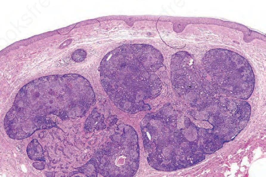
圖 31-109:Trichoblastoma:低倍視野,顯示一個假包膜化 (pseudoencapsulated) 的多結節性基底樣細胞群。注意無收縮人工假象 (retraction artifact)。間質成分在視野中央最易見到。
Fig. 31.109 Trichoblastoma: low-power view of a pseudoencapsulated multinodular basaloid cell population. Note the absence of a retraction artifact. The stromal component is best seen in the center of the field.
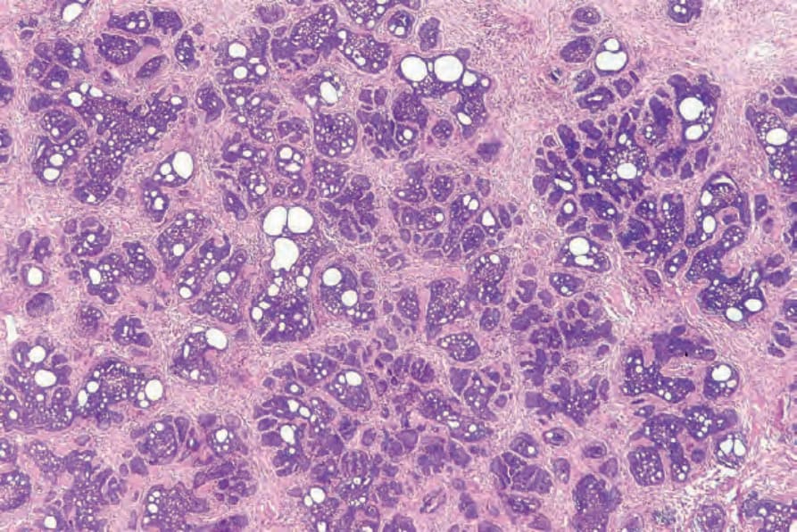
圖 31-110:Trichoblastoma:類 trichoepithelioma 變異型(trichoblastic fibroma)的掃描視野,顯示嗜鹼性腫瘤小葉與腺樣變化 (adenoid change) 病灶的混合。注意豐富、緻密細胞性的間質。
Fig. 31.110 Trichoblastoma: scanning view of a trichoepithelioma-like variant (trichoblastic fibroma) showing an admixture of basophilic tumor lobules and foci of adenoid change. Note the abundant densely cellular stroma.
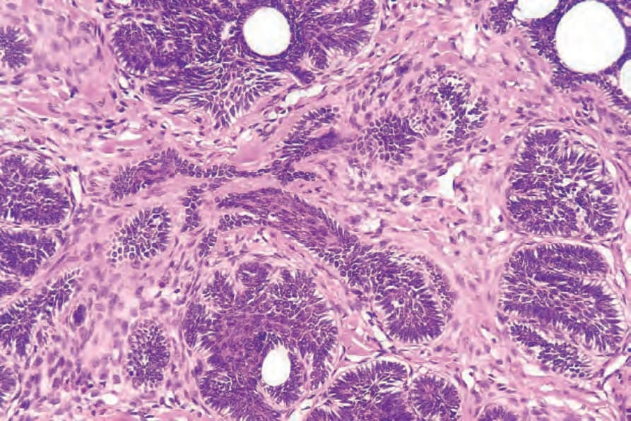
圖 31-111:Trichoblastoma:高倍視野,顯示上皮性條索與間質。
Fig. 31.111 Trichoblastoma: high-power view showing epithelial strands and stroma.
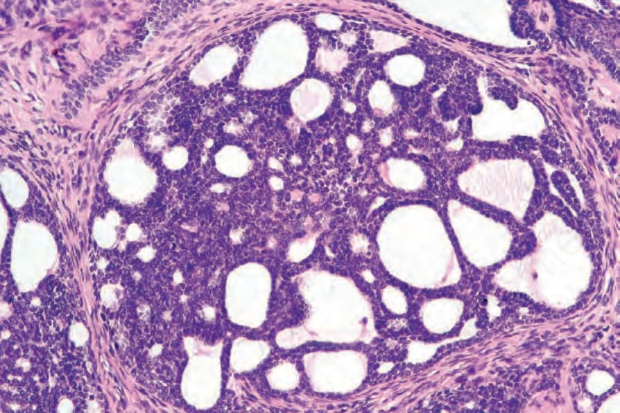
圖 31-112:Trichoblastoma:間質黏液沉積導致腺樣病灶 (adenoid foci),如本視野所示。此特徵可能造成與腺樣型基底細胞癌 (adenoid basal cell carcinoma) 的混淆。
Fig. 31.112 Trichoblastoma: stromal mucin deposition results in adenoid foci as shown in this field. This feature may cause confusion with adenoid basal cell carcinoma.
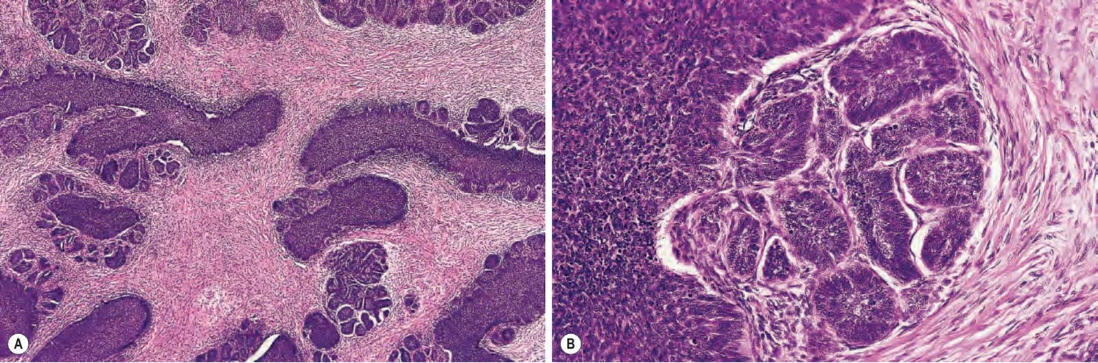
圖 31-113:(A, B) Trichoblastoma:在此例中有一明顯的間質成分。注意周邊柵欄狀排列 (peripheral palisading)。
Fig. 31.113 (A, B) Trichoblastoma: in this example, there is a conspicuous stromal component. Note the peripheral palisading.
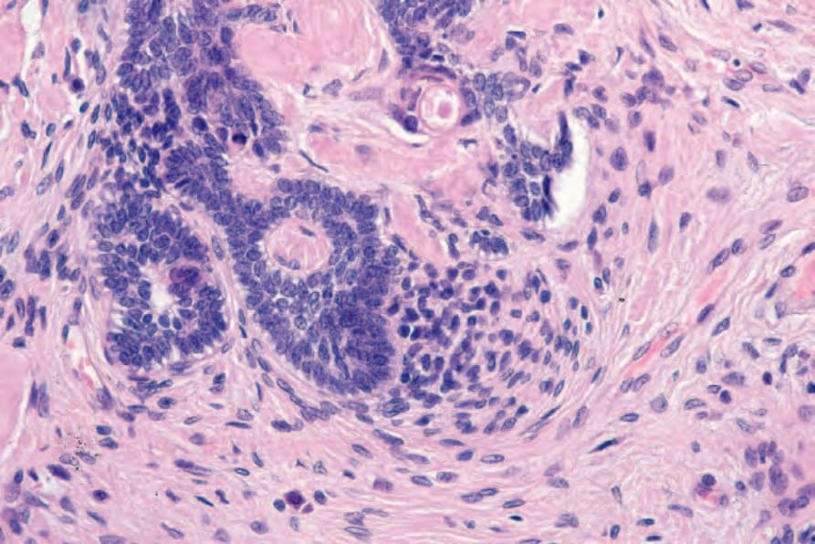
圖 31-114:Trichoblastoma:間質凝聚導致原始毛乳頭形成(乳頭狀間質小體 papillary mesenchymal body)。
Fig. 31.114 Trichoblastoma: condensation of the stroma has resulted in primitive hair papilla formation (papillary mesenchymal body).
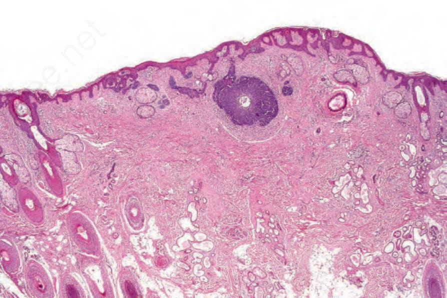
圖 31-115:Trichoblastoma:此例發生於皮脂腺痣 (nevus sebaceous) 之內,僅由上皮成分組成。間質誘導甚少。在過去,此類病灶曾被視為基底細胞癌。
Fig. 31.115 Trichoblastoma: this example, which arose in a nevus sebaceous, consists solely of an epithelial component. There is little stromal induction. In the past, such lesions were regarded as basal cell carcinoma.
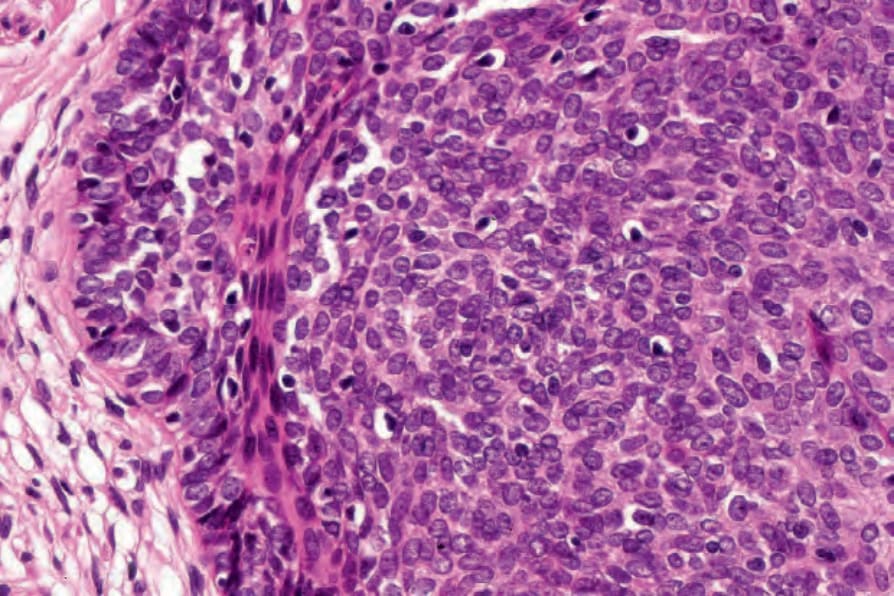
圖 31-116:Trichoblastoma:高倍視野。
Fig. 31.116 Trichoblastoma: high-power view.
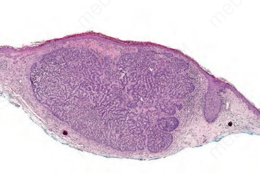
圖 31-117:Trichoblastoma:rippled-pattern 變異型,顯示突出的柵欄狀排列。
Fig. 31.117 Trichoblastoma: rippled-pattern variant showing prominent palisading.
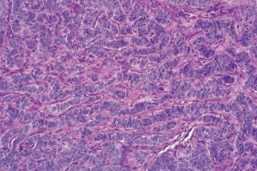
圖 31-118:Trichoblastoma:高倍視野。
Fig. 31.118 Trichoblastoma: high-power view.
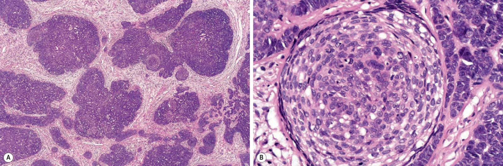
圖 31-119:(A, B) Trichoblastoma(trichogerminoma):此例主要由生發性成分組成,包含基底樣細胞與明顯的淡染微結節(Zellballen)混雜。
Fig. 31.119 (A, B) Trichoblastoma (trichogerminoma): this example consists predominantly of a germinative component comprising basaloid cells admixed with distinct pale micronodules (Zellballen).
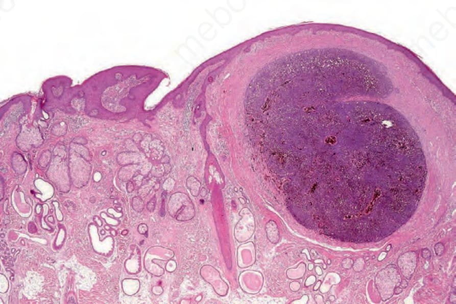
圖 31-120:Pigmented trichoblastoma:此病灶發生於皮脂腺痣 (nevus sebaceous) 的背景之中。有重度黑色素 (melanin) 色素沉著。
Fig. 31.120 Pigmented trichoblastoma: this lesion developed in a background of nevus sebaceous. There is heavy melanin pigmentation.
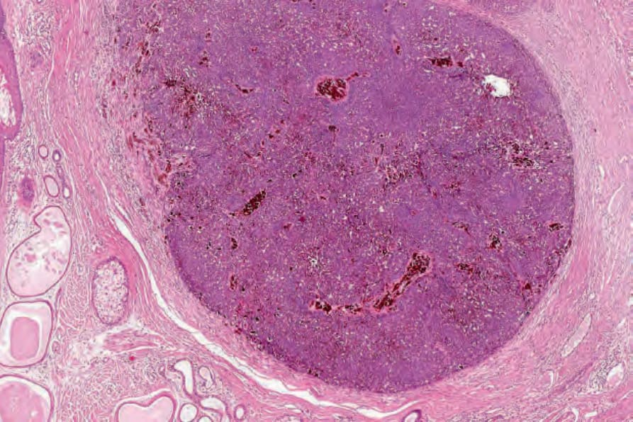
圖 31-121:Pigmented trichoblastoma:中倍視野。有大量色素。
Fig. 31.121 Pigmented trichoblastoma: medium-power view. There is abundant pigment.
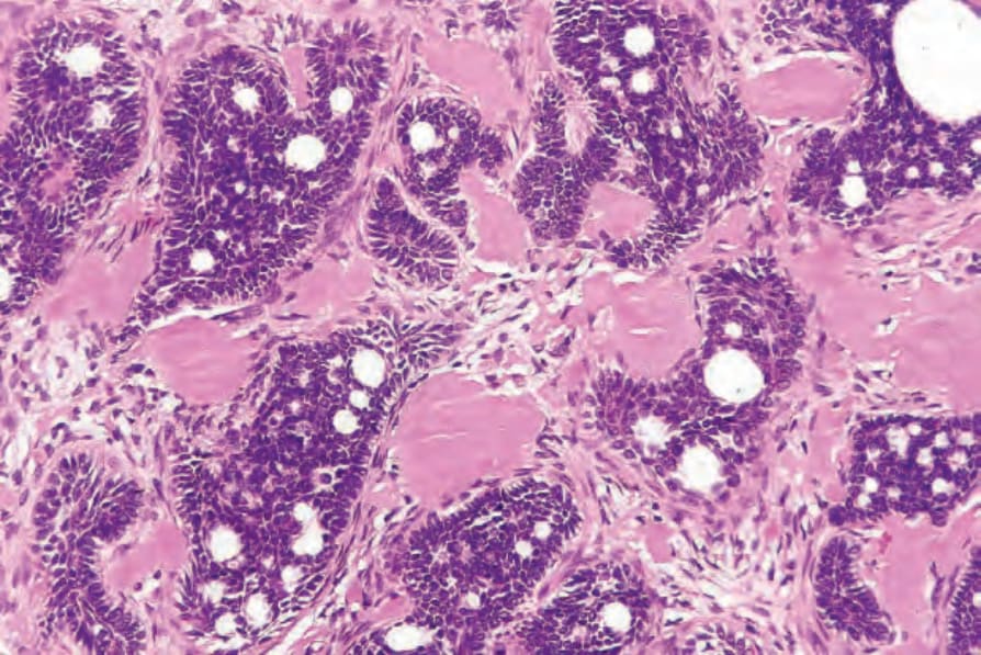
圖 31-123:Trichoblastoma:在此例中有明顯的類澱粉沉積 (amyloid deposits)。
Fig. 31.123 Trichoblastoma: in this example, there are conspicuous amyloid deposits.
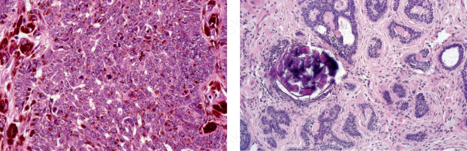
圖 31-124:Trichoblastoma:局部鈣化 (focal calcification) 為一不算罕見的特徵。
Fig. 31.124 Trichoblastoma: focal calcification is a not uncommon feature.