Trichoblastoma
Trichoblastoma
Trichogenic tumors are neoplasms recapitulating the germinative hair bulb and its associated mesenchyme.1 Their nomenclature is, however, exceedingly confusing. Analogous to odontogenic neoplasms, Headington classified these tumors according to the relative amounts of epithelial and mesenchymal components and the presence of stromal inductive change.2 Purely epithelial tumors were labeled trichoblastoma while mixed epithelial and mesenchymal tumors were referred to as trichoblastic fibroma or trichogenic trichoblastoma in the presence of hair follicle differentiation.2,3 Individual neoplasms are, however, capable of showing varying degrees of differentiation, and the originally proposed classification is too strict and limited.4 In addition, subsequent reports of trichogenic neoplasms have used a variety of designations, confusing classification of these tumors even further. For example, tumors resembling trichoblastic fibroma have been referred to as giant solitary trichoepithelioma, subcutaneous trichoepithelioma, or immature trichoepithelioma. More recently, entities such as trichogerminoma, rippled-pattern trichoblastoma, rippled-pattern trichomatricoma, and melanotrichoblastoma have also been described. These tumors display some unique histologic features but also share features of the traditional trichogenic tumors. While it is important to recognize distinct histologic patterns within a neoplasm, it is equally important that classification is comprehensible and meaningful. Since the clinical presentation and behavior of the aforementioned entities are similar and a variety of histologic patterns may be seen in any one tumor, trichogenic tumors in this text are unified under the single heading trichoblastoma.
Cutaneous lymphadenoma has been shown to represent a variant of trichoblastoma and is likely synonymous with trichoblastoma with
Trichoblastoma typically behaves in a benign fashion. Malignant change has, however, been rarely described (see below).13,43–54
Histologic features Trichoblastoma is a well-circumscribed but unencapsulated nodular tumor spanning the entire dermis, characteristically extending into subcutaneous tissue (Fig. 31.109). A purely subcutaneous location may rarely be seen.1,3,15–18,55,56 It is devoid of epidermal or follicular derivation and characterized by variably sized epithelial nests closely resembling basal cell
1577 Trichoblastoma
carcinoma. Peripheral palisading is conspicuous but there is stromal condensation around tumor lobules, and cleft artifact is not a prominent feature. The amount of surrounding stroma as well as ‘stromal induction’ is variable between different tumors as well as within the same lesion.
eddies around a central space or keratinized core, and occasionally focal glycogen-rich clear cell change is evident.1
The fibromyxoid stroma is an important, integral component of the tumor (Fig. 31.113). It comprises both stellate and spindled cell fibroblasts and characteristically is associated with primitive hair papilla formation – so-called papillary mesenchymal bodies, which often indent the adjacent epithelium (Fig. 31.114).1
The tumor described as trichoblastic fibroma is characteristically biphasic, being composed of lobules of basaloid cells intimately associated with a conspicuous fibromyxoid stroma (Fig. 31.110).1,5,19 Larger lobules and their stroma are often arranged in a mosaic pattern, while the smaller islands of tumor cells are set in close clusters with little intervening stroma. Tumor cells are small and basophilic with minimal cytoplasm. They often show peripheral palisading. Pleomorphism is not a feature, but mitotic activity is frequently brisk and apoptotic bodies may be evident. Some of the lobules have associated narrow epithelial strands, giving rise to ‘antler-like’ patterns (Fig. 31.111).15,17 Occasionally, a cribriform appearance is evident (Fig. 31.112).1 Sometimes the large lobules are associated with keratin cyst formation although much less frequently than seen in trichoepithelioma. Keratinization is usually epidermoid in nature, but pilar type may also be represented. The small tumor lobules sometimes form whorls or squamous
At the opposite end of the spectrum, some tumors are predominantly composed of large basaloid epithelial lobules showing peripheral palisading and only scant sclerotic intervening stroma with no or only little evidence of stromal induction (Figs 31.115 and 31.116).9,11,12,18,21–25 Within the tumor lobules, cells may take on a spindle appearance and align to form nuclear palisading reminiscent of Verocay body formation. This pattern can be focal or extensive, and tumors such as this have been referred to as rippled-pattern trichoblastoma (rippled-pattern trichomatricoma) (Figs 31.117 and 31.118).9,21–25 Sebaceous differentiation is sometimes observed in this variant.
Tumors described as trichogerminoma show additional distinctive histologic features.13,14,57 They are composed of small basaloid epithelial lobules and nests separated by thin fibrous strands. The lobules are composed of
1578 Tumors of the hair follicle
A
B
1579 Trichoblastoma
densely packed basaloid cells reminiscent of ‘Zellballen’ and closely resemble the hair bulb (Fig. 31.119). A peripheral palisade surrounds them.
Trichoblastoma is rarely heavily pigmented and contains abundant intralesional dendritic melanocytes (Figs 31.120–31.122). This variant has been referred to as pigmented trichoblastoma or melanotrichoblastoma.11,12,58
Occasionally, abundant clear cell change (clear cell trichoblastoma) may be evident, and ductal as well as sebaceous differentiation has been described.13,24,25,59–65
Nodular trichoblastoma with adamantinoid features is a distinctive variant thought to be synonymous with cutaneous lymphadenoma.37
Panfolliculoma is an exceedingly rare and unusual tumor.36,66 It falls into the spectrum of trichoblastoma but shows unique histologic features with differentiation toward all elements of the hair follicle. It is well demarcated and symmetrical, and both solid and cystic with a wide range of differentiation toward germinal hair bulb and papilla as well as follicular matrix and inner and outer root sheath.67,68 Focal sebaceous differentiation has also been reported.69 Rarely, the tumors are intraepidermal.70,71
A rare subset of tumors is characterized by an infundibular cyst with additional germinative and matrical follicular differentiation. These tumors have been referred to as trichoblastic infundibular cysts or cystic trichoblastoma.72,73
A
B
1580 Tumors of the hair follicle
in 11% of sporadic trichoblastomas.96,97 In contrast, no mutations in the PTCH, KRAS, or BRAF gene have been detected.97,98
Differential diagnosis Trichoblastoma is most commonly mistaken for conventional trichoepithelioma and nodular basal cell carcinoma.
It is much larger than conventional trichoepithelioma and is situated within deep dermis and subcutaneous tissue, while ‘conventional’ trichoepithelioma is centered in mid-dermis. Trichoblastoma shows less keratinization and is devoid of epidermal or follicular origin.
Lack of epidermal origin, more conspicuous stroma with prominent papillary mesenchymal bodies, and absence of retraction artifact are useful diagnostic features in excluding basal cell carcinoma. It is, however, often extremely difficult to make this distinction with confidence, especially on a small biopsy specimen. Complete excision is therefore the treatment of choice.
A mild chronic inflammatory cell infiltrate is sometimes present, and mast cells are often conspicuous. Amyloid deposits are commonly found within the stroma, and focal calcification is sometimes evident (Figs 31.123 and 31.124).11,33 Merkel cells frequently populate trichoblastoma.74,75
Trichoblastoma may arise within a nevus sebaceous and rarely occurs in a poroma (see Fig. 31.115).58,61,76–87
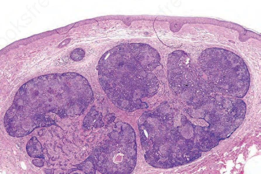
Fig. 31.109 Trichoblastoma: low-power view of a pseudoencapsulated multinodular basaloid cell population. Note the absence of a retraction artifact. The stromal component is best seen in the center of the field.
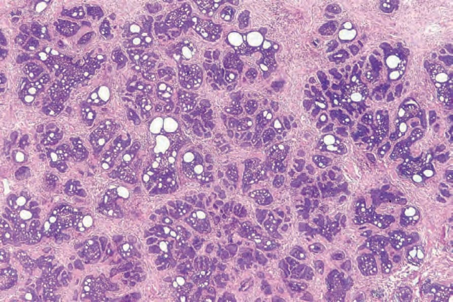
Fig. 31.110 Trichoblastoma: scanning view of a trichoepithelioma-like variant (trichoblastic fibroma) showing an admixture of basophilic tumor lobules and foci of adenoid change. Note the abundant densely cellular stroma.
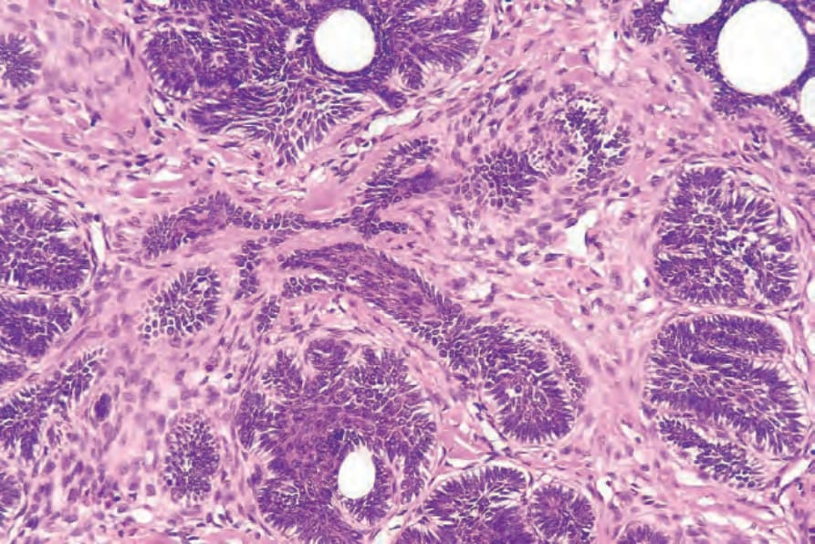
Fig. 31.111 Trichoblastoma: high-power view showing epithelial strands and stroma.
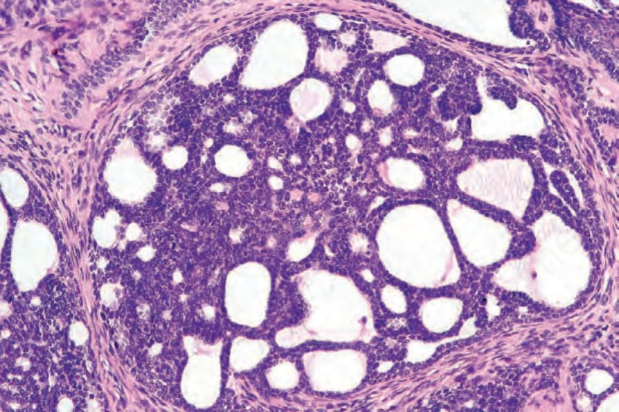
Fig. 31.112 Trichoblastoma: stromal mucin deposition results in adenoid foci as shown in this field. This feature may cause confusion with adenoid basal cell carcinoma.
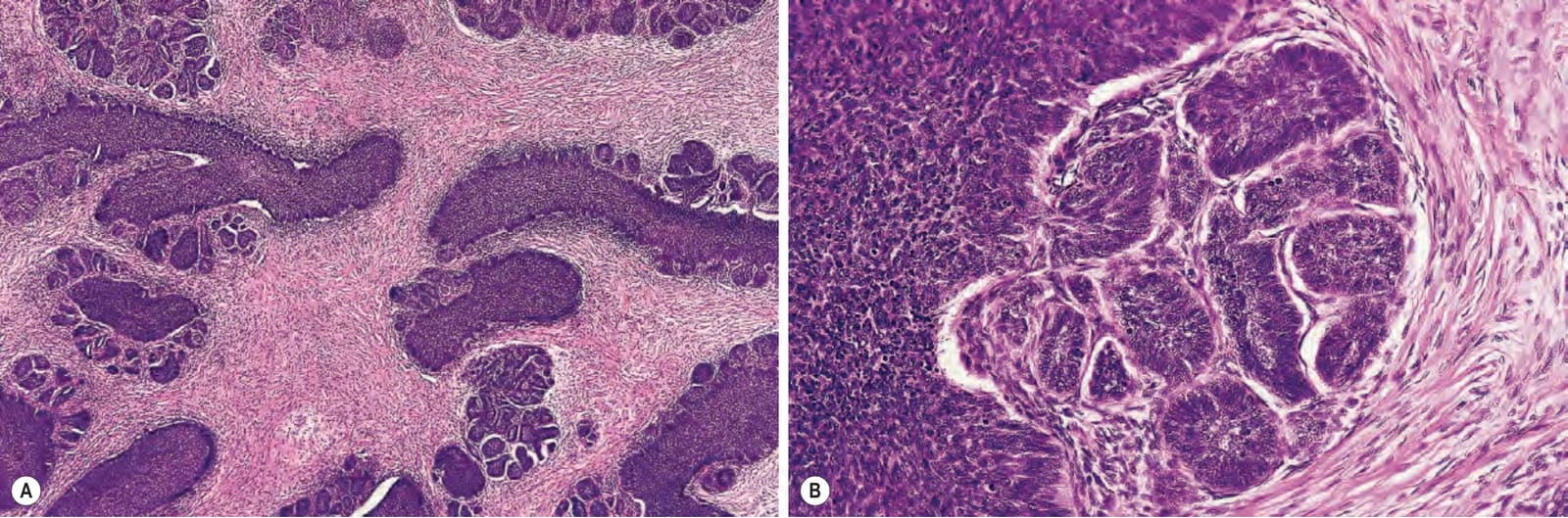
Fig. 31.113 (A, B) Trichoblastoma: in this example, there is a conspicuous stromal component. Note the peripheral palisading.
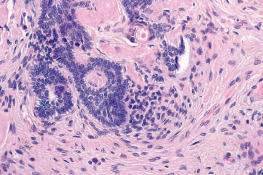
Fig. 31.114 Trichoblastoma: condensation of the stroma has resulted in primitive hair papilla formation (papillary mesenchymal body).
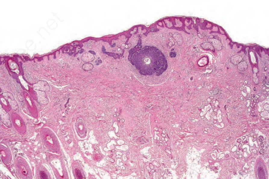
Fig. 31.115 Trichoblastoma: this example, which arose in a nevus sebaceous, consists solely of an epithelial component. There is little stromal induction. In the past, such lesions were regarded as basal cell carcinoma.
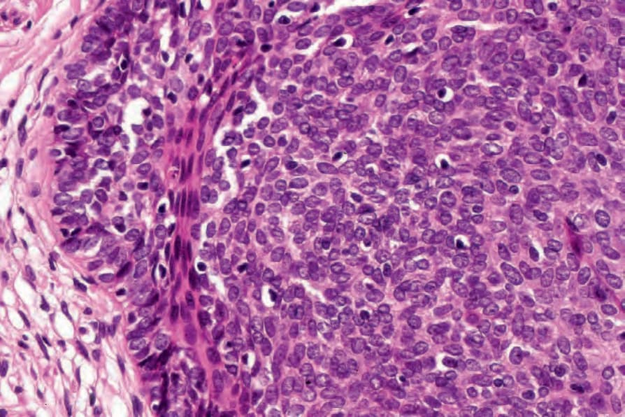
Fig. 31.116 Trichoblastoma: high-power view.
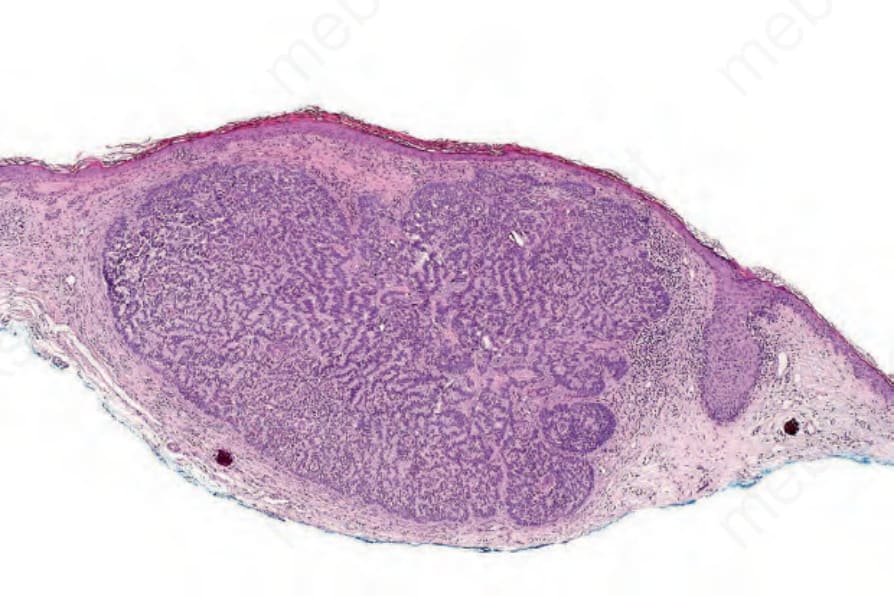
Fig. 31.117 Trichoblastoma: rippled-pattern variant showing prominent palisading.
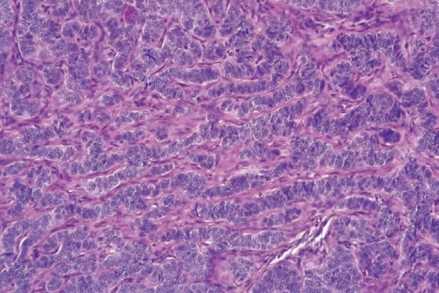
Fig. 31.118 Trichoblastoma: high-power view.
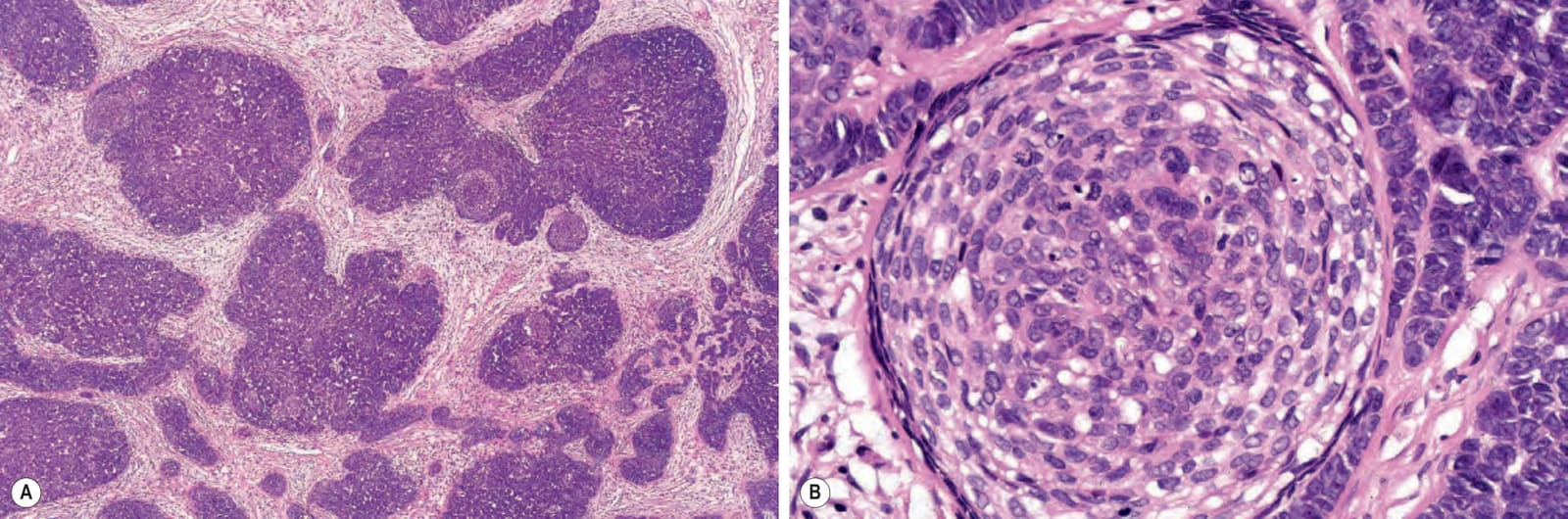
Fig. 31.119 (A, B) Trichoblastoma (trichogerminoma): this example consists predominantly of a germinative component comprising basaloid cells admixed with distinct pale micronodules (Zellballen).
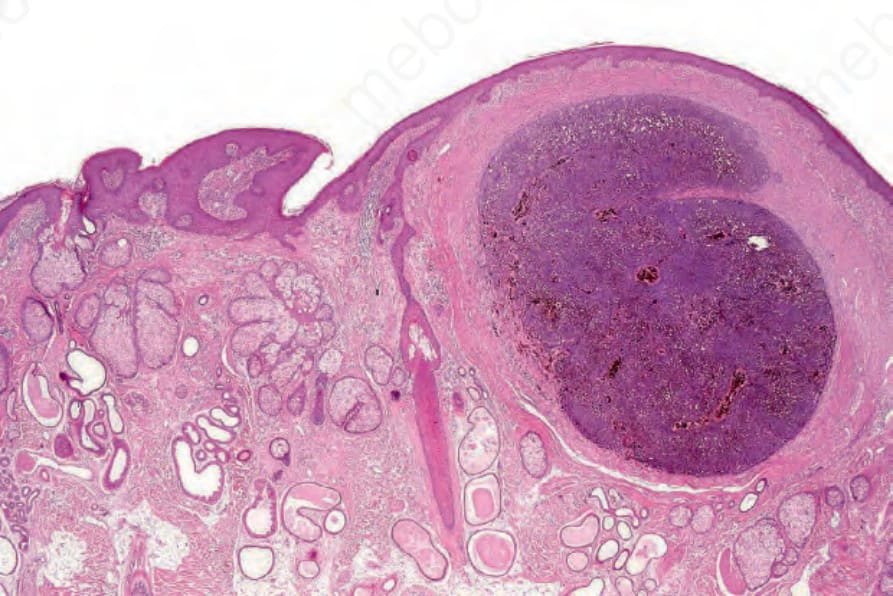
Fig. 31.120 Pigmented trichoblastoma: this lesion developed in a background of nevus sebaceous. There is heavy melanin pigmentation.
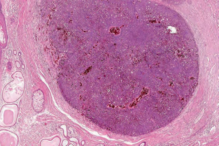
Fig. 31.121 Pigmented trichoblastoma: medium-power view. There is abundant pigment.
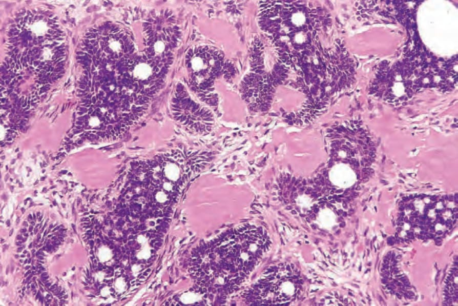
Fig. 31.123 Trichoblastoma: in this example, there are conspicuous amyloid deposits.
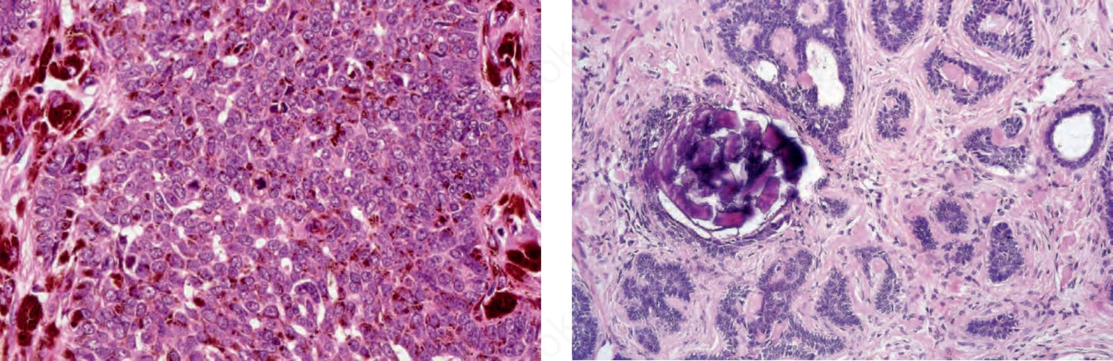
Fig. 31.124 Trichoblastoma: focal calcification is a not uncommon feature.