血管內假性T細胞淋巴瘤 (Intravascular pseudo-T-cell lymphoma)
臨床特徵 (Clinical Features)
- 在因外傷或各種發炎及腫瘤性病變(包括 lichen sclerosus、hidradenitis suppurativa 與 hemangioma)而進行的皮膚切片中,偶爾可見到淋巴管內聚集的 CD30 陽性、或較少見地 CD30 陰性的母細胞性 (blastic) 細胞。
- 其行為表現似為良性,目前所報告的所有病例皆無任何不良影響。
組織病理特徵 (Histopathology)
- 數量不等的真皮淋巴管因母細胞性細胞的增生而擴張,這些細胞經常(但並非總是)為 CD30 陽性 (Figs 29.171–29.173)。
- 通常也表現其他 T 細胞標記,包括 CD3 與 CD4。
- B 細胞標記為陰性。
- 此增生細胞通常為多株性 (polyclonal)。
鑑別診斷 (Differential Diagnosis)
- 鑑別診斷包括 intravascular T、NK/T 或 B cell lymphoma 以及 intravascular histiocytosis。
- 臨床病理對照、免疫表型分析 (immunophenotyping) 與株系分析 (clonality studies) 對於確立正確診斷至關重要。
於 pseudolymphomatous angiokeratoma 中的內皮細胞似乎表現 WT1。
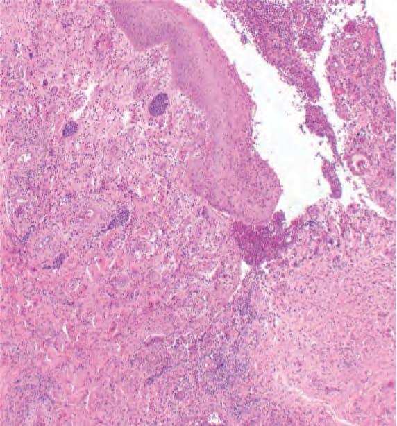
圖 29-171:血管內假性T細胞淋巴瘤:眾多擴張的淋巴管腔內含有母細胞性淋巴樣細胞 (Intravascular pseudo-T-cell lymphoma: numerous dilated lymphatic channels containing blastic lymphoid cells)。
Fig. 29.171 Intravascular pseudo-T-cell lymphoma: numerous dilated lymphatic channels containing blastic lymphoid cells.
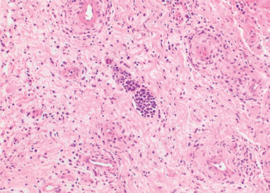
圖 29-172:血管內假性T細胞淋巴瘤:擴張淋巴管腔內含母細胞性淋巴樣細胞的近觀 (Intravascular pseudo-T-cell lymphoma: closer view of a dilated lymphatic vessel containing blastic lymphoid cells)。
Fig. 29.172 Intravascular pseudo-T-cell lymphoma: closer view of a dilated lymphatic vessel containing blastic lymphoid cells.
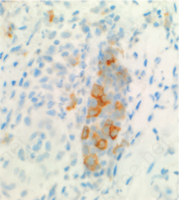
圖 29-173:血管內假性T細胞淋巴瘤:許多但並非全部的母細胞性 T 細胞對 CD30 呈陽性 (Intravascular pseudo-T-cell lymphoma: many but not all of the blastic cells T cells are positive for CD30)。
Fig. 29.173 Intravascular pseudo-T-cell lymphoma: many but not all of the blastic cells T cells are positive for CD30.
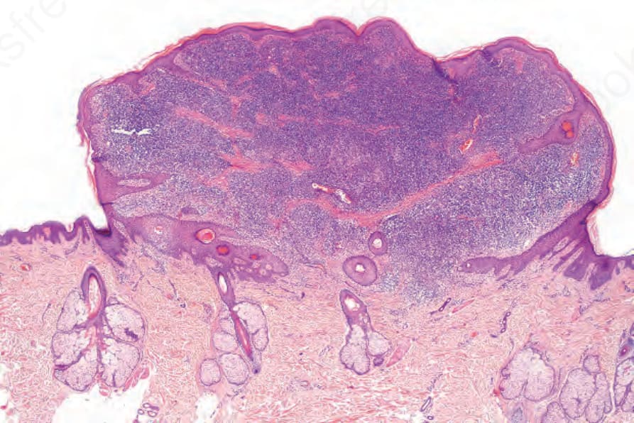
圖 29-174:富含T細胞的血管瘤樣息肉狀假性淋巴瘤 (TRAPP):息肉狀結節的掃描視野 (T-cell rich angiomatoid polypoid pseudolymphoma (TRAPP): scanning view of polypoid nodule)。
Fig. 29.174 T-cell rich angiomatoid polypoid pseudolymphoma (TRAPP): scanning view of polypoid nodule.
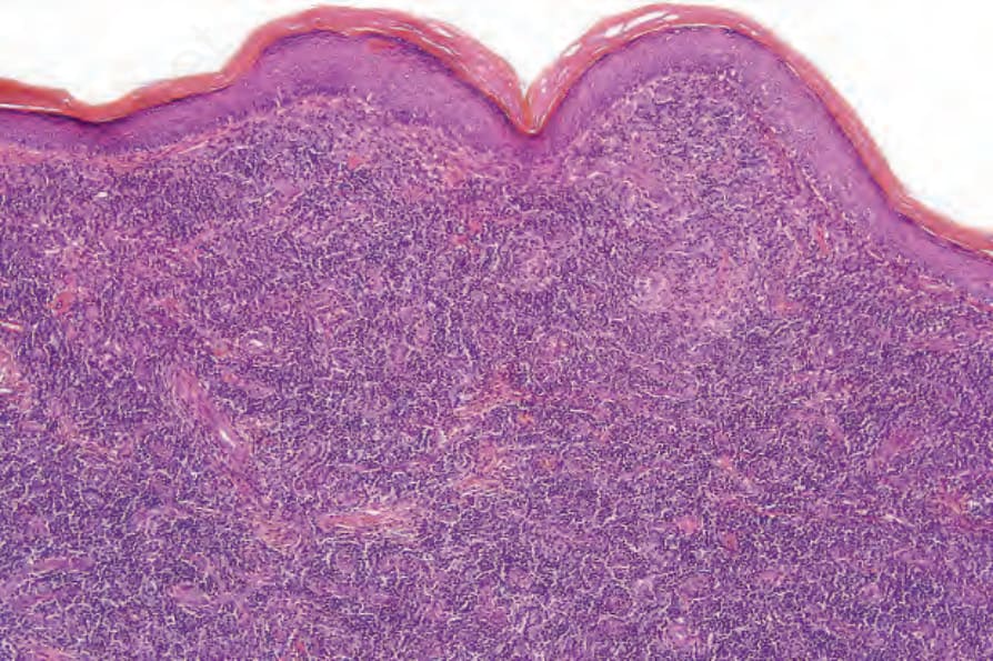
圖 29-175:富含T細胞的血管瘤樣息肉狀假性淋巴瘤 (TRAPP):中倍視野顯示無表皮侵犯 (T-cell rich angiomatoid polypoid pseudolymphoma (TRAPP): medium-power view showing lack of epidermal involvement)。
Fig. 29.175 T-cell rich angiomatoid polypoid pseudolymphoma (TRAPP): medium-power view showing lack of epidermal involvement.