臨床特徵 (Clinical Features)
- 在高達 4% 的 mycosis fungoides (MF) 病例中可見肉芽腫性浸潤 (granulomatous infiltrate)。其臨床表現與典型 MF 無法區分;病人表現為丘疹 (papules)、斑塊 (plaques) 與腫瘤 (tumors),且可能呈階梯式進展 (stepwise progression)(圖 29.58)。
- 大多數報告的病例描述於成人,但罕見情況下亦發生於兒童。
- 它偶可與 sarcoidosis 或泛發性 granuloma annulare 相關,並曾有類似魚鱗癬樣 (ichthyosis-like) 之表現的報告。
- 肉芽腫被認為代表對腫瘤的免疫媒介性 (immunologically mediated) 反應,最初被認為意味著預後較佳。然而,本病亦有快速進展的型態,因此其他因素在決定預後上可能更為重要。
- 肉芽腫性發炎 (granulomatous inflammation) 在 folliculotropic MF 中亦常見,可見於高達四分之一的病例。在此情況下,肉芽腫性發炎代表對破裂毛囊 (ruptured hair follicles) 的反應。
組織病理特徵 (Histopathology)
- 在所有病例中,皆有 MF 的底層特徵。相關的肉芽腫通常為類肉瘤型 (sarcoid type),常伴隨異物巨細胞 (foreign body giant cells) 與 Langhans giant cells(圖 29.59–29.61)。
- 雖然此成分可能僅佔浸潤的一小部分,但在許多病例中可佔 25% 或更多,因而有被誤診為發炎性或感染性病變的可能性。
- 有時可見噬淋巴細胞現象 (lymphophagocytosis),而噬彈力纖維現象 (elastophagocytosis) 則常見。
- 在某些情況下,可見類似 granuloma annulare 或 necrobiosis lipoidica 的柵狀組織球性肉芽腫 (palisading histiocytic granulomata)(圖 29.62 與 29.63),而在其他情況下肉芽腫則呈結核樣 (tuberculoid)。應進行微生物特殊染色 (special stains for microorganisms) 以排除感染。
鑑別診斷 (Differential Diagnosis)
- 次發性侵犯皮膚者,以及原發性皮膚 B 細胞淋巴瘤 (primary cutaneous B-cell lymphoma, PCBCL)。
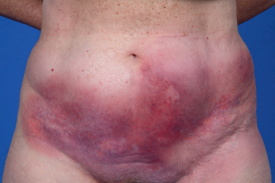
圖 29.58:肉芽腫性蕈樣肉芽腫 (granulomatous mycosis fungoides):下腹部硬結性融合斑塊。承蒙 M. Duvic 醫師惠予提供,美國德州休士頓 MD Anderson Cancer Center。
Fig. 29.58 Granulomatous mycosis fungoides: indurated coalescing plaques on lower abdomen. By courtesy of M. Duvic, MD, the MD Anderson Cancer Center, Houston, Texas, USA.
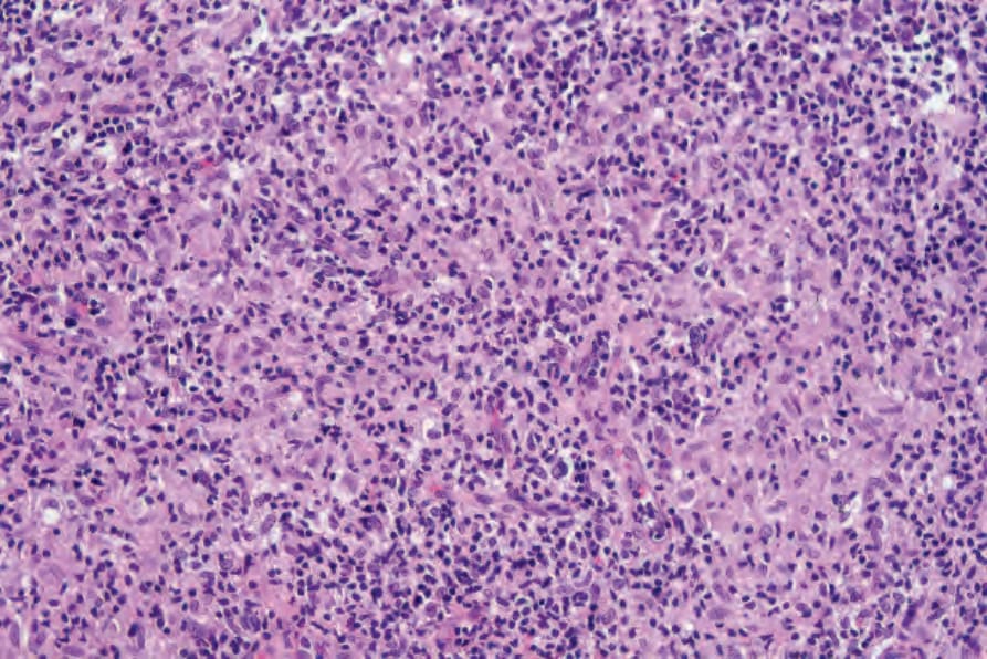
圖 29.59:肉芽腫性蕈樣肉芽腫 (granulomatous mycosis fungoides):此視野中可見發育良好的肉芽腫性發炎浸潤,背景為異型淋巴細胞 (atypical lymphocytes)。
Fig. 29.59 Granulomatous mycosis fungoides: in this field there is a well-developed granulomatous inflammatory infiltrate with a background of atypical lymphocytes.
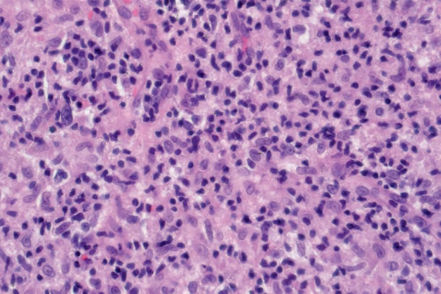
圖 29.60:肉芽腫性蕈樣肉芽腫 (granulomatous mycosis fungoides):高倍視野。
Fig. 29.60 Granulomatous mycosis fungoides: high-power view.
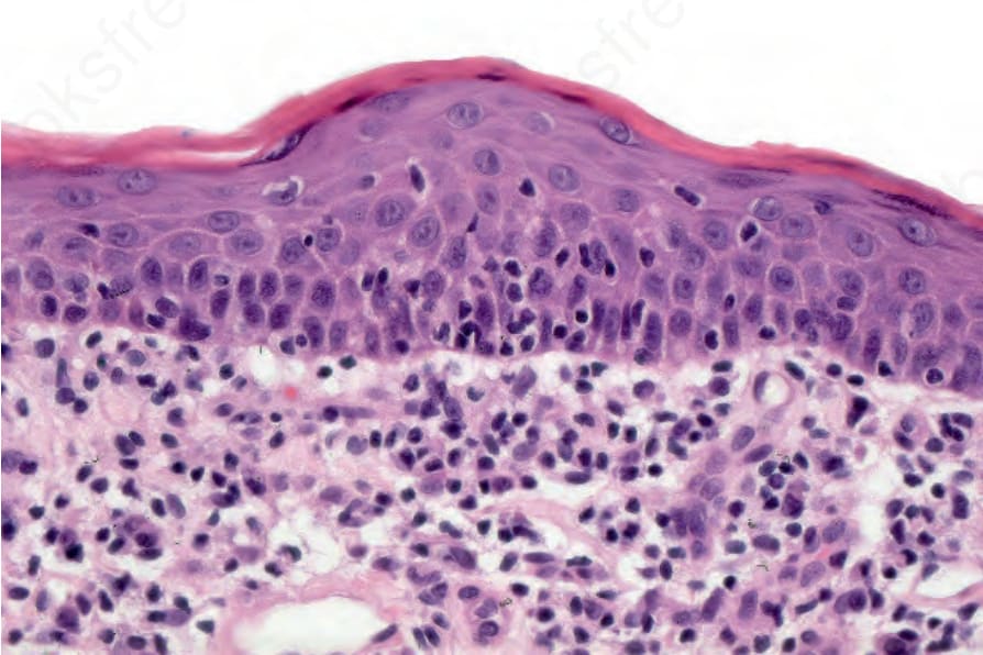
圖 29.61:肉芽腫性蕈樣肉芽腫 (granulomatous mycosis fungoides):表皮趨向性 (epidermotropism) 伴淋巴細胞貼附 (lymphocyte tagging)。
Fig. 29.61 Granulomatous mycosis fungoides: epidermotropism with lymphocyte tagging.
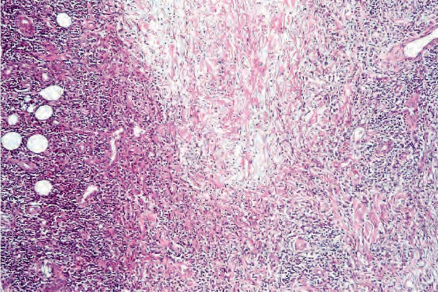
圖 29.62:肉芽腫性蕈樣肉芽腫 (granulomatous mycosis fungoides):此病例呈現類膠原變性 (necrobiosis-like) 特徵。膠原纖維 (collagen fibers) 腫脹且呈嗜伊紅性 (eosinophilic)。
Fig. 29.62 Granulomatous mycosis fungoides: this case shows necrobiosis-like features. The collagen fibers are swollen and eosinophilic.
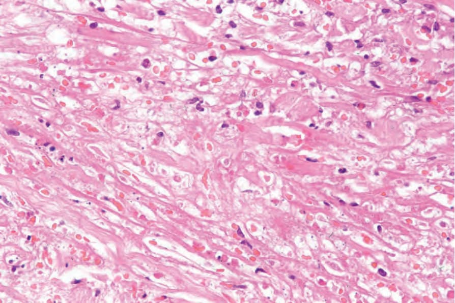
圖 29.63:肉芽腫性蕈樣肉芽腫 (granulomatous mycosis fungoides):膠原變性 (necrobiosis) 的近距離視野。
Fig. 29.63 Granulomatous mycosis fungoides: close-up view of necrobiosis.
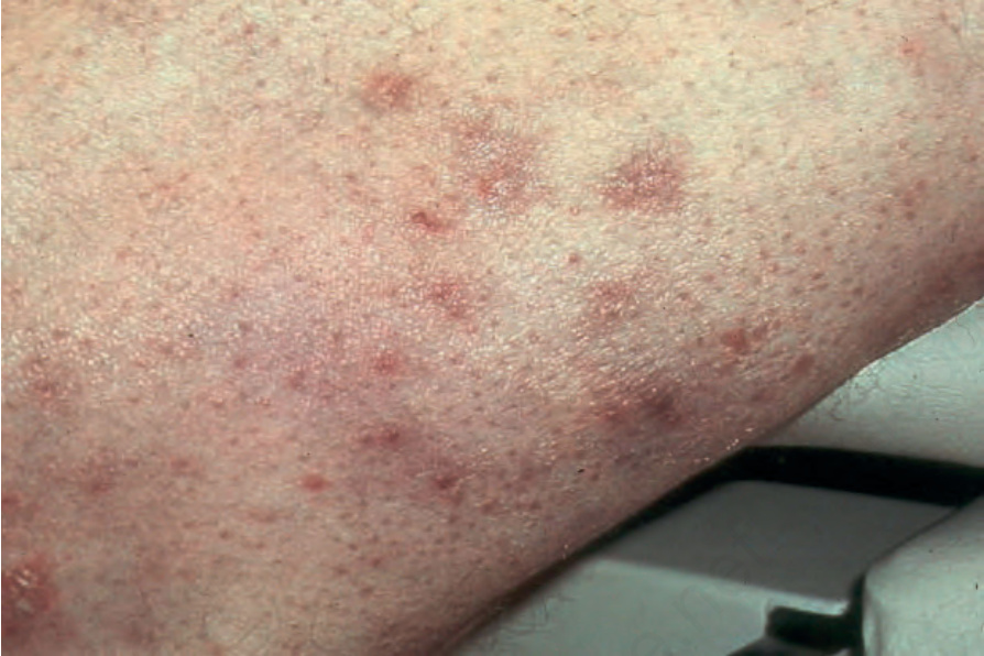
圖 29.64:汗管趨向性蕈樣肉芽腫 (syringotropic mycosis fungoides):此病人手臂上可見明顯的丘疹 (papules)。承蒙 H. Naeem 醫師惠予提供,美國波士頓 Harvard Medical School。
Fig. 29.64 Syringotropic mycosis fungoides: distinct papules are present on this patient’s arm. By courtesy of H. Naeem, MD, Harvard Medical School, Boston, USA.