Granulomatous mycosis fungoides
Granulomatous mycosis fungoides
Clinical features A granulomatous infiltrate is seen in up to 4% of cases of MF.1–16 The clinical features are indistinguishable from those of typical MF; patients present with papules, plaques, and tumors, and may run a stepwise progression (Fig. 29.58).13 Most reported cases have been described in adults, but exceptional cases occur in children.7,17 It may rarely be seen in association with sarcoidosis or generalized granuloma annulare, and an ichthyosis-like presentation has been reported.10,11,16,18–21 Granulomas are thought to represent an immunologically mediated reaction to the tumor, and was initially supposed to indicate a favorable prognosis.22 However, there are examples of rapidly progressing forms of the disease, so other factors are likely to be more important in determining outcome.8,14,18,23–26
Granulomatous inflammation is also commonly encountered in folliculotropic MF and may be seen in up to one-quarter of cases.26 In this situation, the granulomatous inflammation represents a reaction to ruptured hair follicles.
Histologic features In all cases, there are underlying features of MF. The associated granulomas are usually sarcoid type, often accompanied by foreign body and Langhans giant cells (Figs 29.59–29.61).13 While this may represent only a minor component of the infiltrate, in many cases it can constitute 25% or more with the
1423 Mycosis fungoides with unusual histologic features
potential for misdiagnosis as an inflammatory or infective condition.5,13,21,27 Lymphophagocytosis is sometimes present, and elastophagocytosis is common.28 In some instances, palisading histiocytic granulomata reminiscent of granuloma annulare or necrobiosis lipoidica are noted (Figs 29.62 and 29.63), whilst in others the granulomata are tuberculoid.2,3,29 Special stains for microorganisms should be performed to exclude an infection.
secondarily involving the skin, and primary cutaneous B-cell lymphoma (PCBCL).27,30–32
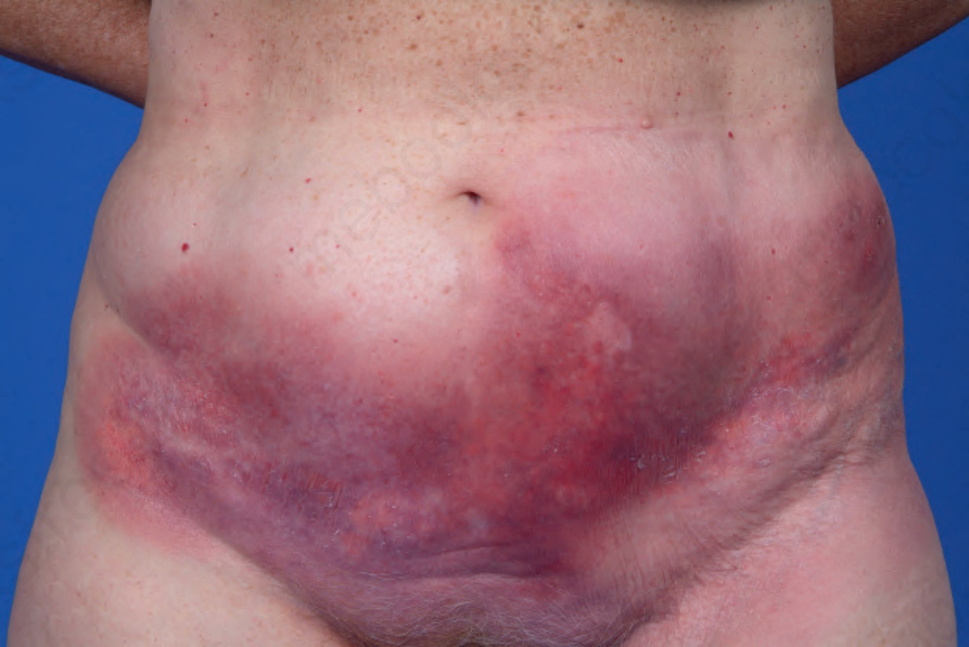
Fig. 29.58 Granulomatous mycosis fungoides: indurated coalescing plaques on lower abdomen. By courtesy of M. Duvic, MD, the MD Anderson Cancer Center, Houston, Texas, USA.
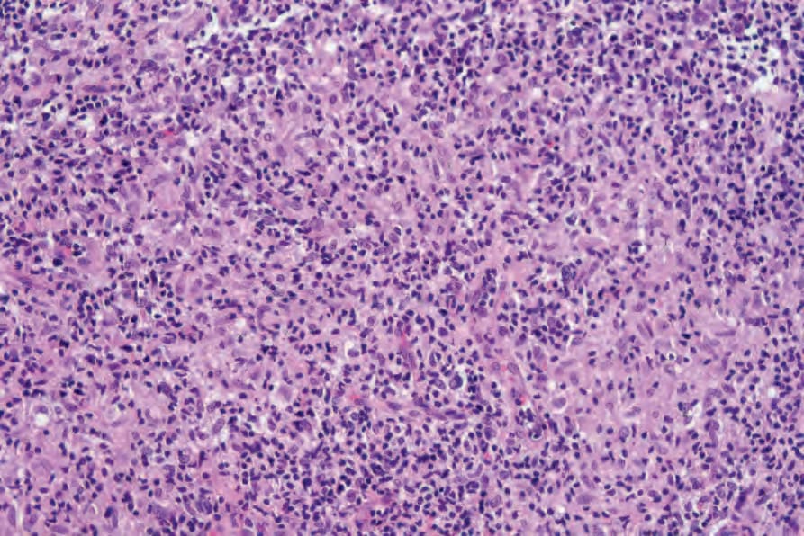
Fig. 29.59 Granulomatous mycosis fungoides: in this field there is a well-developed granulomatous inflammatory infiltrate with a background of atypical lymphocytes.
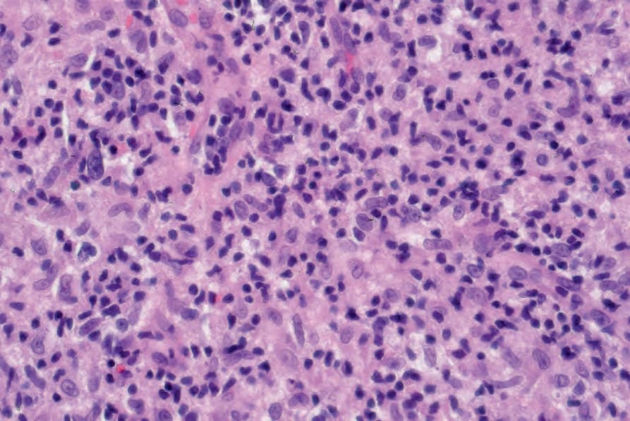
Fig. 29.60 Granulomatous mycosis fungoides: high-power view.
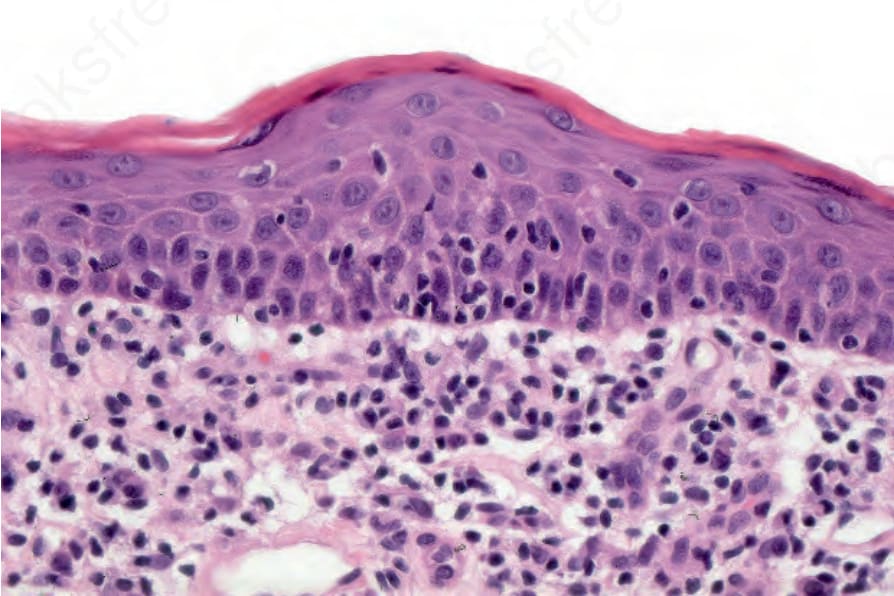
Fig. 29.61 Granulomatous mycosis fungoides: epidermotropism with lymphocyte tagging.
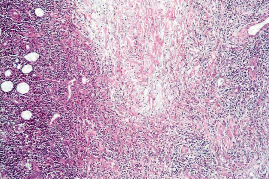
Fig. 29.62 Granulomatous mycosis fungoides: this case shows necrobiosis-like features. The collagen fibers are swollen and eosinophilic.
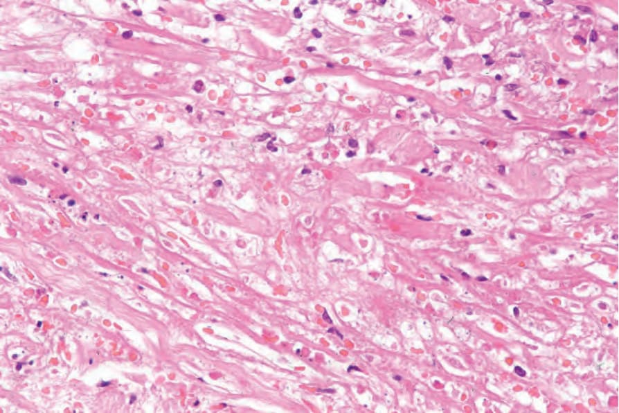
Fig. 29.63 Granulomatous mycosis fungoides: close-up view of necrobiosis.
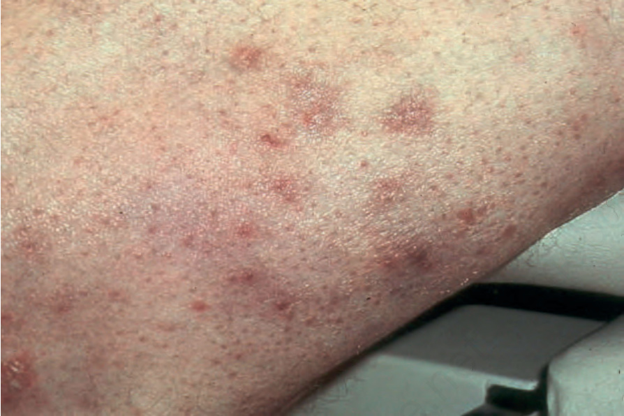
Fig. 29.64 Syringotropic mycosis fungoides: distinct papules are present on this patient’s arm. By courtesy of H. Naeem, MD, Harvard Medical School, Boston, USA.