疾病定義與分類
- 結膜痣 (conjunctival nevus) 是結膜最常見的黑色素細胞性 (melanocytic) 腫瘤,在數個大型系列中佔結膜腫瘤的 23% 至 29%。
- conjunctival nevus 可分為先天型(出生後前 6 個月內出現)與後天型(出生後超過 6 個月才出現)兩類。
臨床特徵 (Clinical Features)
- 在一個包含 410 例 conjunctival nevi 的大型系列中,平均年齡為 32 歲,範圍廣達 2–93 歲。多數病人為白種人 (Caucasian),兩性發生率相當。
- 多數病灶位於球結膜 (bulbar conjunctiva)(72%)(圖 27.26–27.28)、淚阜 (caruncle)(15%)或半月皺襞 (plica semilunaris)(11%)。
- 痣很少見於穹窿 (fornix)、瞼結膜 (tarsal conjunctiva) 或角膜 (cornea) 之內;因此,這些部位的色素性病灶應提高對 PAM 或 melanoma 的懷疑。
- conjunctival nevus 最常發生於角膜緣 (limbus) 附近。最常見的臨床表現是眼睛上出現一個斑點。疼痛非常罕見。
- 臨床上,腫瘤最常呈棕色 (brown),較少呈茶褐色 (tan) 或無色素 (nonpigmented)。病灶內囊腫 (intralesional cysts) 出現於大多數病灶中。供應血管 (feeder vessels) 與內在血管 (intrinsic vessels) 在 conjunctival melanoma 中可能很明顯,在約三分之一的病例中可見。
組織病理特徵 (Histopathology)
- 組織學上,conjunctival nevi 被描述為交界型 (junctional)、複合型 (compound) 或上皮下型 (subepithelial),可視為演化的各個階段。
- 交界痣 (junctional nevi) 僅見於生命早期,痣細胞 (nevus cells) 巢沿著上皮 (epithelium) 與固有層 (substantia propria) 的交界面分布。交界痣細胞通常具有豐富的細胞質。
- 當上皮內 (intraepithelial) 痣細胞開始掉落至固有層、並將表面上皮一同拖入時,痣細胞與上皮內包涵體 (intraepithelial inclusions) 使固有層擴張,痣在臨床上變得更厚。
- 細胞同時存在於上皮與固有層中的痣為複合痣 (compound nevus)(圖 27.29)。複合痣的上皮內成分不應延伸超出上皮下成分外側緣太多。若於上皮下成分外側緣以外甚遠處出現單個或成巢的黑色素細胞 (melanocytes),應提高對上皮內非典型黑色素細胞增生 (intraepithelial atypical melanocytic hyperplasia)(亦稱為 PAM with atypia)的懷疑,後者為 melanoma 的前驅病灶。
- 隨時間進展,痣可能與上覆上皮脫離連接,完全位於固有層內。此型痣稱為上皮下痣 (subepithelial nevus)(圖 27.30),類似皮膚的皮內痣 (intradermal nevus)。
- 與上皮內痣細胞相比,固有層內的痣細胞通常細胞質較少,尤其朝向基底處,反映出成熟現象 (maturation)。某些痣細胞可能含有核內細胞質包涵體 (intranuclear cytoplasmic inclusions)。可辨識出雙核 (binucleated) 與多核 (multinucleated) 細胞。
- 〔Spitz nevi〕由梭形痣細胞 (spindle nevus cells) 構成的束 (fascicles) 所組成,這些細胞通常垂直於表面排列,且呈一致而對稱的排列。Spitz nevi 中的有絲分裂象 (mitotic figures) 或 MIB-1 的旺盛表現反映快速的臨床生長,並不代表惡性。
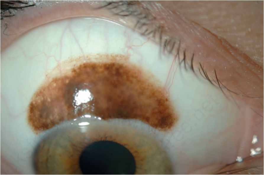
圖 27-26:結膜痣 (conjunctival nevus):上球結膜 (superior bulbar conjunctiva) 上有一色素性病灶。注意病灶內的囊腫 (cysts)。
Fig. 27.26 Conjunctival nevus: there is a pigmented lesion on the superior bulbar conjunctiva. Note the cysts within the lesion.
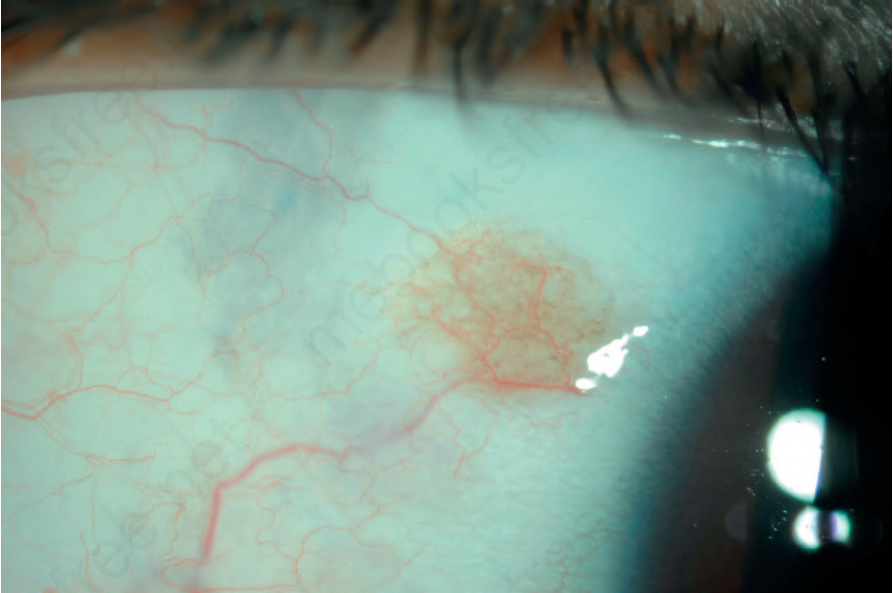
圖 27-28:結膜痣 (conjunctival nevus):球結膜 (bulbar conjunctiva) 上有一界限清楚、輕度色素性、含病灶內囊腫 (intralesional cysts) 的病灶。
Fig. 27.28 Conjunctival nevus: a circumscribed, lightly pigmented lesion with intralesional cysts is on the bulbar conjunctiva.
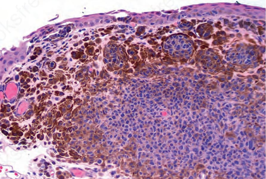
圖 27-29:複合痣 (compound nevus):可見交界巢 (junctional nests) 及固有層 (substantia propria) 受累。
Fig. 27.29 Compound nevus: junctional nests and involvement of the substantia propria is evident.
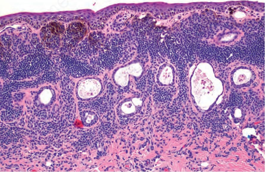
圖 27-30:上皮下痣 (subepithelial nevus):固有層 (substantia propria) 內存在痣細胞,但無交界巢 (junctional nests)。病灶內可見上皮囊腫 (epithelial cysts)。注意淺層痣細胞相較於朝向基底者,其黑色素 (melanin) 色素沉著增加。
Fig. 27.30 Subepithelial nevus: nevus cells are present in the substantia propria without junctional nests. Epithelial cysts are present within the lesion. Note how there is increased melanin pigmentation of the superficial nevus cells compared to those toward the base.
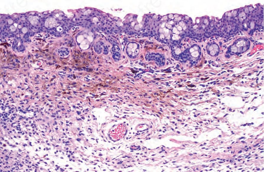
圖 27-31:混合痣 (combined nevus):組織學顯示普通痣細胞 (common nevus cells) 巢以及色素較多的梭形與樹突狀細胞 (spindle and dendritic cells)。
Fig. 27.31 Combined nevus: histology shows nests of common nevus cells and more pigmented spindle and dendritic cells.